Thursday, November 29, 2012

Idiopathic Orbital Inflammatory Syndrome


Orbital inflammatory syndrome (OIS), also known as orbital pseudotumor, is a diagnosis of exclusion when evaluating proptosis. The typical presentation is unilateral, painful proptosis. It is categorized based on region of involvement in the orbit as either diffuse, anterior, lacrimal, myositic, and apical. 

On CECT an enhancing orbital mass is seen.  The mass is hypointense on T1WI and iso- to hyperintense on T2WI. Irregular contrast enhancement is seen with gadolinium. 

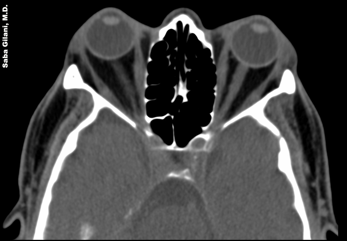
The above image demonstrates an enhancing left orbital apical pseudotumor causing proptosis. This should be differentiated from the imaging appearance of Graves Ophthalmopathy


REFERENCES 
Chaudhry IA, Shamsi FA, Arat YO, et al. Orbital pseudotumor: distinct diagnostic features and management. Middle East Afr J Ophthalmol 2008; 15(1):17-27.
LeBedis CA, Sakai Osamu. Nontraumatic orbital conditions: diagnosis with CT and MR imaging in the emergent setting. Radiographics 2008;28:1741-53.

Wednesday, November 28, 2012

Thoracic Involvement in Amebiasis


  • Amebiasis is an infection due to the protozoan Entamoeba histolytica
  • Most common manifestation is liver abscesses
  • Thoracic involvement is either through direct extension from the liver (most common) or via hematogenous spread
  • Pleural effusions are common, may be an empyema if the effusion is due to a ruptured hepatic abscess
  • Pneumonia with formation of hepatobronchial or bronchobiliary fistula
  • Pericarditis is more common when the left hepatic lobe is involved and can cause cardiac tamponade
  • Invasion of the IVC may cause pulmonary emboli

REFERENCES
Martinez S, Restrepo CS, Carrillo JA, et al. Thoracic manifestations of tropical parasitic infections: a pictorial review. Radiographics 2005;25:135-55.

Tuesday, November 27, 2012

Omental Infarction


The normal appearance of the greater omentum on CT studies is a band of fatty tissue containing vessels that lies anterior to the transverse colon. Omental infarction represents an interruption of the omental arterial supply.  The CT findings of omental infarction are a heterogeneous fatty mass located between the anterior abdominal wall and the colon. Typically, omental infarction occurs in the right abdomen, as in the above case, although in patients with prior abdominal surgery the location is variable.  Associated peri colonic inflammatory changes may be seen.


REFERENCES
Pereira JM, Sirlin CB, Pinto PS, et al. Disproportionate fat standing: a helpful CT sign in patients with acute abdominal pain. Radiographics 2004;24:703-15.

Friday, November 23, 2012

Graves Ophthalmopathy


Graves ophthalmopathy is the most common cause of exophthalmos in adults. The classic imaging finding is enlargement of the extraocular muscles with sparing of their tendinous insertions. These findings tend to be bilateral and symmetric. The above image demonstrates bilateral orbital proptosis and subtle enlargement of the medial and lateral recti muscles bilaterally with sparing of their tendons.

The can be differentiated from orbital pseudotumor which is a cause of unilateral orbital proptosis. In orbital pseudotumor, the extraocular muscles and their tendons are enlarged.


REFERENCES
LeBedis CA and Sakai O. Nontraumatic orbital conditions: diagnosis with CT and MR imaging in the emergent setting. Radiographics 2008;28:1741-53.

Wednesday, November 21, 2012

Serous Atrophy of Bone Marrow

Diffuse serous atrophy of bone marrow occurs in anorexia nervosa, chronic renal insufficiency, thyroid disease, tuberculosis, HIV, and cachexia related to malignancy. It refers to depletion of red and yellow marrow, absence of marrow fat, and accumulation of hyaluronic acid in the marrow spaces. The increased free water content leads to prolonged T1 and T2 relaxation times. T1W images show intermediate marrow signal while T2W images show very high signal intensity. The typical progression of serous atrophy is from the distal extremities to the proximal extremities and axial skeleton.


REFERENCES
Vande Berg BC, Malghem J, Devuyst O, et al. Anorexia nervosa: correlation between MR appearance of bone marrow and severity of disease. Radiology 1994;193:859-64.

Tuesday, November 20, 2012

Splenic Injury in Blunt Abdominal Trauma


The spleen is the most frequently injured organ in blunt abdominal trauma. Injuries can range from subcapsular hematoma to splenic fracture. On imaging studies a subcapsular hematoma will compress the lateral margin of the spleen. A laceration appears as a hypodense linear cleft through the splenic parenchyma. A fracture is a laceration that extends through the splenic hilum, isolating a fragment of splenic tissue.

The above CECT demonstrates a large subcapsular splenic hematoma causing compression and medial deviation of the spleen. A focus of high density within this hematoma represents active contrast extravasation. Angiogram of the splenic artery shows a blush of contrast near the inferior splenic pole which was successfully embolized with Gelfoam.


REFERENCES
Roberts JL, Dalen K, Bosanko CM, et al. CT in abdominal and pelvic trauma. Radiographics 1993;13:735-52.

Monday, November 19, 2012

The Flat Cava Sign

Flattening of the IVC is most commonly described in patients with abdominal trauma leading to hypovolemia and hypoperfusion of the bowel ("shock bowel."). The IVC is considered "flattened" if its AP diameter is less than 9 mm at the level of the renal veins. Associated imaging findings of the "hypovolemia complex" include decreased caliber of the abdominal aorta, increased enhancement of the bowel mucosa, and prolonged intense enhancement of the adrenal glands.

In pediatric patients the constellation of findings seen with post traumatic shock is called the "hypoperfusion complex." Imaging findings include fluid-filled, dilated bowel; increased enhanced of the bowel wall, mesentery, kidneys, pancreas, adrenal glands, aorta, and IVC; decreased caliber of the aorta and IVC. Presence of these findings indicates a poor prognosis.


REFERENCES
Eisenstat RE, Whitford AC, Lane MJ, et al. The "Flat Cava" sign revisited: What is its significance in patients without trauma? AJR Am J Roentgenol 2002;178(1):21-5.
O'Hara SM, Donnelly LF. Intense contrast enhancement of the adrenal glands: another abdominal CT finding  associated with hypoperfusion complex in children. AJR Am J Roentgenol 1999;173(4):995-7.

Friday, November 16, 2012

Meniscal Flounce Revisited


Previously discussed here, meniscal flounce is a normal variant characterized by a single fold along the free edge of a meniscus. By definition it exists in the absence of a meniscal tear. While usually seen in the medial meniscus, the above images demonstrate the finding on the lateral meniscus. 


REFERENCES
Park JS, Kyung NR, Yoon KH. Meniscal flounce on knee MRI: correlation with meniscal locations after positional changes. AJR Am J Roentgenol 2006;187:364-70.
Yu JS, Cosgarea AJ, Kaeding CC. Meniscal flounce MR imaging. Radiology 1997;203:513-5.

Thursday, November 15, 2012

Post Thrombotic Syndrome


Post thrombotic syndrome (PTS) is a late complication of deep venous thrombosis (DVT) characterized by chronic pain, heaviness, and leg swelling in the effected limb. On physical exam edema, telangectasia, and hyperpigmentation may be seen and in severe cases venous ulceration may develop. The pathophysiology is felt to be an interplay between the presence of an acute thrombus and attempts at vein recanalization leading to valvular incompetence in the weeks following a DVT. This leads to venous hypertension which causes edema and ulceration.

Traditionally, conservative management with compression stockings has been the mainstay of therapy in most patients. Newer endovascular techniques such as iliocaval stenting are becoming more popular especially due to poor patient compliance with traditional therapy. 

Endovascular thrombolysis is evolving as a therapy for DVTs with the hope of preventing the development of PTS. Some of the first endovascular methods were catheter directed thrombolysis (CDT) and percutaneous mechanical thrombectomy (PMT) which were determined to have safety limitations precluding their widespread use. Yet a newer technique called pharmacomechanical catheter directed thrombolysis combines CDT and PMT and is currently being studied in the ATTRACT clinical trial. In this method, intravenous thrombolytic agents are administered and combined with mechanical maceration of clot which further disperses the fibrinolytic drug and accelerates thrombolysis. This helps dissolve clot fragments which would otherwise embolize to the lungs. 

The use of endovascular DVT therapy as first line treatment may improve patient outcomes by prevention of development of PTS.


REFERENCES
Kahn SS. The post-thrombotic syndrome: the forbidden morbidity of deep venous thrombosis. J Thromb Thrombolysis 2006;21(1):41-8.
Vedantham S. Deep venous thrombosis: the opportunity at hand. AJR Am J Roentgenol 2009;193(4):922-7.

Wednesday, November 14, 2012

Enlarged Parietal Foramina


Enlarged parietal foramina are considered a benign normal variant in calvarial ossification. They are associated with anomalies of cerebral venous development, variations in occipital cortical infolding, scalp defects, and cleft palate. Although sometimes palpable, these defects are usually discovered incidentally. For patients with prominent foramina, cerebral vascular imaging may be considered for work up of aforementioned anomalies.


REFERENCES
Fink AM and Maixner W. Enlarged parietal foramina: MR imaging features in the fetus and neonate. AJNR 2006;27:1379-81.
Reddy AT, Hedlund GL, Percy AK. Enlarged parietal foramina: association with cerebral venous and cortical anomalies. Neurology 2000;54(5):1175-8.

Tuesday, November 13, 2012

Red Bone Marrow Reconversion

Bone marrow is primarily red (composed of hematopoietic elements) at birth and converts to yellow with skeletal maturation. The two types of marrow differ in their composition: red marrow is approximately 40% water and 40% microscopic fat, while yellow marrow is approximately 80% fat. This lends to the differing MR imaging appearances of red and yellow marrows.

Reversal of a normal marrow conversion pattern (marrow reconversion) can occur with various stressors. Specifically, reconversion occurs in healthy individuals with changing metabolic demands, for example those enlisting in an athletic training program or altitude climbers. Slightly different is the idea of marrow repopulation which happens in patients with a chronic illness such as sickle cell disease or thalassemia. A third cause of increased red marrow in adults is marrow stimulation which occurs in patients who are receiving red cell and/or granulocyte stimulating factors.

The differential diagnosis for increased red marrow in adults includes marrow deposition disease (Gaucher's), myelofibrosis, leukemia and lymphoma.


REFERENCES
Poulton TB, Murphy WD, Durek JL, et al. Bone marrow reconversion in adults who are smokers: MR imaging findings. AJR Am J Roentgenol 1993;161(6):1271-21.
Zajick DC Jr, Morrison WB, Schweitzer ME, et al. Benign and malignant processes: normal values and differentiation with chemical shift MR imaging in vertebral bodies. Radiology 2005;237(2):590-6.

Monday, November 12, 2012

Penetrating Neck Injuries


Penetrating neck injuries are usually evaluated in zones. 
  • Zone I: 
    • extends from the sternal notch to the cricoid cartilage
    • contents - branches of the inominate artery and brachiocephalic veins, branches of subclavian arteries and veins, common carotid and vertebral arteries, trachea, esophagus, thyroid
  • Zone II:
    • extends from the cricoid cartilage to the angle of the mandible
    • contents - common, internal, and external carotid arteries, larynx, upper esophagus, pharynx
    • most commonly surgically explored
  • Zone III: 
    • extends from the angle of the mandible to the base of the skull
    • contents - internal cartoid, vertebral, and branches of the external carotid arteries, internal jugular vein, pharynx
Due to the various vascular structures in each of the zones, CTA is often used to evaluate neck trauma. 

The above images are from a patient with bilateral penetrating neck injuries (note the subcutaneous and intramuscular emphysema). The second image shows the bifurcation of the left common carotid artery (arrowhead). In more cephlad images, the left internal carotid artery is not opacified (arrowhead, third image). Surgical exploration demonstrated a Zone II injury to the left common carotid artery as well as to the left internal jugular vein.


REFERENCES
Nunez DB, Torres-Leon M, Munera F. Vascular injuries of the neck and thoracic inlet: helical CT-angiographic correlation. Radiographics 2004;24:1087-98.
Steenburg SD, Sliker CW, Shanmuganathan K, et al. Imaging evaluation of penetrating neck injuries. Radiographics 2010;30:869-86.

Thursday, November 8, 2012

Lady Windermere Syndrome


An 88 year old Caucasian woman presents to the ER with hemoptysis. CT scan reveals bronchiectasis in the medial segment of the right middle lobe with scattered peripheral nodules. Findings are typical for  Mycobacterium avium intracellulare complex (MAIC). This is an atypical mycobacterial infection seen in elderly females. It is thought that the imaging findings may be due to chronic suppression of a cough leading to retained secretions in the dependent portions of the ventral lung fields.


REFERENCES
Rossi SE, Franquet T, Volpacchio M, et al. Tree-in-bud pattern at thin-section CT of the lungs: radiologic-pathologic overview. Radiographics 2005;25:789-801.

Wednesday, November 7, 2012

Gastrointestinal Manifestations of Behcet Disease


Behcet disease (BD) is an idiopathic vasculitis affecting multiple organs. Involvement of the GI tract is seen in up to 50% of cases. Specific GI tract manifestations include:

  • ulceration: most common finding
    • localized - usually in the ileocecal region
    • diffuse - usually in the colon
    • ulcers are deep, often penetrating to the serosal layer 
    • recur at surgical anastamotic sites
  • thickened mucosal folds in the small bowel
  • increased incidence of fistulas and perforations
    • microperforations are common due to deep, penetrating nature of ulcers

REFERENCES
Chae EJ, Do KH, Seo JB, et al. Radiologic and clinical findings of Behcet disease: comprehensive review of multisystemic involvement. Radiographics 2008;28:e31.
Chung SY, Ha HK, Kim JH, et al. Findings of Behcet syndrome involving the gastrointestinal tract. Radiographics 2001;21:911-24.

Tuesday, November 6, 2012

Lower Extremity Arterial Trauma


A middle aged male presented with a femur fracture sustained after a tree fell on his leg. After closed reduction of the fracture, peripheral pulses were absent. Diagnostic conventional angiogram demonstrated a dissection of the distal superficial femoral artery at the level of the fracture with no distal reconstitution of the popliteal artery.

Imaging recommendations:
  • perform CT angiography in patients who are stable and have suspected (but not confirmed) arterial injury
    • faster than conventional angiography
    • limitations include poor timing of contrast bolus, streak and motion artifact
  • conventional angiography in patients with strong indication of arterial injury
    • prolongs ischemic time 
    • radiologic intervention is possible and preferred over surgical exploration.


REFERENCES
Miller-Thomas MM, West OC, Cohen AM. Diagnosing traumatic arterial injury in the extremities with CT angiography: pearls and pitfalls. Radiographics 2005;25:S133-42.
Rieger M, Mallouhi A, Tauscher T, et al. Traumatic arterial injuries of the extremities: initial evaluation with MDCT angiography. AJR Am J Roentgenol 2006;186(3):656-64.

Monday, November 5, 2012

Menetrier Disease


  • rare, idiopathic hypertrophic gastropathy
  • patients usually present with epigastric pain and hypoalbuminemia (from loss of albumin into the gastric lumen)
  • bimodal age distribution
    • childhood form thought to be linked to cytomegalovirus infection
  • diagnosis is made by combination of endoscopic and radiologic findings
    • Upper GI series: thickened, lobulated folds in the gastric fundus which trap barium; hypersecretion may dilute barium which may prevent mucosal coating
    • CECT: thickening of mucosa and submucosa which projects into the gastric lumen
  • differential diagnosis
    • Zollinger Ellison Syndrome - rugal fold thickening is not as pronounced, gastric ulcers, pancreatic involvement
    • Gastritis - thickened folds usually in the antrum, H. pylori positive
    • Lymphoma - thickened folds due to soft tissue masses not due to edema

REFERENCES
Friedman J, Platnick J, Farruggia S, et al. Menetrier Disease. Radiographics 2009;29:297-301.
Kanne JP, 

Friday, November 2, 2012

Gonadal Vein Thrombosis


Gonadal vein thrombosis can be a cause of acute pelvic pain. It is most commonly seen in post partum females but may also present in patients after pelvic surgery, pelvic trauma, and pelvic inflammatory disease. There is a predilection for this finding on the right side as retrograde flow through the left gonadal vein prevents stasis. 

On CECT, an enlarged gonadal vein that parallels the course of the psoas muscles with an enhancing wall and an intraluminal filling defect will be seen. Perivenous inflammatory changes may be seen. The patient above is post cesarean section. A filling defect is seen in the right gonadal vein (adjacent to the IVC) consistent with thombosis.


REFERENCES
Bennett GL, Slywotzky CM, Giovanniello G. Gynecologic causes of acute pelvic pain: spectrum of CT findings. Radiographics 2002;22:785-801.

Thursday, November 1, 2012

AV Fistula Planning

In patients with end stage renal disease (ESRD), the ideal venous access for hemodialysis should fulfill three requirements: 1. indefinite life, 2. high flow, 3. no complications such as thrombosis or aneurysm formation. The native arteriovenous (AV) fistula comes closest to fulfilling these requirements and is the best option for venous access. 

In planning for creation of an AV fistula several factors have to be taken into considerations including the patient's vascular history. Patients who have had prior central venous catheters, pacemakers may be poor candidates for AV fistula creation because these procedures are associated with venous stenosis.


REFERENCES
Patel AA, Tuite CM, and Trerotola SO. K/DOQI guidelines: what should an interventionalist know? Semin Intervent Radiol 2004;21(2):119-24.