Wednesday, December 23, 2015

Management of Pancreatic Cystic Lesions

Ryan Schwope
Thick slab MRCP image showing massive dilatation of the main pancreatic duct
Incidental cystic pancreatic lesions found on 13% of MRI abdomens
  • Wide variety of pathology both benign and malignant
  • Imaging findings and demographics are the key to diagnosis

Cystic Pancreatic Neoplasms (Four major categories)

  1. Serous cystadenoma: Benign (very low malignant potential)
  2. Mucinous cystic neoplasm (MCN): Premalignant or malignant
  3. Intraductal papillary mucinous neoplasm (IPMN): Malignant potential (Main Duct >> Branch Duct)
  4. Unusual cystic neoplasms:
    • Solid pseudopapillary neoplasm (SPN): Low grade malignancy
    • Cystic forms of more common neoplasms (neuroendocrine)

Nonneoplastic Pancreatic Cysts

  • Pseudocyst
  • Retention cyst
  • Lymphoepithelial cyst
  • Localized ductectasia

Major Imaging Features Guiding Management

  • Number and size of cystic components: Risk of malignancy increases when size ≥ 3 cm
  • Septations and solid components: Mural nodule has a 87% Sp and 56% Sn for malignancy
  • Main pancreatic duct (MPD) dilatation and communication with the cystic lesion: MPD > 10 mm has a 77% Sp and 67-92% Sn for malignancy

Sendai Criteria

High Risk Stigmata
  • Jaundice
  • MPD ≥ 10 mm
  • Enhancing solid component
Worrisome Features
  • Size ≥ 3 cm
  • MPD 5-9 mm
  • Non-enhancing mural nodules
  • Thick enhancing cystic wall
  • Lymphadenopathy
  • Abrupt Duct Termination

Management

  • Any worrisome features present = Endoscopic Ultrasound (EUS) and Cyst aspiration with fluid analysis
  • Any high risk stigmata present or suspicious cytology on EUS = Surgical resection
  • MCN or SPN = Surgical resection
  • Serous cystadenoma
    • 2-3 cm: F/U every 2 years
    • ≥ 4 cm: consider resection
  • IPMN:
    • Main duct and combined type: Surgical resection (but depends on location, pt. age/clinical status)
    • Branch duct type = follow if < 3 cm and contains no solid components
      • If < 2cm F/U q1yr; if growth FU q6mo
      • If 2-3 cm F/U q6mo x 2 years, then q1yr
      • Consider EUS (if mucinous then resect)
      • If growth ≥ 3 cm, resect

What the Clinician/Surgeon wants to know

  1. Number of cystic lesions
  2. Largest cystic lesion
    • Unilocular
    • Multilocular: Microcystic (<2cm) or Macrocystic (> 2cm)
  3. Lesion size
  4. Lesion location: Head/Body/Tail
  5. Septations: None/Thin/Thick (> 2mm)
  6. Solid components: Present/Absent
  7. Calcifications: None/Coarse/Rim/Central
  8. Communication with MPD: Present/Absent
  9. Main pancreatic duct diameter: > 5 mm/Not dilated

References

Wednesday, December 9, 2015

Bisphosphonate-related Osteonecrosis of the Jaw


Ryan Schwope BROJN1
Figure 1: Axial CT of the facial bones. There is a mixed sclerotic and lytic lesion within the mandible (Figure 1, arrows) with foci of cortical interruption (Figure 2, arrowheads). 

Ryan Schwope BROJN 2
Figure 2: Sagital CT of the facial bones. There is a mixed sclerotic and lytic lesion within the mandible (Figure 1, arrows) with foci of cortical interruption (Figure 2, arrowheads).

Bisphosphonate-related osteonecrosis of the jaw (BRONJ) is associated with the use of bisphosphonates to treat severe osteoporosis, and metabolic and oncologic bone conditions, including hypercalcemia associated with malignancy. These agents inhibit osteoclasts, reducing bone resorption and osteolysis, and also possess antiangiogenic properties, reducing blood flow and necrosis. Patients with BRONJ present with pain and exposed, nonvital bone involving the maxillofacial structures. The incidence of BRONJ increases with the duration of treatment, especially with the intravenous route and concomitant steroid therapy. The osteonecrosis usually is participated by dental extraction. When spontaneous, it commonly occurs along the mylohyoid ridge. BRONJ can mimic osteomyelitis and osteoradionecrosis. A history of dental caries and the presence of periosteal elevation can help direct one towards the diagnosis of osteomyelitis. Osteoradionecrosis can be excluded if the patient has not received oropharyngeal radiation therapy. Jaw neoplasm, primary or metastatic, can potentially also mimic BRONJ. Treatment of BRONJ consists of cessation of bisphosphonate drug therapy, antibiotics for secondary infection, and surgical debridement of necrotic sequestra.

References:


Thursday, November 19, 2015

Lipoleiomyoma


Ryan Schwope
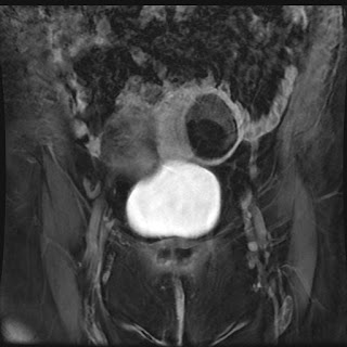
CT shows a predominantly fat attenuation, intramural uterine mass. There is rim of surrounding calcification as well as a few foci of internal soft tissue density at the superior aspect of the mass 

Ryan Schwope
   MR imaging showed the majority of the uterine mass as hyperintense on T1 weighted imaging (isointense to subcutaneous fat)
Ryan Schwope
T2 weighted image with fat saturation MRI shows the uterine mass markedly hypointense (isointense to subcutaneous fat)
Ryan Schwope
Post contrast T1 weighted MRI image with fat saturation show mld enhancement of the soft tissue component  along the superior margin of the mass; the majority of the mass is markedly hypointense (isointense to subcutaneous fat)
Uterine lipoleiomyomas are rare, benign tumors with a variable reported incidence ranging from 0.03% to 0.2%. The exact etiology of these lesions is unclear.  It is postulated lipoleiomyomas either arise from fatty metaplasia of the smooth muscle cells of leiomyomas, or from misplaced embryonic fat cells in the uterus. CT is highly specific for the diagnosis when an intrauterine mass is seen containing both macroscopic fat and soft tissue density. MR can also be confirmatory as the mass will have high T1 weighted signal which can be confirmed as fat by using a fat suppression. The role of imaging is also to differentiate lipoleiomyoma from an ovarian teratoma, a much more common entity presenting as a fat-containing pelvic mass. Lipoleiomyomas require no treatment or follow-up whereas teratomas are frequently resected. 


References


Monday, September 28, 2015

Giant Cell Containing Lesions of Bone

Reactive Benign Malignant
  • Osteosarcoma
  • Clear cell chondrosarcoma
  • Metastatic carcinoma
  • Malignant fibrous histiocytoma
  • Solitary fibrous tumor metastasis

Saturday, June 20, 2015

ILAR Classification of Juvenile Idiopathic Arthritis

Juvenile idiopathic arthritis (JIA) is an umbrella term for a group of abnormalities characterized by chronic articular inflammation and association with HLA alleles. The International League Against Rheumatism (ILAR) has classified JIA into seven subtypes, including an unclassifiable group. These include:
  • Systemic arthritis: Arthritis in one or more joints with or preceded by fever of at least 2 weeks' duration documented to be daily for at least 3 days, and accompanied by one or more of: evanescent erythematous rash, lymphadenopathy, hepatomegalyor splenomegaly, or both, serositis
  • Oligoarthritis: Arthritis in 4 or fewer joints in the first 6 months. Subtypes include persistent (no more than 4 joints throughout the course of the disease) and extended (more than 4 joints after the first 6 months).
  • Polyarthritis, RF negative: Arthritis affecting 5 or more joints in the first 6 months of disease. RF is negative
  • Polyarthritis, RF positive: Arthritis affecting 5 or more joints in the first 6 months of disease. Two or more tests for RF, conducted at least 3 months apart during the first 6 months, are positive. Considered the pediatric version of adult rheumatoid arthritis.
  • Psoriatic: Arthritis plus psoriasis OR Arthritis plus at least two of the following: dactylitis, nail pitting or onycholysis, psoriasis in a first-degree relative.
  • Enthesis-related: Arthritis plus enthesitis OR Arthritis or enthesitis, plus at least two of the following: presence of or a history of sacroiliac joint tenderness and/or inflammatory lumbosacral pain‡, presence of HLA-B27 antigen, onset of arthritis in a male over 6 years of age, acute (symptomatic) anterior uveitis, history of AS, ERA, sacroiliitis with IBD, reactive arthritis, or acute anterior uveitis in a first-degree relative
  • Unclassified: Arthritis that fulfills criteria in none of the above categories, or fulfills criteria in two or more of the above categories

References

Petty RE, Southwood TR, Baum J, Bhettay E, Glass DN, Manners P, Maldonado-Cocco J, Suarez-Almazor M, Orozco-Alcala J, Prieur AM. Revision of the proposed classification criteria for juvenile idiopathic arthritis: Durban, 1997. J Rheumatol. 1998 Oct;25(10):1991-4.

Sunday, January 25, 2015

Subcutaneous Panniculitis-Like T-Cell Lymphoma


Subcutaneous panniculitis-like T-cell lymphoma is a rare cutaneous T-cell lymphoma (SPTCL) that is characterized by subcutaneous nodules and plaques. The similarity of presentation to that of panniculitis is the reason for the long name. The disease most commonly involves the legs, but can also affect the trunk. It can be differentiated from the cutaneous T-cell lymphomas (e.g., mycosis fungoides and Sézary syndrome) by the fact that the former are cutaneous, and SPTCL is subcutaneous.

References

Levine BD, Seeger LL, James AW, Motamedi K. Subcutaneous panniculitis-like T-cell lymphoma: MRI features and literature review. Skeletal Radiol. 2014 Sep;43(9):1307-11.

Friday, January 16, 2015

Costochondritis

Costochondritis, as its implies, is a nonspecific term for inflammation of the costal cartilage. The problem with this definition is that it implies a non-specific process, where inflammation can be caused by any number of pathological processes, including trauma, infection, or radiation.

The clinical entity of costochondritis, on the other hand, has specific features:
  • Self-limited
  • Usually multiple affected levels
  • Can occur at the costochondral junction or at the chondrosternal joints
  • No swelling or induration on physical examination
  • Pain that is reproduced by palpation and may radiate on the chest wall
This definition differentiates costochondritis from the much rarer Tietze syndrome (TS). In TS, the inflammatory process causes visible enlargement of the costochondral junction(s) and is most commonly isolated to a single rib (usually the 2nd, but can also involve the 3rd rib). TS can be caused by infectious, rheumatologic, and neoplastic processes. The infection is usually associated with trauma (e.g., stab wounds, iatrogenic) or with intravenous drug use.

Costochondritis is relatively common, and can be seen is as many as 14% of adolescents and 13 to 36% of adults with chest pain.

References

Proulx AM, Zryd TW. Costochondritis: diagnosis and treatment. Am Fam Physician. 2009 15;80(6):617-20.