Showing posts with label Interventional radiology. Show all posts

Thursday, March 1, 2018

Persistent Sciatic Artery

Axial and coronal CT images with intravenous contrast demonstrate a left persistent sciatic artery (long arrows) coursing through the greater sciatic notch and deep to the gluteus maximus muscle. Note the asymmetrically diminutive left external iliac artery (short arrows)

  • A persistent sciatic artery (PSA) is a rare vascular anomaly in which the internal iliac artery courses through the greater sciatic notch and in to the thigh
    • During embryonic development, the sciatic artery usually involutes as the femoral artery develops
    • Recognition of an unusually enlarged internal iliac artery and a diminutive external iliac artery are some clues in diagnosing a PSA
    • The PSA course runs close to the sciatic nerve, and can run within the nerve sheath in some cases
    • Distally, the PSA runs deep to the gluteus maximus muscle, coursing along the adductor magnus muscle
  • Reported in up to 0.03-0.06% of the population and can be bilateral in 20% of cases
  • PSA is considered complete when it is the dominant blood supply to the popliteal artery and incomplete with the femoral artery is the dominant blood supply to the popliteal artery
  • 5 types have been described:
    • type 1 is a complete PSA with a normal femoral artery
    • type 2 is a complete PSA with a incompletely developed femoral artery
    • type 3 is a incomplete PSA (only the cephalic portion is present) and normal femoral artery
    • type 4 is a incomplete PSA (only the caudal portion is present) and normal femoral artery
    • type 5 is when the PSA arises from the median sacral artery
  • Majority (80%) become symptomatic at some point presenting with intermittent claudication, ischemia, pulsatile mass or neurologic symptoms
  • Susceptible to repetitive trauma from sitting and hip flexion/extension
    • Results in premature atherosclerosis and aneurysm formation
    • Aneurysm found in 48%, and stenosis and occlusion of the PSA in 7% and 9%, respectively
References:
  1. Mcquaid M, Gavant ML. Posttraumatic pseudoaneurysm of a persistent sciatic artery. AJR Am J Roentgenol. 1995;164 (6): 1514-5. 
  2. Pillet J, Albaret P, Toulemonde JL, Cronier P, Raimbeau G, Chevalier JM. Tronc arteriel ischiopoplite, persistance de l’artere axiale. Bull Assoc Anat 1980;64:109e22.
  3. Pillet J, Cronier P, Mercier Ph, Chevalier JM. The ischio popliteal arterial trunk: a report of two cases. Anat Clin 1982; 3:329e31. 
  4. Gauffre S, Lasjaunias P, Zerah M. Sciatic artery: a case, review of literature and attempt of systematization. Surg Radiol Anat 1994;16(1):105e9.
  5. Bower EB, Smullens SN, Parke WW. Clinical aspect of persis- tent sciatic artery: report of two cases and review of the literature. Surgery 1977;81(5):588e95.

Monday, April 10, 2017

False Perpetuations: Main Portal Vein Size and Portal Hypertension

Perpetuation: A main portal vein (MPV) diameter >13 mm is "consistent with portal hypertension" (pHTN)

This cutoff of 13 mm is based on weak literature (mainly from the 1980's), some of which did not include comparison values of normal patients

  • One comparative study using ultrasound found (Radiology 1982; 142: 167-172):
    • In 79 patients with pHTN
      • 36 had a MPV diameter of <13 mm 
      • 33 had a MPV diameter >/= 13 mm
      • The MPV was not visualized in 10 patients
    • In the 45 control patients
      • The MPV diameter was < 13 mm in 41 cases
      • The MPV was not visualized in 4 patients. 

More recent studies have found that there is no significant difference in MPV diameters when comparing patients without cirrhosis to patients with cirrhosis, and the normal MPV diameter is significantly larger than the 13 mm cutoff

  • A study (Eur J Gastroenterol Hepatol 2004; 16:147-155) from King's College using ultrasound (49 controls and 14 cirrhotics) found: 
    • the average MPV diameters were 9.6 cm and 10.8 cm in patients without and with cirrhosis, respectively.
  • A second study (JCAT 2008; 32: 198-203) from UCSF using CT (59 controls and 67 cirrhotics) found:
    • The average MPV diameters were 14.5 cm and 14.8 cm in patients without and with cirrhosis, respectively.
  • Using CT, the MPVs in healthy renal donor patients were measured before and after the administration of intravenous contrast, and in the axial and coronal planes (Abdom Radiol 2016; 41:1931-1936). This study found:
    • The average MPV diameter was 15.5 +/- 1.9 mm
      • This value was significantly different than 13 mm
    • Post-contrast MPVs were 0.56 mm larger compared to non-contrast
    • A positive correlation between BMI and height versus MPV diameter
In fact, the MPV size can be reduced in portal hypertension and has been described as a sign of hepatofugal MPV flow (AJR 2003; 181: 1629-1633). This study found:
  • A MPV diameter of less than 1 cm is a highly sensitive (but not very specific) for MPV flow reversal in patients with cirrhosis

Sunday, March 19, 2017

Chondroblastoma

X-ray (radiograph), CT, and MRI of chondroblastoma of the femur.

General

  • Terminology: “giant cell variant” (1927) → epiphyseal chondromatous giant cell tumor → calcifying giant cell tumor → chondroblastoma
  • 1% to 2% of all primary bone tumors
  • 9% of all benign bone tumors
  • Mean age of 15-18 years
  • M >>F
  • Mean duration of symptoms 8.7 months
  • Trivia: Most common benign neoplasm of the patella

Imaging Features

  • Epiphysis/apophysis +/- metaphyseal/diaphyseal involvement
  • Metaphyseal/diaphyseal occurrence without epiphyseal/apophyseal involvement exceptionally rare
  • Proximal tibia >> proximal femur > distal femur > proximal humerus
  • Well-defined, sclerotic margins on radiographs
  • Can involve the cortex, resulting in expansion, thinning, or disruption.
  • Stippled matrix calcification seen in minority of cases
  • Periosteal reaction seen in majority of cases
  • Extensive peri-lesional edema on MRI is common
  • Homogeneously hypointense on T1
  • Variable on T2: can be diffusely hypointense, or have small cystic areas of increased T2 signal or fluid-fluid levels
  • Heterogeneous and moderate enhancement in solid portions. Less commonly, homogeneous and marked enhancement

Differential Diagnosis

Management/Prognosis

  • Curettage or resection
  • RFA (small lesions, small series, not common)
  • Local recurrence rate: 5.0% after curettage
  • Local recurrence rate: 0% after resection
  • Recurrence most frequent in the proximal humerus
  • Malignant transformation and benign pulmonary metastases extremely rare

References

Wednesday, January 6, 2016

Endologix Stent-graft

Ryan Schwope
Arterial phase CT shows smoothly marginated contrast extending beyond the confines of the metal struts of the stent
Ryan Schwope
Delayed CT shows the smoothly marginated contrast extending beyond the confines of the metal strut unchanged in size and configuration when compared to arterial phase imaging
Most endostent devices used for endovascular repair of aortic aneurysms have the graft material sutured on the luminal side of the stent. The AFX endostent device (Endologix, Irvine, California) is different from other devices in that the graft material is sutured on the outside of the metal component, attached only at the proximal and distal ends.

By intent, the graft material of the AFX stent-graft can separate from the metal struts where it is not directly apposed to the aortic wall.  During surveillance with contrast-enhanced imaging of patients who have reviewed this device, contrast can be detected outside of the metallic construct. Although this finding can imply a type 1 or 3 endoleak when identified with other endostents, it is a normal finding with the AFX device.

Avoiding misinterpretation as an endoleak can be achieved by confirmation of the type of endostent device used for aneurysm repair. In addition, the contrast extending beyond the metal struts does not change in configuration or size on delayed imaging.

References:

J Vasc Interv Radiol. 2012 Nov;23(11):1544-6.

Thursday, February 21, 2013

To Stent or Not to Stent?


The above CECT axial images are part of a trauma work up in a young patient involved in an MVA with multiple abdominal injuries. Several liver lacerations are seen with subtle active extravasation of contrast. The third axial image shows a focal dissection at the origin of the celiac artery (red arrow). Sagittal reconstruction demonstrates absence of the celiac origin (the SMA origin is seen). Conventional angiogram shows a dissection at the origin of the celiac artery with opacification of its distal branches.

Typically, this dissection would be treated with a stent requiring short term anti-coagulation therapy (at least 6 months). However, a patient with multiple liver lacerations is not a candidate for anti-coagulation. Thus, despite injury to a major abdominal visceral artery, no intervention could be performed. The patient was monitored for hemodynamic stability and received several units of packed RBCs with no further drop in hematocrit.

Tuesday, January 22, 2013

Uterine Artery Embolization: Complications

Uterine artery embolization (UAE) is increasingly performed as a minimally invasive alternative to hysterectomies and myomectomies in women with leiomyomas. As with any procedure, there are associated complications which include:
  1. Fibroid passage: usually 3-6 months after procedure
  2. Pulmonary embolus: most common cause of death after UAE
  3. Deep venous thrombus: usually involving pelvic veins
  4. Infectious disease: endometritis (most common), pelvic inflammatory disease, pyometria
  5. Inadvertent embolization of adjacent organs or of a malignant leiomyosarcoma
  6. Ovarian dysfunction due to inadvertent embolization of the ovarian artery

REFERENCES
Kandarpa K and Machan L. Handbook of Interventional Radiologic Procedures. 4th ed.
Kitamura Y, Ascher SM, Cooper C, et al. Imaging manifestations of complications associated with uterine artery embolization. Radiographics 2005;25:S119-32.

Monday, January 14, 2013

Vertebral Artery Transection


The unfortunate consequence of a rapid acceleration-deceleration injury. Sagittal image shows dissociation of the cervical spine at C6-C7. Coronal reformatted image from the CTA demonstrates loss of the right vertebral artery from it's origin to the level of C5 with presumable retrograde filling of its distal portion via collaterals. 

Thursday, December 27, 2012

Endovascular Management of TIPS-related Hepatic Encephalopathy

Hepatic encephalopathy is a common complication that occurs after creation of a TIPS shunt. It is typically managed conservatively with modifications in diet and medication. In patients who do not respond to conservative management, liver transplantation may be an option. Some endovascular techniques aimed at reducing the amount of blood shunted away from the liver may also be employed.
  • shunt occlusion via embolic agents
    • has risk of variceal rebleeding
    • sudden changes in hemodynamics (cardiac output, hypotension, metabolic acidosis) can be fatal
    • reversible shunt occlusion using short term balloon occlusion of the TIPS can prevent complications from variceal rebleeding (occlusive balloon can be deflated if life threatening bleeding occurs and thus TIPS can be reopened)
  • shunt reduction
    • using constrained stents to reduce the lumen of the shunt
    • makes it difficult to regulate blood flow through the shunt, especially around the constrained portions
      • adjunct embolization of the dead space around the shunt can be performed
      • use of constrained covered stent grafts has allowed better control of flow through the shunt while reducing its lumen
  • retrograde embolization of a splenorenal shunt with ethanolamine oleate
    • can only be done when a spontaneous splenorenal shunt is present
    • maintains patency of TIPS
    • severe renal dysfunction and pulmonary edema are among some of the side effects of ethanolamine usage
REFERENCES
Madoff DC, Wallace MJ, Ahrar K, et al. TIPS-related hepatic encephalopathy: management options with novel endovascular techniques. Radiographics 2004;24:21-36.

Thursday, December 20, 2012

Mesenteric Hematoma Revisited


The above CECT images show hazy stranding of the mesentery in a patient with blunt abdominal trauma. Findings are consistent with a mesenteric hematoma, previously discussed here.

Friday, December 14, 2012

Dialysis Catheter Placement

The Kidney Disease Outcomes Quality Initiative (K/DOQI) outlines vascular access management for hemodialysis patients. Of these guidelines, the following should be taken into consideration by interventional radiologists involved in the placing catheters for access in hemodialysis patients

  1. patient evaluation including history of prior central venous catheters, pacemakers, prior vascular access failure, coagulation disorders, valvular heart disease
  2. tunneled catheters should be placed when dialysis access is needed for greater than 3 weeks
  3. right internal jugular vein access is preferred with the tip of the catheter in the right atrium
  4. catheter related infections should be managed as follows:
    • catheter site infection with negative blood cultures - treat with topical antibiotics; do not remove catheter unless there is no response to topical treatment in which case catheter should be exchanged over a wire to preserve venous access
    • bacteremia - treat with systemic antibiotics appropriate for cultured organism; if patient is clinically unstable remove the catheter; if the patient is clinically stable the catheter should only be removed if the patient is symptomatic (fever, elevated WBC) after 36 hours of treatment with antibiotics

REFERENCES
Patel AA, Tuite CM, Trerotola SA. K/DOQI Guidelines: what should an interventionalist know? Semin Intervent Radiol 2004;21(2):119-24.

Thursday, December 13, 2012

Traumatic Fracture of an Axillofemoral Bypass Graft


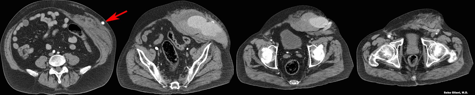
A man presented to the emergency room after a motor vehicle accident. CECT showed an axillofemoral bypass graft (red arrow) that was fractured due to blunt trauma (last image) with an expanding anterior abdominal wall hematoma and active extravasation of contrast (higher density in second and third images).

Tuesday, December 4, 2012

Endovascular Management of Placenta Accreta


The placenta accreta spectrum involves abnormal placental implantation. Placenta accreta is implantation of the chorionic villi in the myometrium while increta and percreta refer to implantation through the myometrium and in the serosa respectively. The incidence of placenta accreta is one in 2500 deliveries. Prior history of cesarean section and advanced maternal age are considered risk factors. Placenta accreta is associated with massive blood loss during delivery which could be fatal.

Techniques to minimize intraoperative blood loss focus on reducing arterial flow to the uterus and include extraluminal ligation of the pelvic arteries or endovascular occlusion of the iliac arteries with or without embolization.

Endovascular iliac artery occlusion is performed prior to planned cesarean section and involves puncture of the bilateral femoral arteries with placement of balloon catheters in each internal iliac artery, preferably in its anterior trunk. After the umbilical cord is clamped, the balloons are inflated and remain so until skin closure, occluding the internal iliac arteries and reducing blood flow to the uterus. If endovascular balloon occlusion is not successful at controlling intraoperative hemorrhage, embolization with gelatin sponge particles can be performed. The latter is associated with greater radiation doses to the fetus.

To present date, there are few studies (each with small sample sizes) showing the efficacy of endovascular balloon occlusion techniques in the perioperative management of placenta accreta. However, the existing data demonstrates it to be a safe technique to prevent blood loss in these patients.


REFERENCES
Tan CH, Tay KH, Sheah K, et al. Perioperative endovascular internal iliac artery occlusion balloon placement in the management of placenta accreta. AJR Am J Roentgenol 2007;189:1158-63.

Tuesday, November 20, 2012

Splenic Injury in Blunt Abdominal Trauma


The spleen is the most frequently injured organ in blunt abdominal trauma. Injuries can range from subcapsular hematoma to splenic fracture. On imaging studies a subcapsular hematoma will compress the lateral margin of the spleen. A laceration appears as a hypodense linear cleft through the splenic parenchyma. A fracture is a laceration that extends through the splenic hilum, isolating a fragment of splenic tissue.

The above CECT demonstrates a large subcapsular splenic hematoma causing compression and medial deviation of the spleen. A focus of high density within this hematoma represents active contrast extravasation. Angiogram of the splenic artery shows a blush of contrast near the inferior splenic pole which was successfully embolized with Gelfoam.


REFERENCES
Roberts JL, Dalen K, Bosanko CM, et al. CT in abdominal and pelvic trauma. Radiographics 1993;13:735-52.

Thursday, November 15, 2012

Post Thrombotic Syndrome


Post thrombotic syndrome (PTS) is a late complication of deep venous thrombosis (DVT) characterized by chronic pain, heaviness, and leg swelling in the effected limb. On physical exam edema, telangectasia, and hyperpigmentation may be seen and in severe cases venous ulceration may develop. The pathophysiology is felt to be an interplay between the presence of an acute thrombus and attempts at vein recanalization leading to valvular incompetence in the weeks following a DVT. This leads to venous hypertension which causes edema and ulceration.

Traditionally, conservative management with compression stockings has been the mainstay of therapy in most patients. Newer endovascular techniques such as iliocaval stenting are becoming more popular especially due to poor patient compliance with traditional therapy. 

Endovascular thrombolysis is evolving as a therapy for DVTs with the hope of preventing the development of PTS. Some of the first endovascular methods were catheter directed thrombolysis (CDT) and percutaneous mechanical thrombectomy (PMT) which were determined to have safety limitations precluding their widespread use. Yet a newer technique called pharmacomechanical catheter directed thrombolysis combines CDT and PMT and is currently being studied in the ATTRACT clinical trial. In this method, intravenous thrombolytic agents are administered and combined with mechanical maceration of clot which further disperses the fibrinolytic drug and accelerates thrombolysis. This helps dissolve clot fragments which would otherwise embolize to the lungs. 

The use of endovascular DVT therapy as first line treatment may improve patient outcomes by prevention of development of PTS.


REFERENCES
Kahn SS. The post-thrombotic syndrome: the forbidden morbidity of deep venous thrombosis. J Thromb Thrombolysis 2006;21(1):41-8.
Vedantham S. Deep venous thrombosis: the opportunity at hand. AJR Am J Roentgenol 2009;193(4):922-7.

Monday, November 12, 2012

Penetrating Neck Injuries


Penetrating neck injuries are usually evaluated in zones. 
  • Zone I: 
    • extends from the sternal notch to the cricoid cartilage
    • contents - branches of the inominate artery and brachiocephalic veins, branches of subclavian arteries and veins, common carotid and vertebral arteries, trachea, esophagus, thyroid
  • Zone II:
    • extends from the cricoid cartilage to the angle of the mandible
    • contents - common, internal, and external carotid arteries, larynx, upper esophagus, pharynx
    • most commonly surgically explored
  • Zone III: 
    • extends from the angle of the mandible to the base of the skull
    • contents - internal cartoid, vertebral, and branches of the external carotid arteries, internal jugular vein, pharynx
Due to the various vascular structures in each of the zones, CTA is often used to evaluate neck trauma. 

The above images are from a patient with bilateral penetrating neck injuries (note the subcutaneous and intramuscular emphysema). The second image shows the bifurcation of the left common carotid artery (arrowhead). In more cephlad images, the left internal carotid artery is not opacified (arrowhead, third image). Surgical exploration demonstrated a Zone II injury to the left common carotid artery as well as to the left internal jugular vein.


REFERENCES
Nunez DB, Torres-Leon M, Munera F. Vascular injuries of the neck and thoracic inlet: helical CT-angiographic correlation. Radiographics 2004;24:1087-98.
Steenburg SD, Sliker CW, Shanmuganathan K, et al. Imaging evaluation of penetrating neck injuries. Radiographics 2010;30:869-86.

Tuesday, November 6, 2012

Lower Extremity Arterial Trauma


A middle aged male presented with a femur fracture sustained after a tree fell on his leg. After closed reduction of the fracture, peripheral pulses were absent. Diagnostic conventional angiogram demonstrated a dissection of the distal superficial femoral artery at the level of the fracture with no distal reconstitution of the popliteal artery.

Imaging recommendations:
  • perform CT angiography in patients who are stable and have suspected (but not confirmed) arterial injury
    • faster than conventional angiography
    • limitations include poor timing of contrast bolus, streak and motion artifact
  • conventional angiography in patients with strong indication of arterial injury
    • prolongs ischemic time 
    • radiologic intervention is possible and preferred over surgical exploration.


REFERENCES
Miller-Thomas MM, West OC, Cohen AM. Diagnosing traumatic arterial injury in the extremities with CT angiography: pearls and pitfalls. Radiographics 2005;25:S133-42.
Rieger M, Mallouhi A, Tauscher T, et al. Traumatic arterial injuries of the extremities: initial evaluation with MDCT angiography. AJR Am J Roentgenol 2006;186(3):656-64.

Thursday, November 1, 2012

AV Fistula Planning

In patients with end stage renal disease (ESRD), the ideal venous access for hemodialysis should fulfill three requirements: 1. indefinite life, 2. high flow, 3. no complications such as thrombosis or aneurysm formation. The native arteriovenous (AV) fistula comes closest to fulfilling these requirements and is the best option for venous access. 

In planning for creation of an AV fistula several factors have to be taken into considerations including the patient's vascular history. Patients who have had prior central venous catheters, pacemakers may be poor candidates for AV fistula creation because these procedures are associated with venous stenosis.


REFERENCES
Patel AA, Tuite CM, and Trerotola SO. K/DOQI guidelines: what should an interventionalist know? Semin Intervent Radiol 2004;21(2):119-24.

Wednesday, October 10, 2012

Sclerotherapy for Treatment of Anuerysmal Bone Cysts

Aneurysmal bone cysts (ABCs) are benign expansile lytic bone lesions of unknown origin that occur in the pediatric population. Characteristics of ABCs were previously discussed here.

Typically, the treatment of ABCs involves resection and curettage with or without bone grafting. However, there is a high recurrence rate reported with this treatment method with some studies reporting up to a recurrence rate of greater than 50%.

Surgical resection of ABCs is less favored as an extensive resection may be necessary and may immobilize the patient for a prolonged period. Furthermore, as ABCs are highly vascular lesions, surgical procedures carry the risk of significant blood loss.

Radiotherapy was previously used to treat ABCs but has since been abandoned due to studies demonstrating malignant transformation of the lesions and development of secondary tumors.

The traditional interventional radiology technique of embolization has been used effectively in the treatment of some ABCs. Preoperative selective embolization of the feeding vessel is used to reduce intraoperative blood loss as well as to treat lesions that are surgically inaccessible. Challenges of this therapy include risk of particle embolization to the vertebrobasilar and spinal arterial systems resulting in ischemia to vital structures. Additionally, not all ABCs have a feeding vessel that can be selectively embolized. 

More recently, studies have shown intralesional sclerotherapy to be an effective treatment for ABCs. Sclerosing agents act by damaging the endothelial lining which leads to activation of the coagulation cascade and thrombotic vascular occlusion. Agents that have been used for sclerotherapy include ethanol based solutions, methylprednisone, calcitonin, and doxycycline. Patients usually require several injections of the agent. Post procedure imaging typically shows sclerosis of the lesion with long term complete ossification. Patients have reported a relief of symptoms and low recurrence rates have been reported. This data suggests that image guided sclerotherapy of ABCs may be a safe, minimally invasive method of treating these lesions. 


REFERENCES
Rai AT and Collins JJ. Percutaneous treatment of pediatric aneurysmal bone cyst at C1: a minimally invasive alternative: a case report. AJNR Am J Neuroradiol 2005;26:30-3.
Rastogi S, Varshney MK, Trikha V, et al. Treatment of aneurysmal bone cysts with percutaneous sclerotherapy using polidocanol. J Bone Joint Surg [Br] 2006;88-B:1212-6.


Monday, October 8, 2012

Phantom Calyx


A phantom renal calyx refers to a calyx that does not opacify after administration of contrast due to obstruction or stricture at the infundibulum. The differential diagnosis for this finding includes:
  • neoplasm - most likely transitional cell carcinoma
  • infection - tuberculosis, acute pyelonephritis
  • stricture - due to trauma or passage of stone
  • renal contusion
  • ischemia/renal infarct
  • congenital anomaly


REFERENCES
Dyer RB, Chen MY, Zagoria RJ. Classic signs in uroradiology. Radiographics 2004;24:S247-80.

Friday, October 5, 2012

Traumatic Aortic Transection


The above images are from a patient who suffered a motor vehicle accident. The axial image demonstrates a mediastinal hematoma and an intimal flap in both the ascending and descending aorta. The sagittal image better demonstrates the continuity of the intimal flap along with aortic contour abnormality. These findings are pathognomonic for acute traumatic aortic injury.

A finding that is not demonstrated on these images is active extravasation of intravenous contrast material. Its presence would be concerning for exsanguination. 


REFERENCES
Kuhlman JE, Pozniak MA, Collins JC, et al. Radiographic and CT findings of blunt chest trauma: aortic injuries and looking beyond them. Radiographics 1998;18:1085-1106.
Steenburg SD, Ravenel JG, Ikonomidis JS, et al. Acute traumatic aortic injury: imaging evaluation and management. Radiology 2008;248:748-62.