Tuesday, January 29, 2013

Iatrogenic Pneumocephalus


Axial NECT images from a female patient who presented with a headache 6 hours post partum demonstrate air outlining the anterior horns of the lateral ventricles. The patient received epidural anesthesia during labor which is the most likely cause of this imaging appearance.


REFERENCES
Zak IT, Dulai HS, Kish KK. Imaging of neurologic disorders associated with pregnancy and the post partum period. Radiographics 2007;27:95-108.

Monday, January 28, 2013

Causes of UPJ Obstruction


Congenital
  • Abnormality of collagen muscle causing stenosis at the UPJ
  • High ureteral insertion
  • Aberrant crossing renal vessels: arteries/vein at the renal hilum crossing anterior or posterior to the ureter can cause obstruction
  • Failure of recanalization of the ureter (considered less likely)
  • Presence of valves/kinks (considered less likely)
Acquired
  • Vesicoureteral reflux
  • Stricture or stenosis secondary to trauma, recurrent infection, instrumentation, calculi
  • Neoplasm: benign (polyp) or malignant (transitional cell, squamous cell)

REFERENCES
Lawler LP, Jarret TW, Corl FM, Fishman EK. Adult ureteropelvic junction obstruction: insights with three-dimensional multi-detector row CT. Radiographics 2005;25:121-34.

Friday, January 25, 2013

Focal Nodular Hyperplasia in the Caudate Lobe


Incidentally seen on a trauma CECT was a hyperdense with central low attenuation in the caudate lobe (first image). Further work up with MR re-identified the mass as isointense on T1WI, isointense with a hyperintense central scar on T2WI, and enhancement of the central scar on post contrast T1WI.  The findings are compatible with focal nodular hyperplasia.

Wednesday, January 23, 2013

Name The Device


Axial, coronal, and sagittal CT images show a tubular metallic density inferior to the uterus (arrows) consistent with a vaginal pessary most often used to treat uterine prolapse.

Tuesday, January 22, 2013

Uterine Artery Embolization: Complications

Uterine artery embolization (UAE) is increasingly performed as a minimally invasive alternative to hysterectomies and myomectomies in women with leiomyomas. As with any procedure, there are associated complications which include:
  1. Fibroid passage: usually 3-6 months after procedure
  2. Pulmonary embolus: most common cause of death after UAE
  3. Deep venous thrombus: usually involving pelvic veins
  4. Infectious disease: endometritis (most common), pelvic inflammatory disease, pyometria
  5. Inadvertent embolization of adjacent organs or of a malignant leiomyosarcoma
  6. Ovarian dysfunction due to inadvertent embolization of the ovarian artery

REFERENCES
Kandarpa K and Machan L. Handbook of Interventional Radiologic Procedures. 4th ed.
Kitamura Y, Ascher SM, Cooper C, et al. Imaging manifestations of complications associated with uterine artery embolization. Radiographics 2005;25:S119-32.

Friday, January 18, 2013

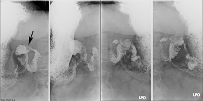
Post Bulbar Duodenal Ulcer


The above images from a double contrast upper GI study show a filling defect in the post bulbar duodenum with radiating folds (arrow) causing distal narrowing/stricture most consistent with a post bulbar ulcer.

Post bulbar ulcers are rare and constitute about 5% of all duodenal ulcers. There is a higher likelihood of life threatening bleeding with post bulbar ulcers than with ulcers in the first portion of the duodenum. While ulcers of the duodenal bulb are almost always associated with H. pylori infection, the prevalence of H. pylori in post bulbar ulcers is significantly less and post bulbar ulcers should be worked up for malignancy and Zollinger-Ellison syndrome. 


REFERENCES
Carucci LR, Levine MS, Rubesin SE, Laufer I. Upper gastrointestinal tract barium examination of postbulbar duodenal ulcers. AJR Am J Roentgenol 2004;182(4):927-30.
Kaufman SA and Levene G. Postbulbar duodenal ulcer. Radiology 1957;69:848-52.

Wednesday, January 16, 2013

The Deep Sulcus Sign


The deep sulcus sign refers to lucency of the lateral costophrenic angle in a supine patient with a pneumothorax. The above images demonstrate the deep sulcus sign at the left costophrenic angle in a pediatric patient. A left lateral decubitus film (second image) confirmed the presence of a pneumothorax.


REFERENCES
Kong A. The deep sulcus sign. Radiology 2003;28:415-6.

Monday, January 14, 2013

Vertebral Artery Transection


The unfortunate consequence of a rapid acceleration-deceleration injury. Sagittal image shows dissociation of the cervical spine at C6-C7. Coronal reformatted image from the CTA demonstrates loss of the right vertebral artery from it's origin to the level of C5 with presumable retrograde filling of its distal portion via collaterals. 

Friday, January 11, 2013

Intestinal Angioedema


Angioedema is a noninflammatory disease characterized by increased capillary permeability with extravasation of intravascular contents leading to edema. The face, limbs, and airways may be involved and angioedema of the latter can cause life threatening airway obstruction. Intestinal involvement is also reported and can present with acute abdomen, or in rare cases, hypovolemic shock.

Angioedema is due to hereditary and idiopathic, or secondary to therapy with angiotensin converting enzyme (ACE) inhibitors in patients with hypertension.

CECT findings include bowel wall and mucosal thickening, enhancement of the mucosa (differentiates from ischemic bowel wall thickening), prominent mesenteric vessels, and ascites. 


REFERENCES
De Backer AI, De Schepper AM, Vandevenne JE, et al. CT of angioedema of the small bowel. AJR Am J Roentgenol 2001;176(3):649-52.
Scheirey CD, Scholz FJ, Shortsleeve MJ, et al. Angiotensin-converting enzyme inhibitor-induced small-bowel angioedema: clinical and imaging findings in 20 patients. AJR Am J Roentgenol 2011;197(2):393-8.

Thursday, January 10, 2013

Isodense Subdural Hematoma


The above NECT images demonstrate a right sided extra-axial crescentic collection that has similar attenuation to gray matter. The collection causes effacement of right sided sulci, and mass effect with midline shift. The isodensity to underlying cortical tissue makes this consistent with an isodense/subacute subdural hematoma indicating that its age ranges from ~3 days to 3 weeks. In patients with anemia, an isodense subdural hematoma can occur in the acute setting.


REFERENCES
Kim KS, Hemmati M, Weinberg PE. Computed tomography in isodense subdural hematoma. Radiology 1978;128:71-4.

Wednesday, January 9, 2013

Causes of Hepatic Capsular Retraction

Peripheral Cholangiocarcinoma
  • malignant tumor arising from interlobular bile ducts
  • peripheral enhancement on arterial phase CECT with gradual centripetal fill in
  • delayed enhancement is common due to fibrotic regions within mass
  • may have dilated bile ducts upstream from the lesion
Confluent Hepatic Fibrosis
  • occurs in the setting of cirrhosis
  • usually in the medial segment of the left lobe and/or anterior segment of the right lobe
  • peripheral wedge shaped regions of low attenuation on CECT that show delayed enhancement
  • biliary ductal dilatation not seen
Metastases
  • treated adenocarcinoma (usually colorectal, breast) can cause hepatic capsular retraction 
  • regions of fibrosis (pseudo-cirrhosis) may also be seen

REFERENCES
Fennessy FM, Moretele KJ, Kluckert T, et al. Hepatic capsular retraction in metastatic carcinoma of the breast occurring with increase or decrease in size of subjacent metastasis. AJR Am J Roentgenol 2004;182(3):651-5.
Lee JW, Kim S, Kwack SW, et al. Hepatic capsular and subcapsular pathologic conditions: demonstration with CT and MR imaging. Radiographics 2008;28:1307-23.
Lipson JA, Qayyum A, Avrin DE, et al. CT and MRI of hepatic contour abnormalities. AJR Am J Roentgenol 2005;184(1):75-81.

Tuesday, January 8, 2013

Achalasia


Achalasia is a disorder of esophageal dysmotility due to failure of the gastroesophageal sphincter to relax because of destruction of its myenteric plexus. Primary achalasia is considered idiopathic while secondary achalasia could be due to destruction of the myenteric plexus by tumor or infection (Chagas disease). The radiologic diagnosis of primary achalasia is best made with barium studies which will show marked dilation of the esophagus with smooth narrowing near the GE junction (bird's beak deformity). Real time imaging will also show absent primary peristalsis and the presence of tertiary contractions. While cross sectional imaging is not the most sensitive in diagnosing achalasia, sometimes the finding is made incidentally as in the above case. The differential diagnosis of achalasia was previously discussed here.


REFERENCES
Noh MH, Fishman EK, Forastiere AA, et al. CT of the esophagus: spectrum of disease with emphasis on esophageal carcinoma. Radiographics 1995;15:1113-34.
Woodfield CA, Levine MS, Rubesin SE, et al. Diagnosis of primary versus secondary achalasia: reassessment of clinical and radiographic criteria. AJR Am J Roentgenol 2000;175(3):727-31.

Friday, January 4, 2013

Killian Jamieson Diverticulum

Killian is usually mentioned when discussing the Killian's dehiscence which is the gap in the posterior hypopharynx where the muscle fibers of the cricopharyngeus muscle and inferior constrictor muscle diverge. This site is where Zenker's diverticula occur.

The Killian-Jamieson space is below the cricopharyngeus muscle and lateral to the longitudinal muscle of the esophagus. This muscular gap is the location of the rarer Killian-Jamieson diverticulum. Fluoroscopy will show an outpouching along the anterolateral esophagus inferior to the cricopharyngeus muscle (as opposed to along the posterior esophagus and superior to the cricopharyngeus muscle in the case of a Zenker's diverticulum). 


REFERENCES
Rubesin SE and Levine MS. Killian-Jamieson diverticula. AJR Am J Roentgenol 2001;177:85-9.

Thursday, January 3, 2013

SATCHMO

The differential diagnosis of a suprasellar mass can be remembered through the SATCHMO mnemonic.

S - suprasellar/sellar adenoma, sarcoid
A - aneurysm, arachnoid cyst
T - teratoma
C - craniopharyngioma
H - hamartoma (of the tuber cinereum), hypothalamic glioma
M - meningioma
O - optic nerve glioma


The above image is from a patient who presented to the ER several times after multiple motor vehicle accidents. On clinical exam he was found to have visual field defects. NECT demonstrates a hyperattenuating sellar mass. Post operative diagnosis was pituitary macroadenoma.

Wednesday, January 2, 2013

Gossypiboma

Stemming from the Latin gossypium, meaning cotton, gossypiboma refers to retained cotton products such as surgical sponges. A gossypiboma can elicit an inflammatory response leading to formation of an intraabdominal abscess, or cause obstructive symptoms due to mass effect.


REFERENCES
Manzella A, Filho PB, Albuquerque E, et al. Imaging of gossypibomas: pictorial review. AJR Am J Roentgenol 2009;193:S94-101.