Sunday, March 26, 2017

Ulnar Dimelia

Ulnar dimelia is a rare congenital disorder characterized by duplication of the ulna, absence of the radius, and polydactyly. Patients can also have arterial anomalies, such as absence of the radial artery, duplication of the ulnar artery, and abnormal arterial arches in the hand. Nerve anomalies may also be present and include shortening of the radial nerve and duplication of ulnar nerve (with or without connections to the median nerve)

Radiopaedia has some great images of type I ulnar dimelia

Distinguishing features of the two types of ulnar dimelia
Feature Type I Type II
Index finger 1 2
Lunate 1 2
Trapezoid 1 2

References

  • Afshar A. Ulnar dimelia without duplicated arterial anatomy. J Bone Joint Surg Br. 2010 Feb;92(2):293-6. doi: 10.1302/0301-620X.92B2.23057.
  • Tomaszewski R, Bulandra A. Ulnar dimelia-diagnosis and management of a rare congenital anomaly of the upper limb. J Orthop. 2015 Feb 18;12(Suppl 1):S121-4.

Sunday, March 19, 2017

Chondroblastoma

X-ray (radiograph), CT, and MRI of chondroblastoma of the femur.

General

  • Terminology: “giant cell variant” (1927) → epiphyseal chondromatous giant cell tumor → calcifying giant cell tumor → chondroblastoma
  • 1% to 2% of all primary bone tumors
  • 9% of all benign bone tumors
  • Mean age of 15-18 years
  • M >>F
  • Mean duration of symptoms 8.7 months
  • Trivia: Most common benign neoplasm of the patella

Imaging Features

  • Epiphysis/apophysis +/- metaphyseal/diaphyseal involvement
  • Metaphyseal/diaphyseal occurrence without epiphyseal/apophyseal involvement exceptionally rare
  • Proximal tibia >> proximal femur > distal femur > proximal humerus
  • Well-defined, sclerotic margins on radiographs
  • Can involve the cortex, resulting in expansion, thinning, or disruption.
  • Stippled matrix calcification seen in minority of cases
  • Periosteal reaction seen in majority of cases
  • Extensive peri-lesional edema on MRI is common
  • Homogeneously hypointense on T1
  • Variable on T2: can be diffusely hypointense, or have small cystic areas of increased T2 signal or fluid-fluid levels
  • Heterogeneous and moderate enhancement in solid portions. Less commonly, homogeneous and marked enhancement

Differential Diagnosis

Management/Prognosis

  • Curettage or resection
  • RFA (small lesions, small series, not common)
  • Local recurrence rate: 5.0% after curettage
  • Local recurrence rate: 0% after resection
  • Recurrence most frequent in the proximal humerus
  • Malignant transformation and benign pulmonary metastases extremely rare

References

Sunday, March 12, 2017

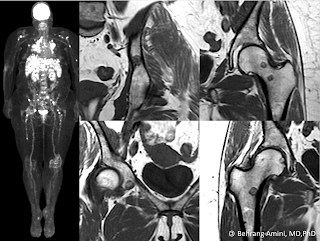
Fat-containing bone lesions

FDG PET and MRI of a patient with sarcoidosis and fat-containing bone lesions.

The presence of fat within a bone lesion is almost always reassuring, although rare exceptions exist. The following differential diagnosis is based on a combination of published papers and my own anecdotal experience:
  • Hemangioma:
  • Intra-osseous lipoma and lipoma variants, including fibrolipoma, angiolipoma and myelolipoma.
  • Enchondroma:
  • Liposclerosing myxofibrous tumor (LSMFT): Nearly all occur in the intertrochanteric region. Now felt to represent a variant of fibrous dysplasia.
  • Osteoporosis: Can give the appearance of lucent bone lesions on CT. These won't have defined margins, and measurement of internal attenuation will reveal the fatty nature of the lesion.
  • Bone infarction: Trivial, but included for the sake of completeness
  • Paget disease of bone:
  • Focal red marrow rest: Ill-defined, intermediate T1 signal. May contain subtle areas of internal fat.
  • Lymphoma: Not truly a fat-containing lesion, but can entrap fat as the tumor infiltrates marrow.
  • Sarcoid: The case above shows a patient with sarcoid and nodal, hepatic, and osseous involvement. Can have fuzzy margins ("brush border").
  • Treated metastasis: One of the ways metastases respond to therapy is by developing internal fat. Myeloma lesions can even entirely "disappear" due to fatty replacement.
  • Indolent metastases: Medullary thyroid cancer, adenoid cystic carcinoma.
  • Intra-osseous hibernoma: Rare.
  • Solid variant of aneurysmal bone cyst:
  • Nonossifying fibroma:
  • Erdheim-Chester disease:
  • Malignancy arising from a fat-containing lesion: For example, osteosarcoma arising from bone infarction or Paget.

References

Sunday, March 5, 2017

Subperiosteal hemorrhage in neurofibromatosis type 1


Neurofibromatosis type 1 (NF-1), in addition to a neuroectodermal disorder, is accompanied by mesodermal dysplasia that is accompanied by skeletal changes. The typical osseous findings include bowing of the legs, increase in length of long bones, pseudarthrosis, subperiosteal cyst formation, local bony erosions from adjacent lesions, and intramedullary neurofibromas. Except for the last two, which are due to direct involvement by neurofibromas, the remainder are due to dysplastic changes in bones.

A lesser known osseous presentation in bone is the propensity for subperiosteal hemorrhage and hematoma formation. The cause is unknown, but may be related to:
  • Vascular abnormalities: For example, diffuse flat hemangiomas or plexiform dilated veins, which have been described in patients with hypertrophy of the extremities
  • Dysplastic periosteum: The thinking is that mesodermal dysplasia manifests as an abnormally loose periosteum with poor callus response. This would predispose the patient to the formation and propagation of large subperiosteal hematomas.
  • Direct involvement by neurofibromas: Subperiosteal infiltration by neurofibromatous tissues may loosen the periosteum and allow for massive hemorrhage following minor trauma.

References