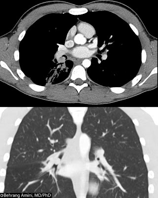
 Bronchial carcinoid tumors are uncommon neuroendocrine neoplasms of the lung, comprising less than 2% of all lung tumors. Carcinoid tumors are considered malignant, with potential for metastasis. They may fall in the spectrum of small cell carcinomas, with low-grade carcinoids on one end and small cell carcinomas on the other. A recently described large cell neuroendocrine variant (intermediate cell neuroendocrine carcinoma) falls between atypical carcinoids and small cell carcinoma of the lung. Like their gastrointestinal counterparts, they can secrete serotonin, adrenocorticotropic hormone, somatostatin, and bradykinin.
Carcinoids can be broadly defined as typical and atypical, with the former having a better prognosis. Imaging features can be similar and depend mainly on location. The vast majority (80%) of bronchial carcinoids are central and are found in the main, lobar, or segmental bronchi, presenting as hilar or perihilar masses with or without obstructive symptoms. The remainder present as peripheral nodules.
Different calcification patterns have been described, with eccentric calcifications more commonly seen. Diffuse calcification of the tumor can simulate broncholithiasis.
Post-contrast images typically (though not invariably) reveal marked, homogeneous enhancement, consistent with the highly vascular nature of these tumors.
Hilar or mediastinal adenopathy can be seen, reflecting either metastasis or reactive adenopathy from obstructive pneumonia.
Central carcinoids
Central carcinoids typically have an endobronchial component, with possible extension into adjacent parenchyma. Tumors with a dominant extraluminal component are referred to "iceberg lesions," while smaller tumors confined completely within the bronchus can also be seen.The typical appearance of a central bronchial carcinoid is a well-defined, round or ovoid hilar or perihilar mass. Lobulated, irregular or ill-defined margins can also be seen. Mediastinal extension and multifocal disease is rare.
Because of the obstructive nature of central carcinoids, patients may present with recurrent atelectasis and pneumonia (this was the presentation of our patient). Contrast-enhanced CT is useful in differentiating an enhancing carcinoid from adjacent atelectasis, consolidation, and mucoid impaction.
Peripheral Carcinoids
A bronchial carcinoid can present as a solitary pulmonary nodule in about 20% of cases. These nodules are usually round or ovoid with smooth or lobulated borders. Atypical carcinoids are more likely to occur in the lung peripheryOther Imaging Findings
Carcinoids are T2-hyperintense, octreotide-avid, and typically negative on FDG. MIBG and octreotide are of similar sensitivity and specificity in detecting liver metastases, and some carcinoids not seen on octreotide imaging may have MIBG uptake.Differential Diagnosis
- Adenoid cystic carcinoma
- Mucoepidermoid carcinoma
- Benign mesenchymal neoplasm
No comments:
Post a Comment
Note: Only a member of this blog may post a comment.