Saturday, April 30, 2011

Grade II Spinal Cord Ependymomas

Ependymomas of the spinal cord originate from the ependymal walls. They have been classified by the World Health Organization into grades I, II, and III. Grade I tumors include myxopapillary ependymomas and subependymomas. Grade II tumors are just called ependymomas and can be further divided as cellular, papillary, clear cell, or tanycytic. Grade III tumors are called anaplastic ependymomas, tend to occur in the brain, and are rare in the spine.

Grade II spinal cord ependymomas (simply called ependymomas) tend to be located somewhat centrally in the cervical spinal cord in association with large satellite cysts.

MRI is the imaging modality of choice. Ependymomas are isointense to slightly hypointense to the spinal cord on T1-weighted images and may have areas of hyperintensity due to hemorrhage. Satellite cysts are T1-hypointense. T2-weighted images reveal a hyperintense mass with or without intramural cysts. Polar (rostral or caudal) cysts and a syrinx may also be seen in the adjacent spinal cord. Hemorrhage may also be seen, sometimes presenting as a "cap sign" (hemosiderin deposits due to chronic hemorrhage at the rostral or caudal margins). Post-contrast images reveal avid and homogeneous enhancement.

Differential considerations for an intramedullary cyst with one or more enhancing nodules includes:
  • Ependymoma: Well-circumscribed mass with hemorrhage. More common in the cervical spine.
  • Myxopapillary ependymoma:
  • Pilocytic astrocytoma: Most common primary spinal cord neoplasm in children. Can be eccentric with indistinct margins. Hemorrhage is uncommon.
  • Hemangioblastoma: Look for a hemangioblastoma in the brain, cysts in the pancreas, and cystic or solid lesions in the kidneys to make the diagnosis of von Hippel-Lindau disease.

References

  • Balériaux DL. Spinal cord tumors. Eur Radiol. 1999;9(7):1252-8.
  • Armstrong TS, Vera-Bolanos E, Gilbert MR. Clinical course of adult patients with ependymoma: Results of the Adult Ependymoma Outcomes Project. Cancer. 2011 Apr 28.

Friday, April 29, 2011

Pleural Masses

Multiple

  • Pleural metastases: Case shown above (Ewing sarcoma)
  • Asbestos related pleural disease: Look for calcifications.
  • Malignant mesothelioma: Diffuse nodular pleural thickening, thicker at lung bases, with a small hemithorax and pleural effusion. Grows along fissures.
  • Invasive thymoma: Pleural metastases rare, but well-described.
  • Lymphoma: Look for other enlarged nodes.
  • Splenosis: Left-sided pleural thickening in a patient with history of distant trauma and an absent spleen.

Solitary

  • Pleural metastases:
  • Solitary fibrous tumor of the pleura: Hypervascular mobile mass displacing rather than invading adjacent structures. No chest wall involvement. Calcifications are more common in malignant lesions, but malignant and benign tumors are indistinguishable on imaging.
  • Lipoma: Like solitary fibrous tumors of the pleura, pleural lipmas may also be pedunculated and mobile.
  • Malignant mesothelioma:

Thursday, April 28, 2011

Cemento-Osseous Dysplasia

Cemento-osseous dysplasias are a group of benign fibro-osseous lesions that arise from periodontal ligament tissues in the tooth-bearing regions of the mandible and maxilla. Cemento-osseous dysplasias can be classified as periapical, focal, and florid based on location and extent (see below).

Early in the course of the disease radiographs and CT reveal a predominantly lucent lesion with multiple closely apposed or confluent, round or ovoid cystic areas. There is a narrow zone of transition with adjacent normal bone. As the disease progresses, the lesions become progressively more opaque internally. There is usually no extension into adjacent bone or cortical expansion. Infection in overlying teeth (spontaneous or post biopsy) may spread across the lesion and evolving into osteomyelitis with sequestration.

Small, discrete lesions may mimic apical periodontitis, while larger lesions can look similar to ossifying fibromas.

Cemento-osseous dysplasia can be classified as:
  • Periapical: Also known as cementoma, periapical cementoma, periapical cemental dysplasia, and periapical fibrous dysplasia. Predominantly involves the apical areas of mandibular incisors.
  • Focal: Essentially the same entity as periapical cemento-osseous dysplasia, but predominantly involving the tooth-bearing areas of the posterior jaws, particularly in sites of former extraction. Some have proposed that both periapical and focal cemento-osseous dysplasia be known as focal cemento-osseous dysplasia.
  • Florid (shown above): A more extensive version of focal cemento-osseous dysplasia that involves two or more quadrants of the jaw. Most cases have bilateral mandibular molar/premolar involvement with or without maxillary involvement. Florid cemento-osseous dysplasia is usually asymptomatic, with pain being the most common symptom in symptomatic patients.
Special thanks to Dr. Carla Ross for the case.

References

  • Eversole R, Su L, ElMofty S. Benign fibro-osseous lesions of the craniofacial complex. A review. Head Neck Pathol. 2008 Sep;2(3):177-202.
  • Scholl RJ, Kellett HM, Neumann DP, Lurie AG. Cysts and cystic lesions of the mandible: clinical and radiologic-histopathologic review. Radiographics. 1999 Sep-Oct;19(5):1107-24.

Wednesday, April 27, 2011

Pineal Region Masses

  • Pineal cyst (shown above): Most common pineal lesion.
  • Pineal tumor: Pineoblastoma (children), germinoma (young adults), pineocytoma (older people).
  • Extrapineal tumor: Meningioma, tectal glioma.
  • Arachnoid cyst: Located in the quadrigeminal plate cistern.
  • Infection: Neurocysticercosis.
  • Congenital: Lipoma.
  • Dermoid/epidermoid cyst:
  • Vein of galen malformation:

Tuesday, April 26, 2011

J-Shaped Sella

The J-shaped sella, seen on the lateral view of the skull, is caused by flattening of the tuberculum sellae, forming the stem of the J. The normal dorsum sellae forms the loop of the J. The extension of the anterior aspect of the sella is distinguished from the true pituitary fossa by the tuberculum sellae.

Differential considerations include:
  • Normal variant: In 5% of normal children. Normalizes later in life.
  • Hypothyroidism:
  • Slowly growing parasellar tumor: Optic glioma (e.g., in neurofibromatosis type 1).
  • Mucopolysaccharidoses: Classically described in Hurler syndrome, but can be seen in the other mucopolysaccharidoses. The case above is from a patient with Hunter syndrome.
  • Mild arrested hydrocephalus:
  • Hajdu-Cheney syndrome: Rare disorder of bone metabolism. The diagnosis is made by finding acroosteolysis with any three of the following: Wormian bones, open skull sutures, platybasia, micrognathia, mid-facial flattening, premature loss of teeth, coarse hair, and short stature.

References

  • Merle P, Georget AM, Goumy P, Jarlot D. Primary empty sella turcica in children. Report of two familial cases. Pediatr Radiol. 1979 Oct;8(4):209-12.
  • Wren MW. Significance of the so-called J-shaped sella in the diagnosis of intracranial aneurysm. Br J Ophthalmol. 1969 May;53(5):307-9.

Monday, April 25, 2011

Gallstones Misbehaving

  • Gallstone ileus: A gallstone that erodes through a biliary-enteric fistula and gets imapcted in the terminal ileum, causing obstruction. Occurs in 15% of patients with a biliary-enteric fistula.
  • Mirizzi syndrome: Gallstone impacted in the cystic duct results in extrinsic compression and obstruction of the extrahepatic bile duct. The cystic duct is usually oriented parallel to the extrahepatic bile duct. CT, ultrasound, or MRI shows a calculus at the junction of the cystic duct and extrahepatic bile duct in conjunction with a dilated bile duct proximal to and a normal-caliber duct distal to the calculus.
  • Bouveret syndrome: Biliary-enteric fistula allows a gallstone to enter the duodenum. Gastric outlet obstruction may occur due to impaction of the gallstone in the distal stomach or proximal duodenum. Occurs most commonly in elderly women.
  • Dropped calculi: Gallstone spillage is fairly common during laparoscopic cholecystectomy (up to 30%). Abscess formation is relatively rare (< 3%), with the calculus serving as the nidus for infection days to years after cholecystectomy. Patients will usually develop recurrent abscesses until the calculus is removed (drainage and antibiotics by themselves are not effective). Calculi may be found anywhere in the peritoneal or even pleural cavity. Dropped calculi may mimic peritoneal metastases and metastatic ovarian cancer. Invasion through the abdominal wall or across tissue planes with fibrosis and draining sinuses can be seen with actinomycosis, and may mimic a malignant process.

References

Sunday, April 24, 2011

The Jatene Procedure: Basics

The Jatene, or arterial switch, procedure corrects D-transposition of the great vessels. Patients usually first receive palliative treatment with prostaglandin E1 and balloon atrial septostomy (Rashkind procedure). The Jatene procedure is then performed in the first month of life

The Jatene procedure involves switching the main pulmonary artery (MPA) and the aorta (Ao) and relocating the ostia of the coronary arteries (RCA and LCA) to the new aorta. The Lecompte procedure is also performed, whereby the main pulmonary artery bifurcation is placed anterior to the ascending aorta. Axial images show the aorta posterior to the main pulmonary artery and between the right and the left pulmonary arteries (RPA and LPA, respectively). Dense contrast is seen in the left brachiocephalic vein (LBCV) on the coronal curved reformatted image.

Complications to watch for include:
  • Left ventricular impairment: Early.
  • Coronary artery issues: Early. Stenosis or occlusion.
  • Supravalvular pulmonary artery stenosis: Most frequent midterm complication. Most commonly occurs at the site of the anastomosis between the former aorta, any interposed graft (used to lengthen the main pulmonary artery), and the main pulmonary artery. Our patient had supravalvular stenosis and underwent pericardial patch augmentation of the main pulmonary artery (not clearly visible on CT).
  • Right or left pulmonary artery stenosis: Tend to occur after a Lecompte procedure, possibly due to stretching of these arteries after extensive dissection. Our patient had narrowing of the right pulmonary artery, which was subsequently stented.

References

Saturday, April 23, 2011

Causes of Polyhydramnios

  • Idiopathic: About 60% of cases.
  • Maternal causes: Diabetes (increases fetal urination), infection (CMV, toxoplasmosis).
  • Placental causes: Placental tumors (e.g., chorioangioma) or vascular malformations.
  • Fetal causes
    • Central nervous system: Decreased swallowing.
    • Gastrointestinal: Atresias at various levels.
    • Cardiovascular: Anemia leading to hyperdynamic circulation and increased fetal urination.
    • Neoplastic: Thoracic lesions with mass effect on esophagus. Renal tumors (mesoblastic nephroma, congenital Wilms). Sacrococcygeal teratoma. Large ovarian cyst.
    • Chromosomal: For example, trisomy 21, 18.

Friday, April 22, 2011

Rauber Sign

The Rauber sign is a radiographic sign of meniscal injury. Originally described before the advent of MRI, more recent work has confirmed it as a specific sign for meniscal tears. The sign is more likely to be seen with older injuries.

On frontal radiographs, there is a projecting traction spur or periosteal deposition along the medial or lateral margin of the tibial plateau. It can be differentiated from an osteophyte by noting the lack of marginal lipping.

Special thanks to Dr. Stephen Bloom for the references.

References

  • Ahn JM, Hwang JY, Lee M, Kim S, Lee SW, Ahn JH. Revisit of Rauber Sign: Radiographic Diagnosis of Meniscal Tear. RSNA 2003.
  • Barucha E. Our experiences on the value of the Rauber roentgen sign in meniscus diagnosis. Monatsschr Unfallheilkd Versicherungsmed. 1960 Oct;63:370-5.
  • Rauber A. Ein wenig bekanntes rontgensymptom bei älteren Meniscusfektionen. Z. Unfallmed. Berufskr. 1944(37): 167-178

Thursday, April 21, 2011

Breast Imaging Bullets

  • Mucinous: Subtype of invasive ductal carcinoma. Dense mass on mammography. Isoechoic to fat on ultrasound. Slow growth with low tendency to metastasize.
  • Tubular: Subtype of invasive ductal carcinoma with the best prognosis. Spiculated mass or architectural distortion on mammography. Hard to see on ultrasound, but may see architectural distortion in a single plane.
  • Lobular neoplasia: New name for lobular carcinoma in situ. ~30% increased risk of cancer (ductal or lobular) in either breast.
  • Roman arch: Micropapillary DCIS (low-grade DCIS).
  • Granular cell tumor: Benign. Derived from Schwann cells.
  • Occult cancers: By MRI, occult ipsilateral cancer in 15%-35% and occult contralateral cancer in 3% of newly diagnosed breast cancer patients.
  • Radial scar: Excisional biopsy due to high incidence of associated proliferative changes with and without atypia, lobular neoplasia, and tubular carcinoma.

  • Focal asymmetry on CC view: Roll views. Superior lesions move in the direction of the roll. Inferior lesions move in the direction opposite to that of the roll.
  • Focal asymmetry on MLO view: Lateral view. Medial lesions move up on the lateral view compared to the MLO (Muffins rise). Lateral lesions move down on the lateral view compared to the MLO (Lead sinks).

  • Shrinking breast: Infiltrating ductal carcinoma or diabetic mastopathy.
  • Isolated unilateral axillary adenopathy: According to the BI-RADS FAQ: "In the absence of known infectious or inflammatory cause, isolated unilateral axillary adenopathy should receive a BI-RADS Category 4 assessment." Concern is for occult breast carcinoma or, much less commonly, metastatic melanoma, ovarian cancer, or other metastatic cancer. If a benign cause can be found (e.g., infection) BI-RADS Category 2 can be used.
  • Isolated bilateral axillary adenopathy: According to the BI-RADS FAQ, in the face of known causes (lymphoma, HIV, sarcoid, systemic lupus erythematosis, psoriasis): BI-RADS Category 2. If no cause is known, then "it may be a sign of lymphoma and a BI-RADS Category 4 assessment is given. "
  • Single dilated duct: Likelihood of being related to breast cancer is exceedingly low. BI-RADS-2 or 3. More recent data (not in the Holy BI-RADS) suggests that a solitary dilated duct "appears to have a greater than 2% likelihood of malignancy, sufficiently high to suggest that a suspicious (BI-RADS 4a) assessment may be appropriate."
  • Multiple filling defects on ductography: BI-RADS 4a.
  • Ductography: 30-G sialogram (blunt tipped) needle. ~1 cc barium (or until patient feels full).
  • Complex mass on ultrasound: BI-RADS-4b
  • Ultrasound-guided core biopsy: 14-G spring-loaded. 3-5 samples. Some places use 11-G.
  • Stereotactic core biopsy: 11-G needle. 12 samples.
  • Solid masses with increased through-transmission: Lymphoma and medullary carcinoma
  • Male breast cancer: Ductal types (men don't have lobules)
  • Architectural distortion: Invasive carcinoma (ductal, lobular), radial scar, biopsy scar.

References

Wednesday, April 20, 2011

Enteric Duplication Cyst

Gastrointestinal duplication cysts are spherical or tubular cystic structures that are lined by gastrointestinal epithelium and muscularis, and are usually attached to the gastrointestinal tract. Enteric duplication cysts most commonly occur in the distal ileum (as in our case), followed by the distal esophagus, stomach, and jejunum. They usually present during the first year of life (our patient was 9 months old).

Fluoroscopic contrast studies usually reveal an intraluminal, intramural, or extrinsic mass. Ultrasound typically shows a bowel signature, with inner hyperechoic and outer hypoechoic rings. In cases of suspected ectopic gastric mucosa (e.g., Meckel diverticulum), 99mTc-pertechnetate can be a helpful problem solving modality.

References

Hur J, Yoon CS, Kim MJ, Kim OH. Imaging features of gastrointestinal tract duplications in infants and children: from oesophagus to rectum. Pediatr Radiol. 2007 Jul;37(7):691-9. Epub 2007 May 31.

Tuesday, April 19, 2011

Radiographic Features of Hyperparathyroidism


Increased parathyroid hormone secretion results in diffuse bone resorption and dimineralization, most apparent at areas of greatest surface area.

  • Periostitis: More commonly seen in secondary hyperparathyroidism. In children periostitis and extensive bone resorption may mimic syphilis or leukemia.
  • Subperiosteal resorption: Classically at the distal phalangeal tufts (acroosteolysis, pink arrows) and along the radial margins of the second and third middle phalanges (blue arrows). Can also be seen along the medial aspect of the humerus, femur, and tibia, the superior and inferior aspects of the ribs and the lamina dura of the teeth (white arrow).
  • Intracortical resorption: Intracortical linear striations (cortical tunneling) can be seen, classically at the index finger metacarpal.
  • Endosteal resorption: Well-seen in the hands. Endosteal scalloping can mimic the endosteal erosions seen in multiple myeloma, and endosteal thinning can mimic cortical thinning seen in osteoporosis.
  • Trabecular resorption: Resorption within medullary bone gives bone a granular appearance, with loss of distinct trabecular detail. In the skull, the diploic space is replaced by connective tissue, leading to a speckled appearance ("salt and pepper" skull).
  • Subchondral resorption: Can mimic inflammatory arthropathy, especially in the sacroiliac (lower 2/3 is articular), acromioclavicular, sternoclavicular, and temperomandibular joints, and at the pubic symphysis (cartilaginous joint).
  • Subligamentous resorption: Most commonly seen at the trochanters, ischial tuberosities, sacroiliac joints (upper 1/3 is syndesmotic), the inferior surface of the calcaneus, the inferior surface of the distal clavicle, and at the elbow.
  • Calcium pyrophosphate dihydrate deposition disease: More commonly seen in primary hyperparathyroidism (up to 40%).
  • Brown tumors (black arrow): Accumulations of osteoclasts and fibrous tissue. Tend to heal after treatment of the underlying disorder. Eccentric/intracortical, lytic, and often expansile. Incidence is greater in primary hyperparathyroidism, but more commonly seen with secondary hyperparathyroidism due to the higher prevalence.
  • Soft tissue calcifications: More commonly seen in secondary hyperpararthyroidism.
  • Bone sclerosis: More commonly seen in secondary hyperpararthyroidism. Can be seen in the metaphyses of long bones, the skull, or the vertebral body endplates (rugger jersey spine). Progressive hypertrophy of the facial and cranial hones can produce "leontiasis ossea" (lion face), and can mimic Paget disease and fibrous dysplasia.

References

  • Gisler DG. Chapter 4: Metabolic bone diseases. in Musculoskeletal Radiology: The Requisites (3rd ed). Mosby (2002): pp 401-404.
  • Resnick D. Chapter 52: Parathyroid Disorders and Renal Osteodystrophy. in Diagnosis of Bone and Joint Disorders (4th ed). Saunders (2002): pp 2043-2111.

Monday, April 18, 2011

Patterns of Delayed Hyperenhancement

Keywords for Dr. Google: ischemic, transmural, subendocardial, midwall, HCM, hypertrophic cardiomyopathy, right ventricular overload, dilated cardiomyopathy, myocarditis, sarcoidosis, Anderson-Fabry, Chagas, global endocardial, amyloidosis, scleroderma, cardiac transplant, epicardial.

References

  • Bluemke DA. MRI of nonischemic cardiomyopathy. AJR Am J Roentgenol. 2010 Oct;195(4):935-40.
  • Mahrholdt H, Wagner A, Judd RM, Sechtem U, Kim RJ. Delayed enhancement cardiovascular magnetic resonance assessment of non-ischaemic cardiomyopathies. Eur Heart J. 2005 Aug;26(15):1461-74.

Sunday, April 17, 2011

Crazy Paving

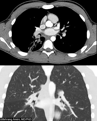
Crazy paving of the lungs refers to scattered or diffuse ground-glass attenuation with superimposed interlobular septal thickening and intralobular lines. While initially thought to be specific for pulmonary alveolar proteinosis, it has since been reported in so many conditions that it is essentially a nonspecific finding. The oculoglossal reflex of PAP persists, however. Clinical presentation can help narrow the differential diagnosis.

Chronic presentation

  • Pulmonary alveolar proteinosis (shown above): Rare, classic condition associated with Crazy paving. Radiographs reveal bilateral, symmetric alveolar consolidation or ground-glass opacity in a perihilar or hilar distribution resembling pulmonary edema. CT reveals diffuse ground-glass attenuation with superimposed intra- and interlobular septal thickening. Patients present with chronic (months to years) progressive dyspnea and dry or minimally productive cough.
  • Nonspecific interstitial pneumonitis: Symmetric, subpleural and basal-predominant ground-glass attenuation is the most common CT finding. Superimposed consolidation and irregular reticular lines can be seen. Patients present with gradually worsening dyspnea over several months, and they often experience fatigue and weight loss.

Subacute presentation

  • Cryptogenic organizing pneumonia (COP): Associated with collagen-vascular diseases, infection, and drugs. Scattered and asymmetric consolidation bilaterally, and classically peripherally. The crazy paving pattern is uncommon. Patients present with mild dyspnea, cough, and fever that have been developing over a few weeks.

Immunocompromised

  • Infection: Pneumocystis jirovecii pneumonia in immunocompromised patients. Ground-glass areas represent foamy alveolar exudates, while the lines represent thickening of the alveolar walls by edema and cellular infiltrates.

Laxative ingestion, use of oil-based nose drops, or fire-breathing

  • Lipoid pneumonia: Due to inhalation of lipid. Consolidation is characteristically fat attenuation. Crazy paving can also be seen, reflecting intraalveolar and interstitial accumulation of lipid-laden macrophages and thickening of alveolar walls.

ICU patient

  • Diffuse alveolar damage:
  • Pulmonary edema:

Miscellaneous

  • Brochoalveolar carcinoma: Ground-glass areas reflect intra-alveolar glycoprotein and the superimposed reticular attenuation is caused by interstitial infiltration by inflammatory or tumor cells.
  • Pulmonary hemorrhage: Multiple causes, including idiopathic pulmonary hemosiderosis, Wegener granulomatosis, Churg-Strauss syndrome, Goodpasture syndrome, collagen-vascular diseases (systemic lupus erythematosus, rheumatoid arthritis, systemic sclerosis, polymyositis, and mixed connective-tissue disease), drug-induced coagulopathy, and hemorrhage associated with malignancy.
  • Inflammatory: Sarcoidosis
  • Pulmonary alveolar microlithiasis (mimic): When extensive, the calcifications can result in interlobular septal thickening. Microliths smaller than 1 mm can produce a ground-glass appearance. Together, these can give an appearance similar to crazy paving.

References

Saturday, April 16, 2011

Widened Diploic Space

Diffuse thickening of the calvarium can be caused by:
  • Normal variant:
  • Chronic shunted hydrocephalus (case shown above):
  • Longstanding anemia: Widening of the diploic space is due to marrow expansion from ineffective and excessive erythropoiesis. Seen in untreated severe congenital hemolytic anaemias (thalassaemia major, sickle cell anaemia), less commonly in patients with severe iron deficiency, cyanotic heart disease, osteopetrosis. The "hair-on-end" appearance is typical.
  • Medications: Phenytoin is classic medication in this regard. Look for cerebellar atrophy. A case of G-CSF treatment causing a "hair-on-end" appearance has also been reported.
  • Paget disease: Regions of sclerosis crossing sutures results in thickening of the diploic space ("Tam-O'-Shanter" skull, named after a Scottish hat that was named after a character in a poem of the same name by Robert Burns).
  • Metabolic disorders: Renal osteodystrophy, hypoparathyroidism, and healing rickets
  • Microcephaly: Skull overgrowth due to small brain.
  • Osteopetrosis: Autosomal recessive form. CT in younger patients may show a high-attenuation inner table; a wide, low-attenuation diploic space, and a less high-attenuation outer table. "Hair-on-end" appearance can also be seen.

References

  • Albert MH, Notheis G, Wintergerst U, Born C, Schneider K. "Hair-on-end" skull induced by long-term G-CSF treatment in severe congenital neutropenia. Pediatr Radiol. 2007 Feb;37(2):221-4. Epub 2006 Dec 21.
  • Elster AD, Theros EG, Key LL, Chen MY. Cranial imaging in autosomal recessive osteopetrosis. Part I. Facial bones and calvarium. Radiology. 1992 Apr;183(1):129-35.
  • Kattan KR. Calvarial thickening after Dilantin medication. Am J Roentgenol Radium Ther Nucl Med. 1970 Sep;110(1):102-5.
  • Lucey BP, March GP Jr, Hutchins GM. Marked calvarial thickening and dural changes following chronic ventricular shunting for shaken baby syndrome. Arch Pathol Lab Med. 2003 Jan;127(1):94-7.
  • Smith SE, Murphey MD, Motamedi K, Mulligan ME, Resnik CS, Gannon FH. From the archives of the AFIP. Radiologic spectrum of Paget disease of bone and its complications with pathologic correlation. Radiographics. 2002 Sep-Oct;22(5):1191-216.

Friday, April 15, 2011

Fetal Ascites

Causes of fetal ascites (urine, meconium, bile, blood, or pus) include:
  • Urinary obstruction: Leading to urinary tract perforation and leakage of urine.
  • Hydrops: Presence of at least two abnormal fetal fluid collections (pleural effusion, pericardial effusion, ascites, or subcutaneous edema).
  • Chromosomal abnormality: Most commonly trisomy 21.
  • Infection: Presence of a thick placenta with fetal ascites and anasarca suggests infection from syphilis or cytomegalovirus.
  • Gastrointestinal atresia/obstruction: Meconium peritonitis from bowel rupture. Look for intraabdominal masses, bowel dilatation and intraabdominal calcifications.
  • Cardiac issues: For example, congestive heart failure.
  • Fetal trauma: Look for presence of abruptio placentae to suggest abdominal trauma.
  • Pseudoascites: Due to hypoechoic abdominal wall muscles. The "fluid" does not extend beyond the anterior margin of the ribs.

References

Thursday, April 14, 2011

Delayed Activity on Dual-Phase Parathyroid Scans

The above images show increased early and delayed uptake inferior to both thyroid glands. SPECT images (not shown) confirmed their position within the parathyroid gland. Ultrasound showed an enlarged parathyroid gland on the left, but nothing on the right.

The principle of dual-phase parathyroid scanning is the clearance of 99mTc-sestamibi or 99mTc-tetrofosmin from the thyroid and its delayed washout from abnormal parathyroid glands. This differential washout allows visualization of abnormal parathyroid glands on delayed images. Dual-phase imaging has a sensitivity of 90% for parathyroid adenomas and a sensitivity 60% for parathyroid hyperplasia.

99mTc-sestamibi and 99mTc-tetrofosmin can both be used and have similar diagnostic accuracy. The former clears from the thyroid gland with a half life of about 30 minutes, while the latter may clear more slowly.

In dual-phase sestamibi imaging, the thyroid and parathyroid glands are imaged at 5 minutes to 15 minutes and 1 hour - 2 hours after tracer injection. SPECT imaging can localize the abnormality in three dimensions. In cases of equivocal lesions, 99mTc-pertechnetate can be injected to see if the equivocal area has increased 99mTc-pertechnetate activity ("concordance" on dual-isotope) and is therefore related to the thyroid. "Discordance" on dual-isotope images indicates parathyroid tissue.

Delayed activity can be due to:
  • Parathyroid adenoma: The classic finding is tracer activity on delayed images; however, this may only be seen in up to 60% of cases. In equivocal cases, dual-isotope technique can be used (see above).
  • Parathyroid hyperplasia:
  • Parathyroid Carcinoma:
  • Thyroid adenoma: Follicular adenoma is the most common false positive. SPECT can be used to localize the lesion to the thyroid gland and dual-isotope technique can be used to assess thyroid origin.
  • Thyroid carcinoma: SPECT can be used to localize the lesion to the thyroid gland and dual-isotope technique can be used to assess thyroid origin.

References

Wednesday, April 13, 2011

Pulmonary Infections in AIDS

Basics

  • Patients with CD4 counts < 500 cells/mm3 are at increased risk for: bacterial pneumonia and pulmonary tuberculosis.
  • Patients with CD4 counts < 200 cells/mm3 are also at increased risk for Pneumocystis jiroveci pneumonia and disseminated tuberculosis.
  • Patients with CD4 counts < 100 cells/mm3 are also at increased risk for fungal and Cytomegalovirus pneumonia.

Bacterial Pneumonia

Findings are similar to those seen in immunocompetent patients: single or multifocal areas of consolidation.

Tuberculosis

In patients with CD4 counts > 200 cells/mm3, findings are similar to those seen in reactivation tuberculosis in immunocompetent patients: Single 1- to 3-cm nodules, consolidation, cavitation (mainly the upper lobes), and tree-in-bud opacities.

In patients with CD4 counts > 100 cells/mm3, findings are similar to those of primary tuberculosis: areas of consolidation, miliary disease, pleural effusion, and enlarged lymph nodes (with necrotizing granulomas).

Pneumocystis jiroveci pneumonia

The most common HRCT finding is confluent, symmetric, bilateral ground-glass opacities, but patients may also present with consolidation, interlobular septal thickening, cystic lesions (~20% of cases, mainly upper lobes), nodules, and crazy paving.

Fungi

Histoplasmosis and coccidioidomycosis: The most common finding is a miliary pattern, but diffuse air-space consolidation can also be seen. Cyst formation can be seen in coccidioidomycosis.

Invasive pulmonary aspergillosis: The most common findings are thick-walled cavitary lesions (pulmonary infarction and abscess formation). Less common findings include single or multiple nodules (with or without ground-glass halos), patchy areas of consolidation, and pleural effusions.

Cryptococcosis: Findings include bilateral nodular or reticular opacities, bilateral consolidation, or miliary nodules. Extensive tissue infiltration with cavitation may be seen in patients with severe immunocompromise.

Cytomegalovirus Pneumonia

Findings are variable and include: bilateral ground-glass opacities, patchy bilateral consolidation, and multiple nodules or mass-like areas of consolidation.

References

Marchiori E, Müller NL, Soares Souza A Jr, Escuissato DL, Gasparetto EL, Franquet T. Pulmonary disease in patients with AIDS: high-resolution CT and pathologic findings. AJR Am J Roentgenol. 2005 Mar;184(3):757-64.

Tuesday, April 12, 2011

Dysmyelinating Disorders (Leukodystrophies)

Dysmyelinating diseases, or leukodystrophies, are inherited neurodegenerative disorders that affect the integrity of myelin in the brain and peripheral nerves. They can be seen in lysosomal storage diseases, peroxisomal disorders, and diseases caused by mitochondrial dysfunction. The most common of these are presented below.
  • Metachromatic leukodystrophy: Late infantile (most common), juvenile, and adult types. Symmetric confluent areas of T2 hyperintensity in the periventricular white matter. Radiating T2-hypointense lines through the demyelinated deep white matter represent sparing of the perivascular white matter and result in the tigroid and leopard skin patterns when imaged in long axis or cross section, respectively. The corpus callosum, internal capsule, corticospinal tracts, and cerebellar white matter may also be involved. Subcortical U fibers are spared.
  • X-linked Adrenoleukodystrophy: Affects the white matter of the central nervous system, adrenal cortex, and testes. Early on, there is symmetric white matter demyelination in the peri-trigonal regions extending across the splenium of the corpus callosum, with sparing of the subcortical white matter. Later, demyelination spreads outward and cephalad to involve most of the cerebral white matter and often the subcortical white matter, as well. Three different zones can be defined in the affected white matter:
    • Inner zone: Irreversible gliosis and scarring. Markedly T2-hyperintense and moderately T1-hypointense.
    • Intermediate zone: Active inflammation and breakdown in the blood-brain barrier. T2-isointense to slightly hypointense and enhancing.
    • Outer zone: Leading edge of active demyelination. Moderately T2-hyperintense without enhancement.
  • Canavan Disease: Spongiform leukodystrophy (extensive vacuolization of subcortical white matter). Symmetric areas of homogeneous, diffuse T2-hyperintensity and T1-hypointensity are seen throughout the white matter. There is preferential involvement of the subcortical U fibers early on.
  • Alexander Disease: Predilection for the frontal lobe white matter early on, with early involvement of the subcortical white matter. Enhancement is seen near the tips of the frontal horns of the lateral ventricles. The disease progresses posteriorly to the parietal white matter and internal and external capsules with cysts in affected regions of the brain.
  • MELAS Syndrome: Mitochondrial Encephalomyopathy with Lactic Acidosis and Stroke-like episodes. Patients are usually normal at birth and during early infancy, then show delayed growth, episodic vomiting, seizures, and recurrent cerebral injuries resembling strokes. MRI shows multiple cortical and subcortical infarct-like lesions that cross vascular boundaries. Varying degrees of generalized cerebral and cerebellar atrophy can also be seen. The parietal and occipital lobes and the basal ganglia are frequently involved.

References

Cheon JE, Kim IO, Hwang YS, Kim KJ, Wang KC, Cho BK, Chi JG, Kim CJ, Kim WS, Yeon KM. Leukodystrophy in children: a pictorial review of MR imaging features. Radiographics. 2002 May-Jun;22(3):461-76.

Monday, April 11, 2011

Umbilical Cord Abormalities

The normal umbilical cord is a spiral of two umbilical arteries and one umbilical vein. The umbilical arteries originate from the fetal internal iliac arteries and pass on either side of the fetal urinary bladder. The umbilical vein drains into the fetal hepatic vein.

Two-Vessel Umbilical Cord

The normal umbilical cord has two arteries and one vein. A two-vessel cord has a single umbilical artery, is seen in up to 1% of pregnancies, and is more common in multiple gestations (especially monozygotics) and in diabetic mothers.

When suspected, the entire umbilical cord must be inspected to look for the second umbilical artery. If the second umbilical artery is seen in any part of the umbilical cord, then it is considered a three-vessel cord.

A two-vessel cord is important because about 30% of fetuses with will have structural anomalies, most commonly cardiovascular and renal. In addition, infants with a two-vessel cord have lower birth weights and higher perinatal mortality rates. Isolated cases of a two-vessel cord are not associated with a risk of aneuploidy.

On prenatal ultrasound, one sees the larger umbilical vein, with flow toward the fetus and the smaller umbilical artery, with flow away from the fetus. Imaging of the fetal pelvis will show an umbilical artery only on one side of the urinary bladder.

The finding of a single umbilical artery should prompt a detailed evaluation of the fetus with special emphasis on the heart and kidneys. If no other anomaly is found, clinical evaluation and/or follow-up ultrasound is recommended to evaluate fetal growth, since there is an association with low birth weight.

Umbilical Knots

Umbilical knots may be true knots or false knots. True knots, which are seen in less than 1% of pregnancies, occur more frequently in long cords, male fetuses, and multiparous women and are associated with a perinatal mortality rate of about 10% . Loose knots early in pregnancy may tighten with fetal movement or during delivery.

Parents should be counseled about the increased risk of intrauterine fetal demise and the pregnancy closely monitored with umbilical artery Doppler until term. Continuous cardiotocography during labor is recommended.

A true knot presents as a localized distention of the umbilical vein on prenatal ultrasound. One side of the distention can be traced into the umbilical vein, while the other side terminates abruptly in the knot. In addition, the vein is very difficult to trace.

True knots can have an appearance similar to small allantoic or omphalomesenteric cysts (see below). In these cases, continuity with the umbilical vein on one side confirms the diagnosis of a knot.

True knots may also have an appearance similar to the rare extra-abdominal umbilical vein varix (see below). An extra-abdominal umbilical vein varix appears as tortuous fusiform dilatations that is in smooth continuity with the umbilical vein on both sides, in contrast to the blind-ending true knot.

False knots are not considered problematic. The term may refer either to focal umbilical venous outpouchings, more appropriately called varices (see below), or exaggerated looping of umbilical cord vessels within the cord.

Umbilical Cord Varices

An umbilical venous varix is a focal dilation of the umbilical vein. It rarely occurs outside the fetus, and is most commonly found in the intra-abdominal portion of the umbilical vein between the anterior abdominal wall and the liver.

Intra-abdominal umbilical vein varices may be associated with fetal abnormalities, such as fetal anemia, and may be an early sign of hydrops. Rarely, a varix of the umbilical vein will be present within the umbilical cord outside the fetus.

On ultrasound, we see focal, usually saccular, dilation of the umbilical vein, just deep to the umbilical cord insertion oriented in the anteroposterior direction at the level of the liver. More fusiform varices are rarely seen and can extend into the intrahepatic portion of the umbilical vein.

An extra-abdominal umbilical vein varix appears as tortuous fusiform dilatations that is in smooth continuity with the umbilical vein on both sides.

Differential considerations for intra-abdominal varices include choledochal cysts (oriented in a craniocaudal direction), hepatic cysts (rare, inside the liver), urachal cysts (extend toward the pelvis), or mesenteric cysts (more rounded and lower in the abdomen).

Allantoic Duct Cyst

Allantoic duct cysts are seen within the umbilical cord and may be seen in association with fetal anomalies (e.g., omphalocele and aneuploidy).

Prenatal ultrasound shows a cyst within the umbilical cord adjacent to the umbilical vessels.

Differential considerations include true knots of the cord (see above) and focal areas of excess Wharton's jelly. The latter are easily differentiated by their echogenicity and lack of well-defined thin walls.

References

  • Hasbun J, Alcalde JL, Sepulveda W. Three-dimensional power Doppler sonography in the prenatal diagnosis of a true knot of the umbilical cord: value and limitations. J Ultrasound Med. 2007 Sep;26(9):1215-20.
  • Hertzberg BS, Bowie JD, Bradford WD, Bolick D. False knot of the umbilical cord: sonographic appearance and differential diagnosis. J Clin Ultrasound. 1988 Oct;16(8):599-602.
  • Jeanty P. Fetal and funicular vascular anomalies: identification with prenatal US. Radiology. 1989 Nov;173(2):367-70.
  • Scioscia M, Fornalè M, Bruni F, Peretti D, Trivella G. Four-dimensional and Doppler sonography in the diagnosis and surveillance of a true cord knot. J Clin Ultrasound. 2011 Mar;39(3):157-9.
  • Van den Hof MC, Wilson RD; Diagnostic Imaging Committee, Society of Obstetricians and Gynaecologists of Canada; Genetics Committee, Society of Obstetricians and Gynaecologists of Canada. Fetal soft markers in obstetric ultrasound. J Obstet Gynaecol Can. 2005 Jun;27(6):592-636.

Sunday, April 10, 2011

Nuchal Translucency

Nuchal translucency is the sonographic appearance of fluid between skin and soft tissue at the back of the fetal neck. Nuchal translucency thickness (NT) is measured at the end of the first trimester, between gestational weeks 11 and 14. The measurement for a normal NT is generally thought to be less than 3 mm. However, this fixed value ignores the normal increase in NT with gestation. More recently, the delta NT has been proposed as a way of accounting for this change in NT with gestation. Delta NT is the difference between the measured NT and the normal median NT at the measured crown–rump length (CRL). The normal median NT can be calculated from the formula:

Log10NT = −0.3599 + 0.0127 CRL − 0.000058 CRL2

A cut-off of 1.5 mm has been proposed for the delta-NT.

Nuchal translucency thickness, when combined with maternal age, is a noninvasive screen for Down syndrome. Combining nuchal translucency measurements with maternal age allows identification of 75% of fetuses with Down syndrome and approximately 70% of fetuses with other chromosome abnormalities. By combining nuchal translucency, maternal age, and maternal serum biochemistry, the detection rate for fetuses with Down syndrome is increased to 89%, with a false-positive rate of 5%.

Fetuses without chromosomal abnormalities also have an increased risk of major anomalies (most commonly cardiac) when the nuchal translucency is thicker than normal.

Nuchal translucency should be measured by properly trained sonographers, otherwise its diagnostic value basically disappears. Detailed guidelines exist for proper measurement of the nuchal translucency:
  • Technique: Either transabdominal or transvaginal
  • Crown–rump length: Should be between 45 mm and 84 mm.
  • Magnification: The fetus should occupy 75% of the image to ensure that each increment in the distance between calipers will be approximately 0.1 mm.
  • Amnion: The fetal skin must be clearly separated from the amnion. This can be achieved by waiting for or encouraging fetal movement (asking the mother to cough or tapping on her lower abdomen).
  • Position of fetal head: The fetal head should be in the neutral position. Hyperextension can increase the measurement by as much as 0.6 mm, and hyperflexion can decrease it by as much as 0.4 mm.
  • Plane: True sagittal scan of the fetus is required. The nasal bone and chin should be visible.
  • Measurement: Finally, we can make the measurement (see figure).
    • The + calipers should be used.
    • The horizontal crossbars of the calipers should not be in the black space.
    • A vertical line connecting the two calipers should be perpendicular to the long axis of fetus.
    • Three measurements must be taken and the highest used for risk stratification.

References

  • Fong KW, Toi A, Salem S, Hornberger LK, Chitayat D, Keating SJ, McAuliffe F, Johnson JA. Detection of fetal structural abnormalities with US during early pregnancy. Radiographics. 2004 Jan-Feb;24(1):157-74.
  • Maymon R, Tercanli S, Dreazen E, Sartorius G, Holzgreve W, Herman A. Comparison of pregnancy outcome of euploid fetuses with increased nuchal translucency (NT) expressed in NT MoM or delta-NT. Ultrasound Obstet Gynecol. 2004 May;23(5):477-81.
  • Snijders RJ, Noble P, Sebire N, Souka A, Nicolaides KH. UK multicentre project on assessment of risk of trisomy 21 by maternal age and fetal nuchal-translucency thickness at 10-14 weeks of gestation. Fetal Medicine Foundation First Trimester Screening Group.Lancet. 1998 Aug 1;352(9125):343-6.
  • Spencer K, Bindra R, Nix AB, Heath V, Nicolaides KH. Delta-NT or NT MoM: which is the most appropriate method for calculating accurate patient-specific risks for trisomy 21 in the first trimester? Ultrasound Obstet Gynecol. 2003 Aug;22(2):142-8.

Saturday, April 9, 2011

Placental Masses and Pseudomasses

  • Chorioangioma: Most common tumor of the placenta. May be a true neoplasm or hamartoma. No metastatic potential. Rare cause of elevated maternal α-fetoprotein. Usually incidental, but large (> 5 cm) or multiple chorioangiomas have high association (up to 50%) with maternal and fetal complications, including polyhydramnios, preterm labor, fetal hemolytic anemia, fetal thrombocytopenia, cardiomegaly, intrauterine growth restriction, toxemia, placental abruption, preeclampsia, and congenital abnormalities. Circumscribed mass with variable echogenicity and anechoic vascular channels. Classically protrudes into the amniotic cavity from the placental surface near the cord insertion.
  • Hydatidiform mole: Heterogeneous mass with multiple cystic structures. Color Doppler will show flow between the cysts, but not within the cysts.
  • Placental teratoma: Very rare. Look for calcifications to suggest diagnosis.
  • Metastases:
  • Placental abruption: Retroplacental abruption can mimic a mass. Color Doppler will not show flow in the hematoma.
  • Leiomyoma: Leiomyoma under a placenta can mimic a placental mass, especially when hemorrhagic or degenerated. Color Doppler may show uterine vessels splayed around the mass.
  • Focal myometrial contraction: Isoechoic to uterine wall. May take up to 30 minutes to resolve and may require follow-up examinations.

References

Friday, April 8, 2011

Duodenum Inversum

Duodenum inversum, also known as duodenum reflexum, is a variation of the duodenal sweep in which the distal duodenum ascends to the right of the spine to the level of the duodenal bulb and then crosses the spine horizontally behind the superior mesenteric artery. The duodenojejunal junction is appropriately located at the "ligament" of Treitz.

Duodenum inversum may be associated with delayed gastric emptying, but is not associated with midgut volvulus.

References

Thursday, April 7, 2011

Cortically Based Brain Neoplasms

  • Astrocytoma May extend to cortex. Minimal or no enhancement or edema in low-grade astrocytomas. Calcification is uncommon.
  • Oligodendrolioma: Slowly growing, well-differentiated neoplasms. Diffusely infiltrating, but appear well-circumscribed. Calcifications are seen in 80% of cases.
  • Ganglioglioma: Well-circumscribed cystic lesion with nodule and minimal edema. Most commonly seen in the temporal and parietal lobes.
  • Desmoplastic infantile glioma (DIG): Variant of ganglioglioma that is usually seen in the first 2 years of life. Low-grade tumor with good prognosis. More commonly seen in the frontal and parietal lobes. Cystic mass with with a cortically based nodule. Nodule is iso- to high-attenuation, T2-hypointense, and enhances. Some may also have a calcified rim.
  • Dysembryoplastic Neuroepithelial Tumor (DNET): Look for "bubbly" appearance on MRI.
  • Pleomorphic Xanthoastrocytoma: Look for a cortically based nodule in a cystic lesion.
See related post on cystic brain masses.

References

Koeller KK, Henry JM. From the archives of the AFIP: superficial gliomas: radiologic-pathologic correlation. Armed Forces Institute of Pathology. Radiographics. 2001 Nov-Dec;21(6):1533-56

Wednesday, April 6, 2011

Congenital Lobar Emphysema

Congenital lobar emphysema refers to progressive hyperinflaction of a lobe due to a check-valve mechanism at the bronchial level. There is no destruction of alveolar walls and the majority of cases are idiopathic. Malacia or stenosis of the bronchial cartilage can be seen in patients and are thought to represent possible etiologies. Cardiovascular anomalies can be seen in about 15% of cases.

The most commonly affected lobe is the left upper lobe (40%), followed by the right middle (35%) and upper (20%) lobes. The lower lobes are less commonly affected.

Most patients present before 6 months of age, most commonly with respiratory distress. Three clinical types can be defined based on the age of presentation: infancy (type I, most common), older children (type II, rare), or incidental in asymptomatic patients (type III, rare).

Chest radiographs obtained during the neonatal period may reveal the affected lobe to be opaque (because of retained fetal lung fluid) or diffusely reticular (due to distended lymphatic channels filled with fetal lung fluid). As the fluid clears by absorption, the affected lung progresses from alveolar opacification to interstitial reticulation to hyperlucency.

Mass effect from the expanded lobe or segment can compress adjacent lobes and structures, sometimes causing ipsilateral or contralateral atelectasis. The adjacent lobe collapses either caudad or cephalad but not medially. The ipsilateral rib spaces may be widened, the ipsilateral hemidiaphragm may be depressed, and there may be contralateral mediastinal shift.

Differential considerations for the hyperlucent lung include pneumothorax, lung cysts (simple or acquired), foreign body with air trapping, and pulmonary sling (only vascular ring associated with asymmetric lung aeration).

The images above show an opacity in the right lung that progressively clears to result in a hyperexpanded right lung that causes compression of the adjacent lung and contralateral mediastinal shift by 6 months. Chest radiograph at 2 years of age reveals a hyperexpanded right hemithorax with widening of the intercostal spaces and leftward mediastinal shift.

References

Berrocal T, Madrid C, Novo S, Gutiérrez J, Arjonilla A, Gómez-León N. Congenital anomalies of the tracheobronchial tree, lung, and mediastinum: embryology, radiology, and pathology. Radiographics. 2004 Jan-Feb;24(1):e17.

Tuesday, April 5, 2011

Gliomatosis Cerebri

Gliomatosis cerebri is a rare diffuse and infiltrative glial neoplasm involving at least two lobes of the brain. It is frequently bilateral and is typically a white matter process.

T2-weighted images typically reveal a homogeneously hyperintense infiltrating mass with mild mass effect on adjacent sulci and ventricles. Minimal or no enhancement is typical (foci of enhancement may represent anaplastic components). Pre-contrast T1-weighted images may reveal an iso- or hypo-intense process. DWI images usually reveal no restricted diffusion. Spectroscopy may reveal marked elevation of the myoinositol peak.

The main differential considerations include:
  • Vasculitis: Usually more patchy; however, biopsy may be needed in some cases.
  • Viral encephalitis: More acute presentation.
  • Anaplastic astrocytoma: May present as an infiltrating process, but is not often diffuse. Enhancement is variable.
Other entities usually included in the differential can be distinguished by their enhancement pattern or distribution.

References

Monday, April 4, 2011

Bronchial Carcinoid Tumor

Bronchial carcinoid tumors are uncommon neuroendocrine neoplasms of the lung, comprising less than 2% of all lung tumors. Carcinoid tumors are considered malignant, with potential for metastasis. They may fall in the spectrum of small cell carcinomas, with low-grade carcinoids on one end and small cell carcinomas on the other. A recently described large cell neuroendocrine variant (intermediate cell neuroendocrine carcinoma) falls between atypical carcinoids and small cell carcinoma of the lung. Like their gastrointestinal counterparts, they can secrete serotonin, adrenocorticotropic hormone, somatostatin, and bradykinin.

Carcinoids can be broadly defined as typical and atypical, with the former having a better prognosis. Imaging features can be similar and depend mainly on location. The vast majority (80%) of bronchial carcinoids are central and are found in the main, lobar, or segmental bronchi, presenting as hilar or perihilar masses with or without obstructive symptoms. The remainder present as peripheral nodules.

Different calcification patterns have been described, with eccentric calcifications more commonly seen. Diffuse calcification of the tumor can simulate broncholithiasis.

Post-contrast images typically (though not invariably) reveal marked, homogeneous enhancement, consistent with the highly vascular nature of these tumors.

Hilar or mediastinal adenopathy can be seen, reflecting either metastasis or reactive adenopathy from obstructive pneumonia.

Central carcinoids

Central carcinoids typically have an endobronchial component, with possible extension into adjacent parenchyma. Tumors with a dominant extraluminal component are referred to "iceberg lesions," while smaller tumors confined completely within the bronchus can also be seen.

The typical appearance of a central bronchial carcinoid is a well-defined, round or ovoid hilar or perihilar mass. Lobulated, irregular or ill-defined margins can also be seen. Mediastinal extension and multifocal disease is rare.

Because of the obstructive nature of central carcinoids, patients may present with recurrent atelectasis and pneumonia (this was the presentation of our patient). Contrast-enhanced CT is useful in differentiating an enhancing carcinoid from adjacent atelectasis, consolidation, and mucoid impaction.

Peripheral Carcinoids

A bronchial carcinoid can present as a solitary pulmonary nodule in about 20% of cases. These nodules are usually round or ovoid with smooth or lobulated borders. Atypical carcinoids are more likely to occur in the lung periphery

Other Imaging Findings

Carcinoids are T2-hyperintense, octreotide-avid, and typically negative on FDG. MIBG and octreotide are of similar sensitivity and specificity in detecting liver metastases, and some carcinoids not seen on octreotide imaging may have MIBG uptake.

Differential Diagnosis

  • Adenoid cystic carcinoma
  • Mucoepidermoid carcinoma
  • Benign mesenchymal neoplasm

References

Jeung MY, Gasser B, Gangi A, Charneau D, Ducroq X, Kessler R, Quoix E, Roy C. Bronchial carcinoid tumors of the thorax: spectrum of radiologic findings. Radiographics. 2002 Mar-Apr;22(2):351-65.

Sunday, April 3, 2011

Upper Extremity Arterial Occlusive Disease

Causes of upper extremity arterial occlusive disease
  • Central embolic disease: Cardiac origin. Most commonly lodge in the brachial artery.
  • Acute trauma:
  • Hypothenar hammer syndrome: Repetitive trauma to the hypothenar area. Angiography may reveal focal ulnar artery aneurysm at the level of the hamate with segmental palmar ulnar artery occlusion and multiple digital artery occlusions. May also see the characteristic corkscrew elongation and ectasia of the ulnar artery. The superficial palmar arch is more often affected, but the deep palmar arch can also be involved.
  • Thoracic outlet syndrome: Due to compression of the subclavian artery at the thoracic outlet, which may result in localized atheroma or aneurysm with embolization.

  • Large and medium vessel vasculitis: Takayasu arteritis and giant cell arteritis. Involve large inflow and medium size proximal outflow arteries.
  • Medium and small vessel vasculitis: Buerger disease (can also involve upper extremity veins), Behcet disease.
  • Small-vessel vasculitis: Rheumatoid arthritis, Sjögren syndrome, Wegener granulomatosis, polyarteritis nodosa, scleroderma, systemic lupus erythematosus, polymyositis, dermatomyositis, mixed connective tissue disorders.

References

  • Hellinger JC, Epelman M, Rubin GD. Upper extremity computed tomographic angiography: state of the art technique and applications in 2010. Radiol Clin North Am. 2010 Mar;48(2):397-421, ix.
  • Maiman MH, Bookstein JJ, Bernstein EF. Digital ischemia: angiographic differentiation of embolism from primary arterial disease. AJR Am J Roentgenol. 1981 Dec;137(6):1183-7.

Saturday, April 2, 2011

Oligodendroglioma

Oligodendrogliomas, the third most common glial neoplasms, are diffusely infiltrating, well-differentiated, slowly growing tumors. They are round or oval and involve the cortex or subcortical white matter. They typically contain calcifications (80% of cases) and most commonly affect the frontal lobe.

on CT, they present as mixed attenuation cortically based lesions with nodular or clumped calcifications. Cystic degeneration is seen in about 20% of cases. Bone windows may reveal expansion, remodeling or erosion of bone. There is variable enhancement and little to no surrounding edema.

The lesions are heterogeneously hypointense to isointense to gray matter on T1-weighted images and demonstrate cortical expansion. T2-weighted images reveal minimal associated edema surrounding a heterogeneously hyperintense mass. Low signal areas correspond to calcium (common) or blood (seen in anaplastic types). No diffusion restriction is typical for these lesions. Oligodendrogliomas typically demonstrate variable and heterogeneous enhancement.

References

Koeller KK, Rushing EJ. From the archives of the AFIP: Oligodendroglioma and its variants: radiologic-pathologic correlation. Radiographics. 2005 Nov-Dec;25(6):1669-88.

Friday, April 1, 2011

Spotted Spleen

A spotted spleen, one with multiple low-attenuation nodules, is an uncommon finding that can be seen with:
  • Tumor: Lymphoma (most common), metastases, hemangioma (most common benign splenic tumor).
  • Infectious: Fungal, mycobacterial (tuberculosis shown above), or parasitic. More frequent in immunosuppressed patients.
  • Inflammatory: Sarcoid is the most common

References

Warshauer DM, Molina PL, Worawattanakul S. The spotted spleen: CT and clinical correlation in a tertiary care center. Comput Assist Tomogr. 1998 Sep-Oct;22(5):694-702.