Tuesday, August 31, 2010

Association of Intraductal Papillary Mucinous Tumor with Autosomal Dominant Polycystic Kidney Disease

There are scattered case reports of autosomal dominant polycystic kidney disease (ADPKD) and polycystic liver disease in association with intraductal papillary mucinous tumors (IPMTs). On the other hand, a cystic pancreatic lesion in a patient ADPKD could be a simple pancreatic cyst, which is seen in up to 10% of patients with ADPKD.

References

  • Sato Y, Mukai M, Sasaki M, Kitao A, Yoneda N, Kobayashi D, Imamura Y, Nakanuma Y. Intraductal papillary-mucinous neoplasm of the pancreas associated with polycystic liver and kidney disease. Pathol Int. 2009 Mar;59(3):201-4.
  • Naitoh H, Shoji H, Ishikawa I, Watanabe R, Furuta Y, Tomozawa S, Igarashi H, Shinozaki S, Katsura H, Onozato R, Kudoh M. Intraductal papillary mucinous tumor of the pancreas associated with autosomal dominant polycystic kidney disease. J Gastrointest Surg. 2005 Jul-Aug;9(6):843-5.

Monday, August 30, 2010

Idiopathic Eosinophilic Esophagitis

Idiopathic eosinophilic esophagitis is thought to develop as an inflammatory response to ingested food allergens. Patients are usually young men (20-40) who present with dysphagia and recurrent food impaction.

The typical fluoroscopic appearance of a ringed esophagus that of multiple, fixed, closely spaced, concentric rings that traverse an area of esophageal stricture. Not all patients, however, have this typical ringed esophagus appearance, which may be a late complication of eosinophilic esophagitis.

References

Sunday, August 29, 2010

The Conotruncus

The conotruncus, also known as the cardiac outflow tract, is formed from two cardiac segments whose borders are imperceptible in mammals: the conus arteriosus and the truncus arteriosus.

The conus arteriosus, also known as the infundibulum (Latin for funnel), is the proximal portion of the outflow tract. It remodels and forms the subpulmonary infundibulum on the right and disappears on the left.

The truncus arteriosus is the distal portion of the developing cardiac outflow tract that divides into the aortic and pulmonic valves.

Here, we limit ourselves to the abnormalities of the conotruncus seen in common types of conotruncal heart defects.
  • Tetralogy of Fallot: Most common cyanotic cardiac defect. Anterior deviation of the conal septum with infundibular hypoplasia, resulting in subpulmonary obstruction. In some cases, the conal septum is absent. On the truncus side of things, there is stenosis or atresia of the pulmonic valve. Less commonly there may be absence of the pulmonary valve leaflets. There is also aortic override.
  • D-loop transposition of the great arteries: Second most common cyanotic congenital heart condition diagnosed in the 1st year of life. There is atrioventricular concordance and ventriculoarterial discordance, meaning the atria and ventricles connect properly, but the ventricles and outflow vessels connect improperly. The truncus abnormality consists the aortic valve situated to the right of and anterior to the pulmonary valve. The Rastelli procedure can be performed in the presence of a ventricular septal defect, connecting the right ventricle to the pulmonary artery.
  • L-loop transposition of the great arteries: Also known as congenitally or physiologically corrected transposition. There is atrioventricular and ventriculoarterial discordance, meaning the atria and ventricles connect improperly, and the ventricles and outflow vessels connect improperly. This results in two parallel circulations with systemic venous return reaching the pulmonary circulation through the right-sided left ventricle, and the pulmonary venous return reaching the aorta through the left-sided right ventricle.
  • Double-outlet right ventricle: Both the pulmonary artery and aorta arise predominantly from the right ventricle. The aorta is usually directly to the right of the pulmonary artery. The mitral and pulmonic valves are not continuous and the majority of patients have pulmonic and mitral valve anomalies.
  • Truncus arteriosus: Single-vessel outflow from the heart. This vessel overrides the ventricular septum and supplies the systemic, pulmonary, and coronary circulation. The conal septum is usually absent. A ventricular septal defect is seen in almost all patients. Classified according to the branching pattern of the pulmonary artery. Associated with DiGeorge syndrome.

    Babies with truncus arteriosus present with a right-sided arch and commonly have cardiomegaly at birth. There are increased pulmonary vascular markings due to increased arterial blood flow.
  • Interrupted aortic arch: As its name suggests, the aortic arch is interrupted between the ascending and descending portions of the thoracic aorta, either completely or incompletely (spanned by a fibrous band).

References

  • Frank L, Dillman JR, Parish V, Mueller GC, Kazerooni EA, Bell A, Attili AK. Cardiovascular MR imaging of conotruncal anomalies. Radiographics. 2010 Jul-Aug;30(4):1069-94.
  • Restivo A, Piacentini G, Placidi S, Saffirio C, Marino B. Cardiac outflow tract: a review of some embryogenetic aspects of the conotruncal region of the heart. Anat Rec A Discov Mol Cell Evol Biol. 2006 Sep;288(9):936-43.

Saturday, August 28, 2010

Simple Asymptomatic Adnexal Cysts

Simple adnexal cysts up to 10 cm have malignancy rates < 1%. In the absence of hard evidence to guide which simple cysts need follow-up, the following guidelines have been adopted:

Premenopausal women:
  • ≤5 cm: Do not need follow-up.
  • 5 cm - 7 cm: Yearly follow-up with ultrasound is recommended.
  • >7 cm: Difficult to assess completely with ultrasound (mural nodules may be obscured). MRI or surgery is recommended.
Postmenopausal women:
  • ≤ 1 cm: Do not need follow-up.
  • 1 cm - 7 cm: 1-year follow-up with ultrasound is recommended. Yearly or less frequent follow-up thereafter based on stability or decrease in size.
  • >7 cm: Difficult to assess completely with ultrasound (mural nodules may be obscured). MRI or surgery is recommended.

References

Levine D, Brown DL, Andreotti RF, Benacerraf B, Benson CB, Brewster WR, Coleman B, Depriest P, Doubilet PM, Goldstein SR, Hamper UM, Hecht JL, Horrow M, Hur HC, Marnach M, Patel MD, Platt LD, Puscheck E, Smith-Bindman R. Management of asymptomatic ovarian and other adnexal cysts imaged at US: Society of Radiologists in Ultrasound Consensus Conference Statement. Radiology. 2010 Sep;256(3):943-54.

Friday, August 27, 2010

Coats Disease

Coats disease is a rare congenital, nonfamilial, idiopathic disease of the vessels of the retina that is characterized by leakage of fluid into and around vessel walls, leading to progressive intraretinal and subretinal leakage and exudative retinal detachment. Weak vessel walls also predispose them to development of telangiectases and aneurysms.

The disease is unilateral in the majority of cases (80%–90%) and has a male predisposition. Diagnosis is usually in childhood, and about 50% of untreated patients will eventually have total retinal detachment, blindness, and painful neovascular glaucoma.

The ophthalmologists handle the diagnosis in the majority of cases. Radiology becomes important in differentiating advanced Coats disease from retinoblastoma because both diseases may present as a non-rhegmatogenous (see below) retinal detachment associated with telangiectases and subretinal collections.

CT and MRI may show partial or total retinal detachment without evidence of an intraocular mass or intraocular calcifications. There is homogeneously increased intraocular attenuation compared to the unaffected eye. The subretinal fluid collection may be seen as hyperintense on all pulse sequences and there may be enhancement along the leaves of the detached sensory retina and at the sites where retina inserts. In contrast, retinoblastoma may show an intraocular mass with calcifications. An additional finding that may help differentiate the two is the observation that the affected globe is smaller than the unaffected globe in Coats disease.

Rhegmatogenous: Comes from the greek rhegma = discontinuity or break. Rhegmatogenous retinal detachment is more common, and occurs when a tear in the retina causes fluid accumulation between the neurosensory retina and retinal pigment epithelium. Non-rhegmatogenous detachments can occur via traction from a contracting vitreous (tractional retinal detachment) or via fluid accumulation due to damage to the retinal pigment epithelium (exudative detachment). Coats disease falls into the latter type of non-rhegmatogenous detachment.

References

Galluzzi P, Venturi C, Cerase A, Vallone IM, Bracco S, Bardelli AM, Hadjistilianou T, Gennari P, Monti L, Filosomi G. Coats disease: smaller volume of the affected globe. Radiology. 2001 Oct;221(1):64-9.

Thursday, August 26, 2010

Portal Hypertensive Gastropathy

Portal hypertensive gastropathy (previously known as "hemorrhagic gastritis," "alcoholic gastritis," "congestive gastropathy," and "gastric mucosal hyperemia") refers to mucosal hyperemia and dilated submucosal vessels in the stomach of patients with chronic portal hypertension. Portal hypertensive gastropathy tends to occur more commonly in cirrhotic patients compared to patients with noncirrhotic causes of portal hypertension.

It is thought that chronic venous congestion and hyperdynamic flow in the splanchnic circulation causes mucosal hyperemia, capillary ectasia, and increased numbers of submucosal arteriovenous communications. This results in dilated intramural arterioles, capillaries, and veins.

It is estimated to cause up to 30% of upper gastrointestinal bleeding in patients with portal hypertension and can occur even in the absence of esophageal or gastric varices. It can also cause persistent bleeding needing medical or surgical treatment. In addition, the presence of portal hypertensive gastropathy may be a risk factor for variceal hemorrhage.

On fluoroscopy, there will be thickened nodular folds in the gastric fundus. Although varices or various forms of gastritis can also produce thickened gastric folds, portal hypertensive gastropathy should be suspected when this finding is detected in patients with known portal hypertension.

Dynamic (e.g., multi-phase liver) CT can also help. A transient gastric perfusion sign has been described as a fairly specific sign for portal hypertensive gastropathy. In normal subjects, the collapsed gastric wall demonstrates homogeneous enhancement during arterial, portal, and delayed CT images. In portal hypertensive gastropathy, on the other hand, there is transient, segmental or subsegmental low-attenuation mucosa in the fundus or body of the stomach on the arterial phase that returns to normal attenuation on portal venous or equilibrium-phase images (see image). This sign has a sensitivity of 75%, specificity of 89%, positive predictive value of 90% and negative predictive value 72%.

Differential considerations include the other causes of gastric wall thickening:
  • Gastritis: For example Helicobacter pylori. The thickened folds are not confined to the fundus. Other abnormalities, such as ulcers, erosions, nodules, or enlarged areae gastricae may also be seen.
  • Lymphoma: Usually much thicker and more lobulated folds.
  • Ménétrier disease: Usually much thicker and more lobulated folds.
  • Gastric varices: May mimic thickened folds on fluoroscopy, but the folds tend to have a serpentine configuration with discrete submucosal nodules (bunch of grapes)

References

  • Chang D, Levine MS, Ginsberg GG, Rubesin SE, Laufer I. Portal hypertensive gastropathy: radiographic findings in eight patients. AJR Am J Roentgenol. 2000 Dec;175(6):1609-12.
  • Kim TU, Kim S, Woo SK, Lee JW, Lee TH, Jeong YJ, Heo J. Dynamic CT of portal hypertensive gastropathy: significance of transient gastric perfusion defect sign. Clin Radiol. 2008 Jul;63(7):783-90.

Wednesday, August 25, 2010

Adenosis in Mammography

Adenosis is hyperplasia of epithelial and myoepithelial cells of the ductules of the terminal ductal lobular unit (TDLU) and the surrounding intralobular stroma. This results in enlargement of the TDLU. Adenosis may be blunt duct type, microglandular, or sclerosing.

Sclerosing adenosis refers to stromal sclerosis involving greater than half of the TDLU that results in enlargement, distortion, and compression of the TDLU. About 50% of cases are associated with calcifications.

Sclerosing adenosis is important because it sometimes presents a diagnostic dilemma for the pathologist by mimicking infiltrating carcinoma, tubular carcinoma in particular. Sclerosing adenosis may also mimic a radial scar by pathology.

On the radiologist's end, sclerosing adenosis more commonly presents with calcifications, but can also present as a spiculated lesion. By itself, sclerosing adenosis is thought to cause a mild (1.7-2.5x) increase in the risk of infiltrating breast cancer. When associated with atypical hyperplasia, the relative risk rises to as much as 6.7.

References

Cyrlak D, Carpenter PM, Rawal NB. Breast imaging case of the day. Florid sclerosing adenosis. Radiographics. 1999 Jan-Feb;19(1):245-7.

Tuesday, August 24, 2010

Patterns of Inflammatory Sinus Disease

Sonkens et al described five patterns of inflammatory paranasal sinus disease, which you should think of when describing paranasal sinus disease.

Osteomeatal complex pattern: The osteomeatal complex is responsible for anterior paranasal sinus drainage. Lesions at the middle meatus of the nasal cavity such as mucosal edema, polypoid lesions, concha bullosa, and septal deviation, can lead to involvement of the maxillary sinus and anterior and middle ethmoid air cells. The frontal sinus may be involved, depending on the insertion of the anterior uncinate process. A limited variant of the osteomeatal complex pattern called the frontal recess inflammatory pattern may occur with isolated involvement of the frontal sinus in cases of direct drainage of the frontal recess into the middle meatus.

Infundibular pattern: Isolated obstruction of the ethmoid infundibulum and/or maxillary sinus. The frontal and ethmoid sinuses are normal. In addition to mucosal edema and polypoid lesions, Haller cells can cause this pattern.

Sphenoethmoidal recess pattern: Involvement of the sphenoid sinus with or without posterior ethmoidal air cells. The site of inflammatory change is the sphenoethmoidal recess.

Sinonasal polyposis pattern: Widespread obstruction (apart from the inferior meatus). Extensive polyps throughout the nasal cavity and the paranasal sinuses, resulting in a mixture of the above patterns.

Sporadic pattern: No clear pattern is seen, with apparently random mucosal thickening, polyps, retention cysts, and mucocoeles.

References

  • Connor SEJ, Hussain S, and Woo EK. Sinonasal imaging. Imaging. 2007; 19(1): 39-54.
  • Eggesbø HB. Radiological imaging of inflammatory lesions in the nasal cavity and paranasal sinuses. Eur Radiol. 2006 Apr;16(4):872-88.
  • Hähnel S, Ertl-Wagner B, Tasman AJ, Forsting M, Jansen O. Relative value of MR imaging as compared with CT in the diagnosis of inflammatory paranasal sinus disease. Radiology. 1999;210(1):171-6.
  • Pruna X. Morpho-functional evaluation of osteomeatal complex in chronic sinusitis by coronal CT. Eur Radiol. 2003;13(6):1461-8.
  • Sonkens JW, Harnsberger HR, Blanch GM, Babbel RW, Hunt S. The impact of screening sinus CT on the planning of functional endoscopic sinus surgery. Otolaryngol Head Neck Surg. 1991 Dec;105(6):802-13.

Monday, August 23, 2010

The Terminal Ductal Lobular Unit

The terminal ductal lobular units (TDLUs) are the functional units of the breast. The TDLU consists of the extralobular terminal ducts and intralobular terminal ducts. The intralobular terminal ducts form the central space of the lobule and have multiple outpouchings called acini or ductules. The ductules differentiate into the secretory units during lactation. Intralobular stroma surrounds the lobules.

Adenosis involves the epithelial and myoepithelial cells of the ductules.

Solitary (central) papillomas originate in the large ducts (segmental or subsegmental), but do not involve the TDLU. On the other hand multiple (peripheral) papillomas have a root in the TDLU and spread into the large ducts.

Cysts arise in the lobule, where the acini dilate, unfold, and then enlarge as fluid-filled cysts.

References

  • Bassett LW et al. Breast Disease. Test and Syllabus. ACR (2000). p 14.
  • Ohuchi N, Abe R, Takahashi T, Tezuka F. Origin and extension of intraductal papillomas of the breast: a three-dimensional reconstruction study. Breast Cancer Res Treat. 1984;4(2):117-28.

Sunday, August 22, 2010

Perimesencephalic Pattern of Hemorrhage

The perimesencephalic pattern of hemorrhage is a specific pattern of subarachnoid hemorrhage that often (95% of the time) signifies a nonaneurysmal etiology, an uneventful clinical course, and an invariably good prognosis. In only about 5% of cases is a perimesencephalic pattern of hemorrhage due to a vertebrobasilar aneurysm.

A subarachnoid hemorrhage is said to be perimesencephalic if:
  • The center of bleeding is located immediately anterior to the midbrain.
  • There is no parenchymal hematoma.
  • Subarachnoid blood can be seen in the posterior part of the anterior interhemispheric fissure, as long as there is not complete filling of the anterior interhemispheric fissure.
  • There may be extension of blood to the basal part of the sylvian fissure, as long as there is no major extension to the lateral sylvian fissure.
  • Small amounts of intraventricular blood may layer dependently, but frank intraventricular hemorrhage should not be seen.

References

Velthuis BK, Rinkel GJ, Ramos LM, Witkamp TD, van Leeuwen MS. Perimesencephalic hemorrhage. Exclusion of vertebrobasilar aneurysms with CT angiography. Stroke. 1999 May;30(5):1103-9.

Saturday, August 21, 2010

Glomus Vagale

Glomus vagale, also known as glomus intravagale and vagal paraganglioma, is a tumor that arises from paraganglia in and around the vagus nerve and has a tendency to affect women in the fifth and sixth decades of life. Unlike paragangliomas elsewhere in the body that secrete epinephrine and norepinephrine, most head and neck paragangliomas are hormonally inactive, earning them the moniker "nonchromaffin paragangliomas."

Glomus vagale tumors are the third most frequent head and neck paragangliomas, after glomus jugulare and carotid body tumors (most frequent). Glomus vagale tumors can arise anywhere along the vagus nerve, but the majority arise along the inferiormost ganglion, the ganglion nodosum, located just below the skull base. The tumors are centered in the parapharyngeal space and may extend superiorly through the jugular foramen into the posterior fossa or inferiorly to the carotid bifurcation. Unlike the carotid body tumors, which splay the internal and external carotid arteries, vagal paragangliomas tend to displace both the external carotid artery and internal carotid artery anteromedially.

CT and MRI reveal a well-defined ovoid parapharyngeal mass with intense contrast enhancement. T1-weighted images may a salt and pepper appearance, with high-signal foci ("salt") within the tumor parenchyma corresponding to subacute hemorrhage and low-signal serpentine or punctate foci ("pepper") corresponding to flow voids.

References

Friday, August 20, 2010

Hemorrhagic Brain Metastases

Most likely brain metastases to present with hemorrhage:
  • Renal cell carcinoma
  • Bronchogenic carcinoma of the lung
  • Melanoma
  • Metastatic choriocarcinoma
  • Medullary thyroid Ca
The most common hemorrhagic metastatic lesion:
  • Bronchogenic carcinoma of lung
Metastases to the brain from prostate cancer are rare and are not typically associated with hemorrhage.

References

Atlas SW, Grossman RI, Gomori JM, Hackney DB, Goldberg HI, Zimmerman RA, Bilaniuk LT. Hemorrhagic intracranial malignant neoplasms: spin-echo MR imaging. Radiology. 1987 Jul;164(1):71-7. Mandybur TI. Intracranial hemorrhage caused by metastatic tumors. Neurology. 1977 Jul;27(7):650-5.

Thursday, August 19, 2010

Filiform Polyps

Filiform (thread-like) polyposis refers to the characteristic radiographic finding of multiple slender, finger-like filling defects seen on contrast studies (from the esophagus to the colon) in patients with inflammatory bowel disease.

Filiform polyps are true polyps consisting of submucosa and mucosa. Also called postinflammatory polyps, they are seen in the healing phase of Crohn disease or ulcerative colitis. The background mucosa is nearly always normal in appearance without evidence of acute inflammation.

References

  • Bray JF. Filiform polyposis of the small bowel in Crohn's disease. Gastrointest Radiol. 1983;8(2):155-6.
  • Cockey BM, Jones B, Bayless TM, Shauer AB. Filiform polyps of the esophagus with inflammatory bowel disease. AJR Am J Roentgenol. 1985 Jun;144(6):1207-8.

Wednesday, August 18, 2010

Solid and Papillary Epithelial Neoplasm of the Pancreas

Solid and papillary epithelial neoplasm (SPEN) of the pancreas is a rare, low-grade malignant tumor that rarely metastasizes and carries a good prognosis.

It occurs predominantly in young women (mean age=25 years), most commonly of African or east Asian heritage.

SPENs are large masses that tend to occur in the body and tail of the pancreas and have areas of hemorrhagic degeneration. Fluid-debris levels may be seen with cystic hemorrhagic degeneration. Peripheral calcifications can be seen in 1/3 of cases.

Differential considerations include:
  • Non-hyperfunctioning islet cell tumors: This is the most difficult to differentiate from SPEN. They may have cystic areas and areas of internal hemorrhage and contain calcification, but tend to occur in an older age group without the female predominance.
  • Serous cystadenoma: Composed predominantly of microscopic cystic spaces and found in older women. Unlike SPEN, they are multilocular and have thin septations. Usually located in the pancreatic head. May have central calcification.
  • Mucinous cystic neoplasm: Can contain large cystic spaces and fluid-debris levels that demonstrate a hematocrit effect. Unlike SPEN, they are multilocular and have thin septations.
  • Pleomorphic carcinoma of the pancreas:
  • Pancreaticoblastoma: Disease of childhood. May have internal necrosis, cystic components. Often seen with liver metastases at the time of diagnosis.
  • Pseudocyst: May calcify peripherally due to internal hemorrhage and mimic SPEN.

References

Tuesday, August 17, 2010

Esophageal Leiomyoma

Leiomyomas of the esophagus are relatively rare, but are by far the most frequent benign tumor of the esophagus. 6o% occur in the lower third of the esophagus, 33% in the middle third and 7% in the upper third, reflecting the smaller amount of smooth muscle in the upper esophagus.

Contrast swallow studies reveal a characteristic appearance:
  • No obstruction to flow of barium
  • Normal motility
  • Mass moves with the esophagus on swallowing
  • Sharp margin between the mass and uninvolved esophagus
  • Smooth, rounded or crescent-shaped filling defect in profile
  • Abrupt "step" or "shelf" in the lumen with on the lateral view.
  • A smooth or lobulated companion soft tissue mass may be seen opposite the filling defect
  • There may be stretching or flattening of overlying mucosal folds by larger masses.
  • Barium column may be split to either side of the mass, producing a "forked-stream" appearance when viewed en face.
  • Esophagus may also appear narrowed near the mass when viewed en face
  • Calcification may be seen, but is unusual

References

Godard JE, McCranie D. Multiple leiomyomas of the esophagus. Am J Roentgenol Radium Ther Nucl Med. 1973 Feb;117(2):259-62.

Monday, August 16, 2010

Mimickers of Esophageal Achalasia

Achalasia may be mimicked on chest radiographs or barium swallow studies by the following conditions:
  • Gastric or esophageal carcinoma: Mass at the esophagogastric junction may mimic achalasia, but tends to involve a longer segment of the distal esophagus with a more irregular tapering of the distal esophagus (as opposed to the smooth beak). Mass effect on adjacent structures can also be seen.
  • Lung cancer: Due to compression by mediastinal lymphadenopathy, direct tumor invasion of mediastinum, radiotherapy, or secondary achalasia (damage to myenteric plexus of esophagus).
  • Scleroderma: Primary stripping wave is absent.
  • Colon adenocarcinoma: Autoimmune gastrointestinal dysmotility due to a paraneoplastic syndrome.
  • Amyloidosis:
  • Chagas disease:
  • Collagen-vascular disease:
  • Lymphoma:

References

  • Dhamija R, Tan KM, Pittock SJ, Foxx-Orenstein A, Benarroch E, Lennon VA. Serologic profiles aiding the diagnosis of autoimmune gastrointestinal dysmotility. Clin Gastroenterol Hepatol. 2008 Sep;6(9):988-92.
  • Makker HK, Chisholm R, Rate AJ, Bancewicz J, Bernstein A. Dysphagia due to secondary achalasia as an early manifestation of squamous cell carcinoma. Postgrad Med J. 1995 Aug;71(838):502-4.

Sunday, August 15, 2010

Esophageal Rings

The A-ring is a muscular ring that results from transient muscular contractions. It is a smooth, broad narrowing that is rarely symptomatic.

The B-ring is a thin mucosal ring at the esophagogastric junction. It is smooth and symmetric. Patients usually present with symptoms when the luminal diameter is less than 13 mm, at which point the B-ring is called a Schatzki ring.

The Z-line is the squamocolumnar junction that is normally near the B-line, but doesn't necessarily correspond to the B-line. It can move proximally in cases of Barrett esophagus. When seen (rare), it is a thin serrated line.

The C-ring is the indentation of the esophagus by the diaphragmatic crura.

Saturday, August 14, 2010

Infundibular Stenosis

The differential consideration for infundibular stenosis.
  • Tumor/mass: Transitional cell carcinoma, renal cell carcinoma, renal sinus lipomatosis, parapelvic cyst.
  • Congenital: Anatomic variant (e.g., crossing vessel). Fraley syndrome (congenital infundibular stenosis).
  • Calculus:
  • Ischemia: Renal infarction.
  • Trauma:
  • Tuberculosis: Uncommon, but characteristic description of an amputated calyx.

References

  • Brennan RE, Pollack HM. Nonvisualized ("phantom") renal calyx: causes and radiological approach to diagnosis. Urol Radiol. 1979;1(1):17-23.
  • Lucaya J, Enriquez G, Delgado R, Castellote A. Infundibulopelvic stenosis in children. AJR Am J Roentgenol. 1984 Mar;142(3):471-4.
  • Wang LJ, Wu CF, Wong YC, Chuang CK, Chu SH, Chen CJ. Imaging findings of urinary tuberculosis on excretory urography and computerized tomography. J Urol. 2003 Feb;169(2):524-8.

Friday, August 13, 2010

Calyceal (Dunbar) Crescents

Calyceal crescents are crescent-shaped collections of contrast seen on the early phase of intravenous urography that fade on delayed images as the contrast is excreted into the pelvicalyceal system.

They are seen in the setting of chronic obstruction with hydronephrosis: Calyceal dilatation causes compression, effacement, and eventual eversion of the papillae, leading to reorientation of the papillary ducts (ducts of Bellini). At the same time, increased pressure in the renal pelvis delays transit of urine, resulting in an increased concentration of the urine in these distorted and reoriented collecting ducts.

References

  • Dunbar JS, Nogrady MB. The calyceal crescent--a roentgenographic sign of obstructive hydronephrosis. Am J Roentgenol Radium Ther Nucl Med. 1970 Nov;110(3):520-8.
  • Khanna G. The calyceal crescent sign. Radiology. 2005 Aug;236(2):653-4.

Thursday, August 12, 2010

Adrenal Myelolipoma

Adrenal myelolipoma is a rare benign tumor composed of mature fat and hematopoietic tissues. CT shows a heterogeneously fatty mass with Hounsfield units between -90 and -30. Punctate calcifications may be present.

Ultrasound shows a hyperechoic mass with areas of decreased echogenicity corresponding to the myeloid component.

MRI shows fat signal on T1- and T2-weighted images with areas of intermediate signal intensity (similar to spleen) corresponding to the myeloid components. Macroscopic fat is typical of myelolipoma, but microscopic fat is also present. Fat-saturated images will show signal loss in the fatty components with persistence of high signal corresponding to the myeloid component. Out-of-phase images show slight loss of signal in the fatty components. A lipid-rich adrenal adenoma, on the other hand, would reveal more loss of signal intensity on chemical shift imaging.

Reference

Rao P, Kenney PJ, Wagner BJ, Davidson AJ. Imaging and pathologic features of myelolipoma. Radiographics. 1997 Nov-Dec;17(6):1373-85.

Wednesday, August 11, 2010

Hide-Bound Bowel

"Hide-bound bowel" refers to the close spacing of valvulae conniventes in a dilated small bowel and is characteristically seen with scleroderma. The valvulae conniventes themselves are of normal thickness. The underlying histology is the atrophy and fibrosis of the inner circular layer of the tunica muscularis relative to the outer longitudinal layer. Bowel dilatation causes further packing of the valvulae conniventes, giving us the hide-bound bowel.

There are normally four to seven folds per inch of jejunum.

Reference

  • Levine MS, Rubesin SE, Laufer I. Pattern approach for diseases of mesenteric small bowel on barium studies. Radiology. 2008 Nov;249(2):445-60.
  • Pickhardt PJ. The "hide-bound" bowel sign. Radiology. 1999 Dec;213(3):837-8.

Tuesday, August 10, 2010

Genitourinary Complications of Inflammatory Bowel Disease

Extraintestinal manifestations of inflammatory bowel disease include several affecting the genitourinary system.
  • Fistulas: Seen in 2%-8% of patients. Fistulas most commonly originate from the ileum (60%), but can arise from the colon (20%), and rectum (10%), as well as multiple sites (10%). The vast majority of fistuals terminate in the urinary bladder (90%). Other sites include the urethra (5%) as well as the urachus and ureter.
  • Nephrolithiasis: 2%-6% prevalence. More common in Crohn disease. Calcium-oxalate stones are typical and are due increased intestinal absorption of oxalate. Uric acid calculi may also be seen. These develop in acidic urine and are caused by increased uric acid absorption in injured colon.
  • Acalculus uretheral obstruction: More common on the right. Due to adherence and compression by inflamed terminal ileum
  • Renal amyloidosis: Seen in about 1% of patients, more commonly in Crohn disease. Thought to be due to acute phase reaction proteins
  • Glomerulonephritis: Responsive to therapy for inflammatory bowel disease.

References

Danese S, Semeraro S, Papa A, Roberto I, Scaldaferri F, Fedeli G, Gasbarrini G, Gasbarrini A. Extraintestinal manifestations in inflammatory bowel disease. World J Gastroenterol. 2005 Dec 14;11(46):7227-36.

Monday, August 9, 2010

Megaureter

Megaureter is defined as ureter > 7 mm in diamater and account for 10% of infants with prenatally diagnosed ureteral abnormalities.

Megaureters can be classified as primary or secondary types with subclassifications defined based on the presence of reflux and obstruction. Primary megaureter has been associated with contralateral vesicoureteral reflux, contralateral renal agenesis, contralateral ureteroceles, renal dysplasia, and ureteral duplications.
  • Primary obstructed nonrefluxing megaureter: Due to stenotic, aperistaltic segment of distal ureter at the level of the ureterovesical junction. The normal ureter proximal to the aperistaltic segment dilates because of relative obstruction. Analogous to achalasia and Hirschsprung disease, but ganglion cell issues have not been found. Intervention is indicated in those with progressive hydronephrosis, parenchymal loss, and recurrent infections.
  • Secondary obstructed nonrefluxing megaureter: Due to neurogenic bladder, obstructing ureterocele, or bladder mass.
  • Primary refluxing megaureter: Due to inadequate ureteral tunnel length at the ureterovesical junction and may be obstructed or nonobstructed. More commonly bilateral. Intervention indicated in those with recurrent infections and those with a fixed ureteral orifice abnormality resulting in high-grade reflux.
  • Secondary refluxing megaureter: Due to abnormalities of the bladder or urethra (e.g., neurogenic bladder, posterior urethral valves, or ureterocele).
  • Primary or secondary nonrefluxing nonobstructed megaureter: Diagnosis of exclusion.

References

  • Berrocal T, López-Pereira P, Arjonilla A, Gutiérrez J. Anomalies of the distal ureter, bladder, and urethra in children: embryologic, radiologic, and pathologic features. Radiographics. 2002 Sep-Oct;22(5):1139-64.
  • Mevorach RA. Obstructed Megaureter. eMedicine
  • Rudolph's Pediatrics - 21st Ed. (2003)
  • Vargas B, Lebowitz RL. The coexistence of congenital megacalyces and primary megaureter. AJR Am J Roentgenol. 1986 Aug;147(2):313-6.

Sunday, August 8, 2010

Bilateral Enlarged Kidneys: Differential Diagnosis

Here, we'll consider bilateral enlarged kidneys without focal masses.
  • Diabetic nephropathy: Most common cause of bilateral enlarged kidneys, accounting for 50% of cases. May precede chemical evidence of diabetes by a year or so. The echogenicity of the kidneys is normal.
  • Acute glomerulonephritis: Enlargement is thought to be due to edema. Patients present with renal insufficiency.
  • Vasculitis/Autoimmune: Systemic lupus erythematosus, Wegener (sinusitis and hemoptysis), Goodpasture (pulmonary hemorrhage may be present), Henoch-Shonlein (diffuse purpura), polyarteritis nodosa (multiple aneurysms may be seen on angiography).
  • HIV nephropathy: Enlarged, echogenic kidneys with decreased corticomedullary definition, decreased renal sinus fat, parenchymal heterogeneity, and globular renal configuration. About 10% will have medullary hyperattenuation. Striated nephrograms may also be seen.
  • Leukemia/Lymphoma: Diffuse infiltration of the kidney with preservation of renal architecture and contour and decreased echogenicity seen early. Later, there is an expansile heterogeneous renal mass with loss of normal renal architecture.
  • Autosomal recessive polycystic kidney disease: Massively enlarged hyperechoic kidneys with loss of corticomedullary differentiation.
  • Acute interstitial nephritis: Enlarged echogenic kidneys. Initially reported in association with scarlet fever, acute interstitial nephritis is also known as hypersensitivity nephritis and can be due to humoral and cell-mediated responses to infection or medication.
Mnemonic (courtesy of Dr. J.D. Oldan): HAD LAVA (in the kidneys)

References

  • Di Fiori JL, Rodrigue D, Kaptein EM, Ralls PW. Diagnostic sonography of HIV-associated nephropathy: new observations and clinical correlation. AJR Am J Roentgenol. 1998 Sep;171(3):713-6.
  • Zagoria RJ and Tung GA (1997). Chapter 4. in Genitourinary Radiology: The Requisites. pp 139-140.
See related post on unilateral enlarged kidney.

Saturday, August 7, 2010

Relative Echogenicity of Structures in the Pediatric Brain

After normalizing tissue to bone at the same depth, the relative echogenicity of brain structures can be determined. In order of decreasing echogenicity, they are: Choroid plexus > cerebellar vermis > > basal ganglia and periventricular area.

References

Padilla NF, Enriquez G, Jansson T, Gratacos E, Hernandez-Andrade E. Quantitative tissue echogenicity of the neonatal brain assessed by ultrasound imaging. Ultrasound Med Biol. 2009 Sep;35(9):1421-6.

Friday, August 6, 2010

Triceps Tendon Rupture

Triceps tendon rupture is uncommon and tends to occur in American football players and weight lifters. Olecranon bursitis, osteoarthritis, and steroid injections predispose patients to triceps tendon rupture. Most tears occur near the insertion site.

There is evidence of avulsion injury off the olecranon in 80% of cases. On MRI, the sagittal plane is best for delineating the extent of injury and differentiation partial from complete tears.

The lateral radiograph in this patient reveals posterior soft tissue swelling (white arrow) and bone fragments posteriorly (pink arrows), possibly corresponding to avulsion fragments. Sagittal MRI shows a tendon rupture with retraction from the olecranon (blue arrow). The location of the tendon corresponds roughly to where we saw some of the bone fragments on the lateral radiograph.

Reference

Chung CB. Chapter 11. In Chung CB and Steinbach LS. MRI of the Upper Extremity: Shoulder, Elbow, Wrist, and Hand. Lippincott Williams & Wilkins. 2010. pp454-455.

Thursday, August 5, 2010

Types of Atrial Septal Defects

Atrial septal defects (ASDs) can be divided into four types based on location. From anterior to posterior, they are:
  • Ostium primum atrial septal defect: Second most common type of ASD. Occurs in the lower septum. Associated with complete or partial mitral valve cleft and endocardial cushion defects.
  • Ostium secundum atrial septal defect: Most common type of ASD. Occurs in the central septum. Patent foramen ovale is a variant. Not associated with endocardial cushion defects.
  • Sinus venosus atrial septal defect: Involves the posterior atrial septum near the inferior or superior vena cava (mnemonic vena cava = venosus). May be associated with anomalous drainage of the pulmonary veins to the right atrium.
  • Coronary sinus atrial septal defect: Least common type. The anatomy is important here. On its way to drain into the right atrium, the coronary sinus shares its roof with the posterior wall of the left atrium. Defects in the roof of the coronary sinus allow left atrial blood to be shunted into into the right atrium. For this reason, it is also known as an unroofed coronary sinus. Associated with a left superior vena cava (common) and heterotaxy syndromes.

Wednesday, August 4, 2010

Mesoblastic Nephroma

Mesoblastic nephroma (also known as fetal renal hamartoma or leiomyomatous hamartoma) is the most common neonatal renal tumor and the most frequent benign renal tumor in childhood. Up to 10% of all pediatric renal tumors are mesoblastic nephromas. While classified as benign, local invasion and hematogenous metastases may occur. Despite this tendency, the renal pelvis and vascular pedicle are spared. Additional complications have to do with mass effect and hormonal secretions that may result in polyuria, polyhydramnios in utero, gastrointestinal obstruction, and hypercalcemia.

Two types, the classic variant and the cellular variant, have been described. The cellular type tends to have a poorer prognosis and tends to affect children older than 3 months.

On ultrasound, we'll see a homogeneous echogenic mass arising from the kidney. The classic variant lesions may have a peripheral, hypoechoic, vascular ring on ultrasound or a large solid component, while cellular variant lesions tend to have cystic/necrotic change and central hemorrhage. The vascular ring is similar to those seen in thyroid lesions and represents dilated blood vessels at the periphery of the mass.

On CT, they present as a solid, nonenhancing renal mass with cystic or hemorrhagic components. No calcifications are seen. The collecting system may be enclosed by the mass, leading to apparent contrast excretion within the mass. The renal pelvis, however, is spared. Invasion of the vascular pedicle is also not seen.

T1-weighted images show a lesion with signal intensity characteristics similar to renal cortex and skeletal muscle. T1-weighted images show a hyperintense lesion. Minimal or no contrast-enhancement is seen within the mass, but classic variant lesions may have a peripheral, markedly enhancing ring that may correspond to the vascular ring seen on ultrasound.

The main differential consideration is congenital Wilms tumor. Unfortunately, imaging cannot differentiate the two and biopsy is required. Congenital adrenal neuroblastoma and multicystic dysplastic kidney may also be considered based on imaging findings.

References

Tuesday, August 3, 2010

Hypophosphatasia

Hypophosphatasia is a disease of low or absent serum alkaline phosphatase activity that results in incomplete ossification of cartilage and metaphyses and accumulation of unmineralized osteoid.

At least three types have been described:
  • Neonatal: Diagnosed during the first week of life. Uniformly fatal.
  • Infantile: Manifests by 6 months. Variable survival.
  • Adult: Autosomal recessive.


Radiographically, there is decreased ossification of the skull and vertebrae. The decreased ossification of the skull may appear as isolated segments of thin bone or as a generalized boneless skull (caput membranaceum). Wormian bones may also be seen.

There are also poorly and irregularly ossified short tubular bones that are prone to bowing and fracture and may mimic osteogenesis imperfecta or other causes of florid rickets. Frayed and cupped metaphyses can also be seen.

The accumulation of unmineralized osteoid at the metaphyses is fairly specific and can help differentiate hypophosphatasia from osteogenesis imperfecta and other forms rickets.

References

  • Glass RB, Fernbach SK, Norton KI, Choi PS, Naidich TP. The infant skull: a vault of information. Radiographics. 2004 Mar-Apr;24(2):507-22.
  • States LJ. Imaging of metabolic bone disease and marrow disorders in children. Radiol Clin North Am. 2001 Jul;39(4):749-72. Review.

Monday, August 2, 2010

Cortical Tunneling

Cortical tunneling is the appearance of lucent striations within the cortex along the long axis of the bone. It can be seen as a normal variant (two or fewer in the metacarpal cortex) or in states of rapid bone turnover. Differential considerations include:
  • Normal variant in children: High bone turnover.
  • Hyperparathyroidism: Almost always associated with subperiosteal resorption.
  • Hyperthyroidism:
  • Reflex sympathetic dystrophy:
  • Acromegaly: High bone turnover due to growth hormone
  • Osteoporosis: Commonly seen in the endosteal zone of cortex with sparing of the periosteal cortex. The image shows a cases of cortical tunneling in the distal radius and ulna in a severely osteoporotic patient. When seen en face, the cortical lucencies can mimic medullary lesions.
A grading system for cortical tunneling was initially developed for the metacarpals and metatarsals by Meema and Schatz (1970). I've never heard anyone use this system, but here it is:
  • Grade 0: No striation or only single marginal striation.
  • Grade +: More marked marginal striation than that of grade 0 and/or minimal intracortical striation occupying less than half of the area under observation.
  • Grade ++: Intracortical striae wider than that of grade + and occupying more than half of the area under observation.
  • Grade +++: Marked intracortical striation throughout the entire bone.

References

  • Genant HK, Kozin F, Bekerman C, McCarty DJ, Sims J. The reflex sympathetic dystrophy syndrome. A comprehensive analysis using fine-detail radiography, photon absorptiometry, and bone and joint scintigraphy. Radiology. 1975 Oct;117(1):21-32.
  • Keats TE and Anderson MW. Atlas of Normal Roentgen Variants That May Simulate Disease. 8th edition, page 559; Mosby (2004).
  • Meema HE, Schatz DL. Simple radiologic demonstration of cortical bone loss in thyrotoxicosis. Radiology. 1970 Oct;97(1):9-15.
  • Weiss C. Normal roentgen variant: cortical tunneling of the distal ulna. Radiology. 1980 Aug;136(2):294.

Sunday, August 1, 2010

Castleman Disease of the Abdomen and Pelvis

Castleman disease is a rare, idiopathic disease characterized by atypical proliferation of lymphocytes. Although almost always benign, a rare, potentially fatal systemic form has been identified. Castleman disease may represent a response to chronic inflammation or a hamartoma of the lymph system, depending on who you believe.

It is most commonly found in the mediastinum (70% of case), but can be found in the neck (10%), pelvis (5%), or axilla (2%). It most commonly manifests as a localized mass or masses.

Three types have been defined histologically:
  • Hyaline-vascular type: 90% of cases. 70% younger than 30. Usually asymptomatic
  • Plasma cell type: 50% symptomatic (fever, elevated ESR, anemia, hypergammaglobulinemia, and splenomegaly).
  • Mixed type.
Imaging findings in the mediastinum differ by histology, but all forms show enhancement. The hyaline-vascular type may present as a solitary, noninvasive mass; a dominant infiltrative mass with associated lymphadenopathy; or a matted lymphadenopathy without a dominant mass. The plasma cell type presents as a diffuse mediastinal lymphadenopathy.

In the pelvis the lesions may be located in the retroperitoneum, mesentery, porta hepatis, or pancreas. On imaging, there is a well-defined, focal enhancing mass that shows homogeneous contrast enhancement when small (< 5cm) or heterogeneous enhancement when large. Unlike mediastinal lesions, no difference is seen between the different types in the pelvis. A single case of an enhancing retroperitoneal mass with infiltration of the surrounding fat has been reported.

Calcifications may be seen in different patterns: Punctate, coarse, peripheral, or "arborizing."

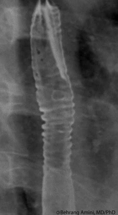
Here we see a right lower quadrant mass, picked up on physical examination, seen on x-ray and further investigated by ultrasound and CT. The differential diagnosis of an enhancing retroperitineal mass such as this is:
  • Lymphoma: Enlarged nodes bilaterally. Confluent soft tissue mantle of nodes surrounding aorta and inferior vena cava. Nodes may displace aorta from spine, unusual for other nodal mets
  • Metastasis
  • Infection: Abscess, tuberculosis.
  • Sarcoma: Large RP mass with or without necrotic or cystic degeneration
  • Schwannoma
  • Paraganglioma
  • Hemangiopericytoma
  • Inflammatory pseudotumor: Heterogeneous mass. Nonenhancing, heterogeneous enhancement, or peripheral enhancement. May have central hypoattenuation due to necrosis in larger lesions. May have Central calcifications.
  • Castleman

References