Saturday, January 16, 2016

Suspected Type I Rotatory Atlatoaxial Subluxation in Asymptomatic Patients

type I Atlantoaxial Rotatory Subluxation
In an earlier post on rotatory atlatoaxial subluxation, we discussed the Fielding and Hawkins classification, and its application to symptomatic patients (e.g., those with torticollis). Here, we discuss the challenge of making a diagnosis of rotatory atlatoaxial subluxation in unselected patients.

That is, what do you do if your neck or c-spine CT scan is obtained with the head turned, and you see what looks like a Type I rotatory atlatoaxial subluxation?

The patient below was referred to our institution in a cervical collar for management of atlantoaxial rotatory subluxation.
normal head rotation
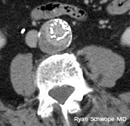
30-year-old man: Thick reconstructions centered at C1-C2 from a CTA obtained with the head rotated to the left. There is apparent type I atlantoaxial rotatory subluxation.
 
Let's take a look at what we know:
  • Normal maximum rotation of the head on body is between 60–80°.
  • Normal maximum rotation of C1 on C2 makes up for about half of that: 30–45° (although some sources say it can be up to 50°).
  • Cutoff for abnormal C1-C2 rotation: >45° or >56°, depending on source.
Looking at our image above, our rotation of C1 on C2 is about 30 degrees, which puts it in the normal range. But, what if the number was higher? How well do we do as radiologists in correctly identifying this as normal?

One study that was helpful came from the forensic field. Pathologists had noted that CTs of cadavers, which are impossible to position "correctly," were resulting in a lot of over-calling of atlantoaxial rotatory subluxation. When they looked at their data they found:
  • 19 cases where the C-spine was stable on autopsy. In those 19, 13 had suspected rotatory subluxation based on on CT (false positives)
  • The C1-C2 angle in those 13 false positives were between 16–47°.
  • All false positives were type I.
  • 2 cases where the C-spine was unstable at autopsy. In those 2 cases, 1 had suspected rotatory subluxation on CT (true positive).
  • The C1-C2 angle in the 1 true positive was 42°.
  • There was a significant association between the false positives and degree of head rotation.

Generalizing this cadaveric study suggests that we need to be careful in calling type I atlantoaxial rotatory subluxation in asymptomatic patients who simply happen to have their head turned in the scanner. This supports the conclusions from a study in living, asymptomatic patients, where the authors showed that incomplete rotational facet displacement on CT was not sufficient to define subluxation.

References

Wednesday, January 6, 2016

Endologix Stent-graft

Ryan Schwope
Arterial phase CT shows smoothly marginated contrast extending beyond the confines of the metal struts of the stent
Ryan Schwope
Delayed CT shows the smoothly marginated contrast extending beyond the confines of the metal strut unchanged in size and configuration when compared to arterial phase imaging
Most endostent devices used for endovascular repair of aortic aneurysms have the graft material sutured on the luminal side of the stent. The AFX endostent device (Endologix, Irvine, California) is different from other devices in that the graft material is sutured on the outside of the metal component, attached only at the proximal and distal ends.

By intent, the graft material of the AFX stent-graft can separate from the metal struts where it is not directly apposed to the aortic wall.  During surveillance with contrast-enhanced imaging of patients who have reviewed this device, contrast can be detected outside of the metallic construct. Although this finding can imply a type 1 or 3 endoleak when identified with other endostents, it is a normal finding with the AFX device.

Avoiding misinterpretation as an endoleak can be achieved by confirmation of the type of endostent device used for aneurysm repair. In addition, the contrast extending beyond the metal struts does not change in configuration or size on delayed imaging.

References:

J Vasc Interv Radiol. 2012 Nov;23(11):1544-6.

Wednesday, December 23, 2015

Management of Pancreatic Cystic Lesions

Ryan Schwope
Thick slab MRCP image showing massive dilatation of the main pancreatic duct
Incidental cystic pancreatic lesions found on 13% of MRI abdomens
  • Wide variety of pathology both benign and malignant
  • Imaging findings and demographics are the key to diagnosis

Cystic Pancreatic Neoplasms (Four major categories)

  1. Serous cystadenoma: Benign (very low malignant potential)
  2. Mucinous cystic neoplasm (MCN): Premalignant or malignant
  3. Intraductal papillary mucinous neoplasm (IPMN): Malignant potential (Main Duct >> Branch Duct)
  4. Unusual cystic neoplasms:
    • Solid pseudopapillary neoplasm (SPN): Low grade malignancy
    • Cystic forms of more common neoplasms (neuroendocrine)

Nonneoplastic Pancreatic Cysts

  • Pseudocyst
  • Retention cyst
  • Lymphoepithelial cyst
  • Localized ductectasia

Major Imaging Features Guiding Management

  • Number and size of cystic components: Risk of malignancy increases when size ≥ 3 cm
  • Septations and solid components: Mural nodule has a 87% Sp and 56% Sn for malignancy
  • Main pancreatic duct (MPD) dilatation and communication with the cystic lesion: MPD > 10 mm has a 77% Sp and 67-92% Sn for malignancy

Sendai Criteria

High Risk Stigmata
  • Jaundice
  • MPD ≥ 10 mm
  • Enhancing solid component
Worrisome Features
  • Size ≥ 3 cm
  • MPD 5-9 mm
  • Non-enhancing mural nodules
  • Thick enhancing cystic wall
  • Lymphadenopathy
  • Abrupt Duct Termination

Management

  • Any worrisome features present = Endoscopic Ultrasound (EUS) and Cyst aspiration with fluid analysis
  • Any high risk stigmata present or suspicious cytology on EUS = Surgical resection
  • MCN or SPN = Surgical resection
  • Serous cystadenoma
    • 2-3 cm: F/U every 2 years
    • ≥ 4 cm: consider resection
  • IPMN:
    • Main duct and combined type: Surgical resection (but depends on location, pt. age/clinical status)
    • Branch duct type = follow if < 3 cm and contains no solid components
      • If < 2cm F/U q1yr; if growth FU q6mo
      • If 2-3 cm F/U q6mo x 2 years, then q1yr
      • Consider EUS (if mucinous then resect)
      • If growth ≥ 3 cm, resect

What the Clinician/Surgeon wants to know

  1. Number of cystic lesions
  2. Largest cystic lesion
    • Unilocular
    • Multilocular: Microcystic (<2cm) or Macrocystic (> 2cm)
  3. Lesion size
  4. Lesion location: Head/Body/Tail
  5. Septations: None/Thin/Thick (> 2mm)
  6. Solid components: Present/Absent
  7. Calcifications: None/Coarse/Rim/Central
  8. Communication with MPD: Present/Absent
  9. Main pancreatic duct diameter: > 5 mm/Not dilated

References

Wednesday, December 9, 2015

Bisphosphonate-related Osteonecrosis of the Jaw


Ryan Schwope BROJN1
Figure 1: Axial CT of the facial bones. There is a mixed sclerotic and lytic lesion within the mandible (Figure 1, arrows) with foci of cortical interruption (Figure 2, arrowheads). 

Ryan Schwope BROJN 2
Figure 2: Sagital CT of the facial bones. There is a mixed sclerotic and lytic lesion within the mandible (Figure 1, arrows) with foci of cortical interruption (Figure 2, arrowheads).

Bisphosphonate-related osteonecrosis of the jaw (BRONJ) is associated with the use of bisphosphonates to treat severe osteoporosis, and metabolic and oncologic bone conditions, including hypercalcemia associated with malignancy. These agents inhibit osteoclasts, reducing bone resorption and osteolysis, and also possess antiangiogenic properties, reducing blood flow and necrosis. Patients with BRONJ present with pain and exposed, nonvital bone involving the maxillofacial structures. The incidence of BRONJ increases with the duration of treatment, especially with the intravenous route and concomitant steroid therapy. The osteonecrosis usually is participated by dental extraction. When spontaneous, it commonly occurs along the mylohyoid ridge. BRONJ can mimic osteomyelitis and osteoradionecrosis. A history of dental caries and the presence of periosteal elevation can help direct one towards the diagnosis of osteomyelitis. Osteoradionecrosis can be excluded if the patient has not received oropharyngeal radiation therapy. Jaw neoplasm, primary or metastatic, can potentially also mimic BRONJ. Treatment of BRONJ consists of cessation of bisphosphonate drug therapy, antibiotics for secondary infection, and surgical debridement of necrotic sequestra.

References:


Thursday, November 19, 2015

Lipoleiomyoma


Ryan Schwope
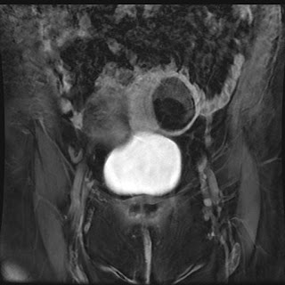
CT shows a predominantly fat attenuation, intramural uterine mass. There is rim of surrounding calcification as well as a few foci of internal soft tissue density at the superior aspect of the mass 

Ryan Schwope
   MR imaging showed the majority of the uterine mass as hyperintense on T1 weighted imaging (isointense to subcutaneous fat)
Ryan Schwope
T2 weighted image with fat saturation MRI shows the uterine mass markedly hypointense (isointense to subcutaneous fat)
Ryan Schwope
Post contrast T1 weighted MRI image with fat saturation show mld enhancement of the soft tissue component  along the superior margin of the mass; the majority of the mass is markedly hypointense (isointense to subcutaneous fat)
Uterine lipoleiomyomas are rare, benign tumors with a variable reported incidence ranging from 0.03% to 0.2%. The exact etiology of these lesions is unclear.  It is postulated lipoleiomyomas either arise from fatty metaplasia of the smooth muscle cells of leiomyomas, or from misplaced embryonic fat cells in the uterus. CT is highly specific for the diagnosis when an intrauterine mass is seen containing both macroscopic fat and soft tissue density. MR can also be confirmatory as the mass will have high T1 weighted signal which can be confirmed as fat by using a fat suppression. The role of imaging is also to differentiate lipoleiomyoma from an ovarian teratoma, a much more common entity presenting as a fat-containing pelvic mass. Lipoleiomyomas require no treatment or follow-up whereas teratomas are frequently resected. 


References


Monday, September 28, 2015

Giant Cell Containing Lesions of Bone

Reactive Benign Malignant
  • Osteosarcoma
  • Clear cell chondrosarcoma
  • Metastatic carcinoma
  • Malignant fibrous histiocytoma
  • Solitary fibrous tumor metastasis

Saturday, June 20, 2015

ILAR Classification of Juvenile Idiopathic Arthritis

Juvenile idiopathic arthritis (JIA) is an umbrella term for a group of abnormalities characterized by chronic articular inflammation and association with HLA alleles. The International League Against Rheumatism (ILAR) has classified JIA into seven subtypes, including an unclassifiable group. These include:
  • Systemic arthritis: Arthritis in one or more joints with or preceded by fever of at least 2 weeks' duration documented to be daily for at least 3 days, and accompanied by one or more of: evanescent erythematous rash, lymphadenopathy, hepatomegalyor splenomegaly, or both, serositis
  • Oligoarthritis: Arthritis in 4 or fewer joints in the first 6 months. Subtypes include persistent (no more than 4 joints throughout the course of the disease) and extended (more than 4 joints after the first 6 months).
  • Polyarthritis, RF negative: Arthritis affecting 5 or more joints in the first 6 months of disease. RF is negative
  • Polyarthritis, RF positive: Arthritis affecting 5 or more joints in the first 6 months of disease. Two or more tests for RF, conducted at least 3 months apart during the first 6 months, are positive. Considered the pediatric version of adult rheumatoid arthritis.
  • Psoriatic: Arthritis plus psoriasis OR Arthritis plus at least two of the following: dactylitis, nail pitting or onycholysis, psoriasis in a first-degree relative.
  • Enthesis-related: Arthritis plus enthesitis OR Arthritis or enthesitis, plus at least two of the following: presence of or a history of sacroiliac joint tenderness and/or inflammatory lumbosacral pain‡, presence of HLA-B27 antigen, onset of arthritis in a male over 6 years of age, acute (symptomatic) anterior uveitis, history of AS, ERA, sacroiliitis with IBD, reactive arthritis, or acute anterior uveitis in a first-degree relative
  • Unclassified: Arthritis that fulfills criteria in none of the above categories, or fulfills criteria in two or more of the above categories

References

Petty RE, Southwood TR, Baum J, Bhettay E, Glass DN, Manners P, Maldonado-Cocco J, Suarez-Almazor M, Orozco-Alcala J, Prieur AM. Revision of the proposed classification criteria for juvenile idiopathic arthritis: Durban, 1997. J Rheumatol. 1998 Oct;25(10):1991-4.