Wednesday, December 21, 2016
M.D. = Makes Decisions (unless you're a radiologist)
In the real world (with the cat), anything other than statement #1 will get you laughed at. In radiology, statement #1 is rare. Instead we teach our residents and fellows, by our own weak examples, to be as non-declarative as possible.
Statements #2 and #3 are as declarative as most radiologists get. "I said most likely. What more do you want from me?!"
Statement #4 just passes the buck to the next radiologist.
Statement #5 combines 2 mild hedge words to produce one super-hedgy sentence.
Statement #6 is the reason Bayes rolls in his grave every time a radiologist signs a report.
Statement #7 is basically saying, "Thanks for the money suckers! This report was useless." We have access to so much patient data these days that it baffles me to see this in reports. Of course, this doesn't apply to cases where we're reading in isolation and when the only history we get from referrings is "pain," or some random ICD code. This negligent absence of data in a requisition borders on (is?) malpractice. I've seen it in imaging referrals my family members get from their doctors and it aggravates me to no end.
Statement #8, I don't even... For a cat/hemangioma?
Look, sometimes we have to hedge. Sometimes we are no better than Plato's cave captives, squinting at shadows with no idea of what's behind us. We know that two or more widely disparate entities can have identical imaging features. But when you know something can only be one thing, just say so. Save the patient some anxiety. And, save the rest of us some money by reducing unnecessary imaging.
What are some of your favorite radiology hedges?
Labels:
Musculoskeletal,
Neuroradiology,
Oncology,
rant,
Terminology
Wednesday, March 2, 2016
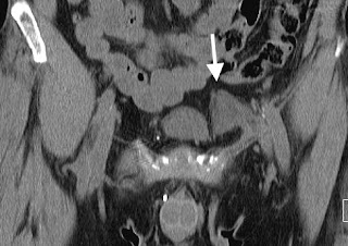
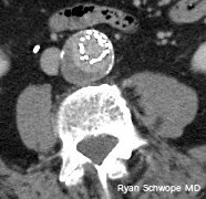
Mimicker: Inguinal Mesh Plug
 |
| Left inguinal mesh plug with a lobular contour |
 |
| Left inguinal mesh plug. Note conical morphology on this coronal MPR. |
- Depending on the inguinal hernia repair method chosen, a mesh plug can be used for reinforcement. One such method uses a polypropylene (Prolene) plug
- The post-operative appearance of an inguinal mesh plug can masquerade as an inguinal mass or lymphadenopathy
Imaging Findings
- On CT, mesh plugs can have a slightly nodular or smooth outline. The density is similar to sightly lower than muscle. Mesh plugs have also been reported as a ring-like density with central fat attenuation, potentially mimicking epiploic appendicitis if on the left
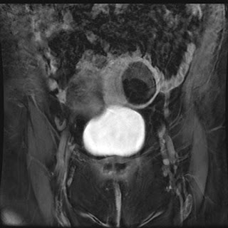
- On MRI, mesh plugs are typically T1 hypointense and demonstrate variable T2 weighted signal
- Due to a granulomatous reaction, mesh plugs can be FDG avid
Helpful hints in preventing misdiagnosis
- Mutliplanar reformations can be useful in demonstrating the conical morphology of the mesh plug, although they can appear ovoid
- Postsurgical changes including skin thickening and suceptabliity artifact can also be helpful imaging features
Saturday, January 16, 2016
Suspected Type I Rotatory Atlatoaxial Subluxation in Asymptomatic Patients
In an earlier post on rotatory atlatoaxial subluxation, we discussed the Fielding and Hawkins classification, and its application to symptomatic patients (e.g., those with torticollis). Here, we discuss the challenge of making a diagnosis of rotatory atlatoaxial subluxation in unselected patients.
That is, what do you do if your neck or c-spine CT scan is obtained with the head turned, and you see what looks like a Type I rotatory atlatoaxial subluxation?
The patient below was referred to our institution in a cervical collar for management of atlantoaxial rotatory subluxation.

30-year-old man: Thick reconstructions centered at C1-C2 from a CTA obtained with the head rotated to the left. There is apparent type I atlantoaxial rotatory subluxation.
Let's take a look at what we know:
One study that was helpful came from the forensic field. Pathologists had noted that CTs of cadavers, which are impossible to position "correctly," were resulting in a lot of over-calling of atlantoaxial rotatory subluxation. When they looked at their data they found:
Generalizing this cadaveric study suggests that we need to be careful in calling type I atlantoaxial rotatory subluxation in asymptomatic patients who simply happen to have their head turned in the scanner. This supports the conclusions from a study in living, asymptomatic patients, where the authors showed that incomplete rotational facet displacement on CT was not sufficient to define subluxation.
That is, what do you do if your neck or c-spine CT scan is obtained with the head turned, and you see what looks like a Type I rotatory atlatoaxial subluxation?
The patient below was referred to our institution in a cervical collar for management of atlantoaxial rotatory subluxation.

30-year-old man: Thick reconstructions centered at C1-C2 from a CTA obtained with the head rotated to the left. There is apparent type I atlantoaxial rotatory subluxation.
Let's take a look at what we know:
- Normal maximum rotation of the head on body is between 60–80°.
- Normal maximum rotation of C1 on C2 makes up for about half of that: 30–45° (although some sources say it can be up to 50°).
- Cutoff for abnormal C1-C2 rotation: >45° or >56°, depending on source.
One study that was helpful came from the forensic field. Pathologists had noted that CTs of cadavers, which are impossible to position "correctly," were resulting in a lot of over-calling of atlantoaxial rotatory subluxation. When they looked at their data they found:
- 19 cases where the C-spine was stable on autopsy. In those 19, 13 had suspected rotatory subluxation based on on CT (false positives)
- The C1-C2 angle in those 13 false positives were between 16–47°.
- All false positives were type I.
- 2 cases where the C-spine was unstable at autopsy. In those 2 cases, 1 had suspected rotatory subluxation on CT (true positive).
- The C1-C2 angle in the 1 true positive was 42°.
- There was a significant association between the false positives and degree of head rotation.
Generalizing this cadaveric study suggests that we need to be careful in calling type I atlantoaxial rotatory subluxation in asymptomatic patients who simply happen to have their head turned in the scanner. This supports the conclusions from a study in living, asymptomatic patients, where the authors showed that incomplete rotational facet displacement on CT was not sufficient to define subluxation.
References
- Fielding JW, Hawkins RJ. Atlanto-axial rotatory fixation. (Fixed rotatory subluxation of the atlanto-axial joint). J Bone Joint Surg Am. 1977 Jan;59(1):37-44.
- Persson A, Falk J, Berge J, Jackowski C. Atlanto-axial rotatory subluxations in postmortem CT: radiologists be aware of a common pitfall. Forensic Sci Int. 2013 Feb 10;225(1-3):9-14.
- Mönckeberg JE1, Tomé CV, Matías A, Alonso A, Vásquez J, Zubieta JL. CT scan study of atlantoaxial rotatory mobility in asymptomatic adult subjects: a basis for better understanding C1-C2 rotatory fixation and subluxation. Spine (Phila Pa 1976). 2009 May 20;34(12):1292-5.
Wednesday, January 6, 2016
Endologix Stent-graft
 |
| Arterial phase CT shows smoothly marginated contrast extending beyond the confines of the metal struts of the stent |
 |
| Delayed CT shows the smoothly marginated contrast extending beyond the confines of the metal strut unchanged in size and configuration when compared to arterial phase imaging |
By intent, the graft material of the AFX stent-graft can separate from the metal struts where it is not directly apposed to the aortic wall. During surveillance with contrast-enhanced imaging of patients who have reviewed this device, contrast can be detected outside of the metallic construct. Although this finding can imply a type 1 or 3 endoleak when identified with other endostents, it is a normal finding with the AFX device.
Avoiding misinterpretation as an endoleak can be achieved by confirmation of the type of endostent device used for aneurysm repair. In addition, the contrast extending beyond the metal struts does not change in configuration or size on delayed imaging.
References:
J Vasc Interv Radiol. 2012 Nov;23(11):1544-6.
Labels:
Devices,
Don't Panic,
Interventional radiology
Wednesday, December 23, 2015
Management of Pancreatic Cystic Lesions
 |
| Thick slab MRCP image showing massive dilatation of the main pancreatic duct |
- Wide variety of pathology both benign and malignant
- Imaging findings and demographics are the key to diagnosis
Cystic Pancreatic Neoplasms (Four major categories)
- Serous cystadenoma: Benign (very low malignant potential)
- Mucinous cystic neoplasm (MCN): Premalignant or malignant
- Intraductal papillary mucinous neoplasm (IPMN): Malignant potential (Main Duct >> Branch Duct)
- Unusual cystic neoplasms:
- Solid pseudopapillary neoplasm (SPN): Low grade malignancy
- Cystic forms of more common neoplasms (neuroendocrine)
Nonneoplastic Pancreatic Cysts
- Pseudocyst
- Retention cyst
- Lymphoepithelial cyst
- Localized ductectasia
Major Imaging Features Guiding Management
- Number and size of cystic components: Risk of malignancy increases when size ≥ 3 cm
- Septations and solid components: Mural nodule has a 87% Sp and 56% Sn for malignancy
- Main pancreatic duct (MPD) dilatation and communication with the cystic lesion: MPD > 10 mm has a 77% Sp and 67-92% Sn for malignancy
Sendai Criteria
High Risk Stigmata- Jaundice
- MPD ≥ 10 mm
- Enhancing solid component
- Size ≥ 3 cm
- MPD 5-9 mm
- Non-enhancing mural nodules
- Thick enhancing cystic wall
- Lymphadenopathy
- Abrupt Duct Termination
Management
- Any worrisome features present = Endoscopic Ultrasound (EUS) and Cyst aspiration with fluid analysis
- Any high risk stigmata present or suspicious cytology on EUS = Surgical resection
- MCN or SPN = Surgical resection
- Serous cystadenoma
- 2-3 cm: F/U every 2 years
- ≥ 4 cm: consider resection
- IPMN:
- Main duct and combined type: Surgical resection (but depends on location, pt. age/clinical status)
- Branch duct type = follow if < 3 cm and contains no solid components
- If < 2cm F/U q1yr; if growth FU q6mo
- If 2-3 cm F/U q6mo x 2 years, then q1yr
- Consider EUS (if mucinous then resect)
- If growth ≥ 3 cm, resect
What the Clinician/Surgeon wants to know
- Number of cystic lesions
- Largest cystic lesion
- Unilocular
- Multilocular: Microcystic (<2cm) or Macrocystic (> 2cm)
- Lesion size
- Lesion location: Head/Body/Tail
- Septations: None/Thin/Thick (> 2mm)
- Solid components: Present/Absent
- Calcifications: None/Coarse/Rim/Central
- Communication with MPD: Present/Absent
- Main pancreatic duct diameter: > 5 mm/Not dilated
References
- Gandhi NS and Hecht E. “Adding Value to Clinical Care: Structured Reporting of Pancreatic Pathology”, Workshop Session Presentation, Society of Abdominal Radiology Annual Meeting, 2015
- Tanaka M,Fernández-del Castillo C, Adsay V,et al. International consensus guidelines 2012 for the management of IPMN andMCN of the pancreas. Pancreatology(2012) May-Jun;12(3):183-97
- DewhurstCE, Mortele KJ. Cystic tumors of the pancreas: Imaging and management. RadiolClin N Am 50(2012)467-86
- SahaniDV, Miller JC, and Fernandez C et al. Cystic pancreatic lesions: Classificationand Management. ACR (2009)
- SahaniDV, Kadavigere R, and Saoker A. Cystic Pancreatic Lesions: A SimpleImaging-based Classification System for Guiding Management. Radiographics (2005)25:1471-1484
Wednesday, December 9, 2015
Bisphosphonate-related Osteonecrosis of the Jaw
 |
| Figure 1: Axial CT of the facial bones. There is a mixed sclerotic and lytic lesion within the mandible (Figure 1, arrows) with foci of cortical interruption (Figure 2, arrowheads). |
 |
| Figure 2: Sagital CT of the facial bones. There is a mixed sclerotic and lytic lesion within the mandible (Figure 1, arrows) with foci of cortical interruption (Figure 2, arrowheads). |
References:
- Am Fam Physician. 2012 Jun 15;85(12):1134-114
- RadioGraphics 2009; 29:1971–1986
- AJNR June 2007 28: 139-1145
Labels:
General,
Head and Neck,
Musculoskeletal,
Neuroradiology,
Oncology
Thursday, November 19, 2015
Lipoleiomyoma
 |
| CT shows a predominantly fat attenuation, intramural uterine mass. There is rim of surrounding calcification as well as a few foci of internal soft tissue density at the superior aspect of the mass |
 |
| MR imaging showed the majority of the uterine mass as hyperintense on T1 weighted imaging (isointense to subcutaneous fat) |
 |
| T2 weighted image with fat saturation MRI shows the uterine mass markedly hypointense (isointense to subcutaneous fat) |
References
- Preito A,Crespo C, Pardo A, Docal I, Calzada J, Alonso P. Uterine lipoleiomyomas: US andCT findings. Abdom Imaging 2000;25:655-7
- MaebayashiT, Imai K, Takekawa Y, et al. Radiologic features of uterine lipoleiomyoma. JComput Assist Tomogr 2003;27:162-5
- AvritscherR, Iyer RB, Ro J, Whitman G. Lipoleiomyoma of the uterus. AJR Am J Roentgenol2001;177:856
- Dodd GD III,Budzik RF. Lipomatous uterine tumors: diagnosis by ultrasound, CT, and MR. JComput Assist Tomogr 1990;14:629-32
Subscribe to:
Posts (Atom)


