Friday, September 7, 2012

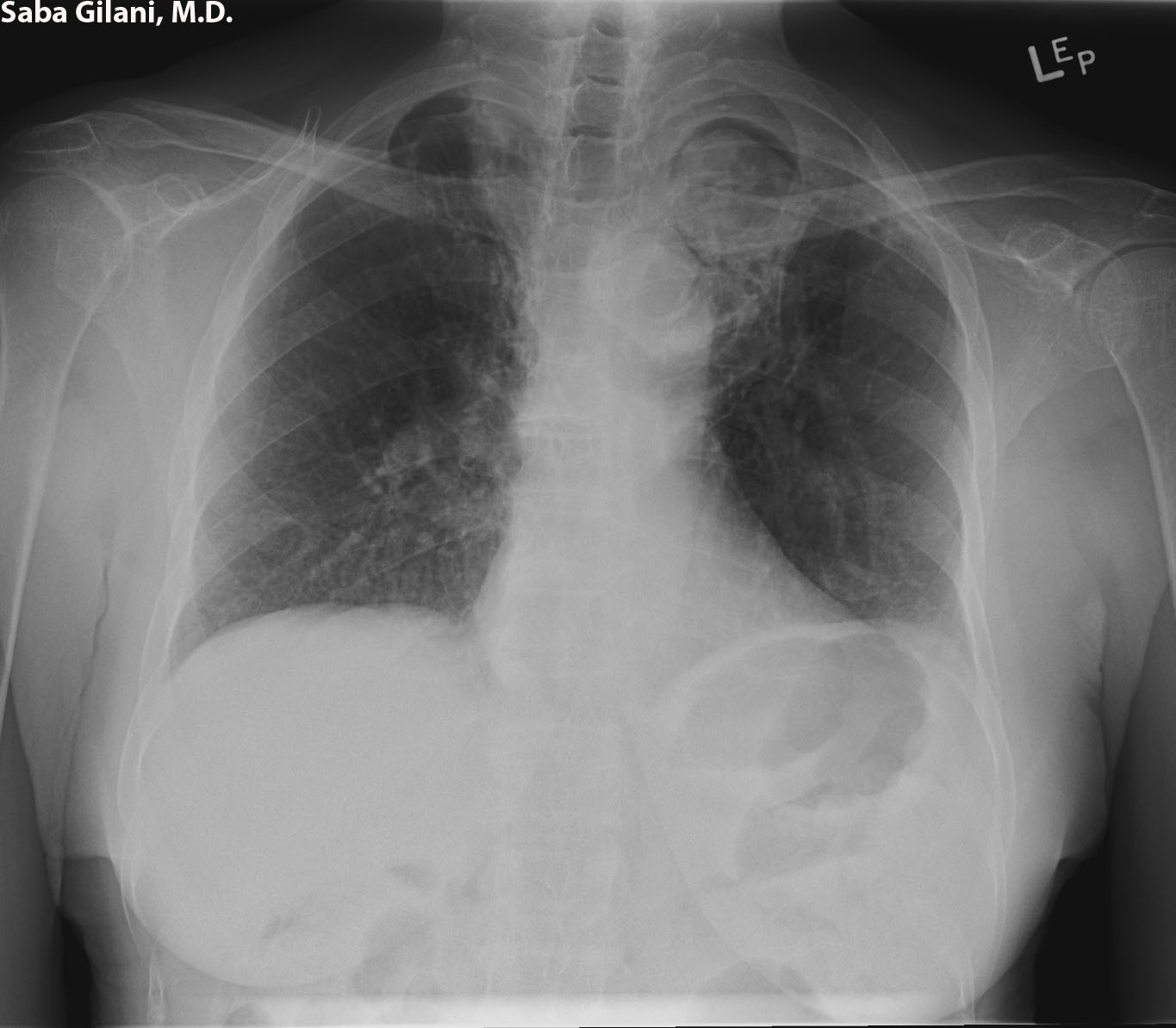
The Air Crescent Sign


The air crescent sign refers to a radiolucent rim around a soft tissue density and is characteristic of invasive pulmonary aspergillosis (or other angioinvasive fungal infections) in immunocompromised patients. It results when Aspergillus invades the pulmonary vasculature and causes hemorrhage with subsequent infarction of pulmonary tissue. Over time, the infarcted tissue retracts and is absorbed along its periphery by leukocytes which is what gives rise to the air crescent appearance. The visualization of the air crescent sign indicates a favorable outcome because patients without leukocyte function cannot form this consolidation.

The image above is from a patient with prior history of tuberculosis who presented with a cough. The air crescent sign is seen in the left upper lobe.


REFERENCES
Abramson S. The air crescent sign. Radiology 2001;18:230-2.

Thursday, September 6, 2012

The Molar Tooth Sign - Joubert Syndrome

Joubert syndrome is an autosomal recessive disorder characterized by cerebellar vermian hypoplasia with a midline cleft and lack of decussation of the superior cerebellar peduncles, the latter causing an enlargement of the peduncles. Other decussation defects of the central pontine and corticospinal tracts as well as dysplasia of the olivary nuclei may also be present. Associated renal, retinal, and hepatic abnormalities are often seen. 

The molar tooth sign refers to the characteristic appearance of the midbrain seen on axial imaging (CT or MR) in patients with Joubert syndrome. Thickened superior cerebellar peduncles surrounding an elongated fourth ventricle give this appearance. A vermian cleft may also be recognized.


REFERENCES
McGraw P. The molar tooth sign. Radiology 2003;29:671-2.

Wednesday, September 5, 2012

Discitis/Vertebral Body Osteomyelitis


Vertebral body osteomyelitis (and associated discitis) is an infection involving the vertebral body endplates that extends to the intervening disc space. While vertebral body endplate destruction may be visible on plain films or bone CT studies, MR is the preferred imaging modality to assess for spread of infection to the disc space and paraspinal tissues, or for development of an epidural abscess. T1W images (A, D) will show endplate destruction and thickening of the paraspinal tissues with ill-defined fat places. T2W images (B) will demonstrate high signal in the intervertebral disc. Gadolinium enhanced images (C) will demonstrate enhancement of the infected vertebral bodies and paraspinal tissues. In the presence of an epidural abscess fluid sensitive sequences will show a collection in the epidural space that demonstrates peripheral enhancement upon gadolinium administration. 


REFERENCES
Dagirmanjian A, Schils J, McHenry M, et al. MR imaging of vertebral osteomyelitis revisited. Am J Roentgenol 1996;167(6):1539-43.
Kowalski TJ, Layton KF, Berbari EF, et al. Follow-up MR imaging in patients with pyogenic spine infections: lack of correlation with clinical features. Am J Roentgenol 2007;28:693-99.

Tuesday, September 4, 2012

Pulmonary Pseudotumors

Nodular Pulmonary Amyloid (Amyloidoma)
  • one of the three presentations of amyloidosis in the chest (the other two are tracheobronchial and nodular septal)
  • solitary or multiple nodules with smooth contours
  • lower lobe, subpleural/peripheral location 
  • may calcify or undergo osseous metaplasia
  • larger nodules may cavitate or become hemorrhagic
  • resection is diagnostic and curative
  • differential - primary or metastatic neoplasms
Exogenous Lipoid Pneumonia
  • caused by aspiration of mineral, vegetable, or animal oil present in food, radiologic contrast media, or oil based medications
  • NECT will show low attenuation, mass-like consolidation, with regions of fat density
  • crazy paving

REFERENCES
Gimenez A, Franquet T, Prats R, et al. Unusual primary lung tumors: a radiologic-pathologic overview. Radiographics 2002;22:601-19.

Monday, September 3, 2012

Happy (Non) Labor Day


Friday, August 31, 2012

Arterial Closure Devices

Initially used by cardiologists during percutaneous coronary interventions, arterial closure devices (ACDs) have made their way into the interventional radiology suite. With the potential to achieve rapid hemostasis and reducing patient time to ambulation, ACDs are increasingly favored over manual compression for arteriotomies especially since preliminary data demonstrates no significant difference in complications between the two methods. 

ACDs can either provide active (or intravascular) closure by engaging the vessel wall via clips, sutures, or plugs, versus passive (or extravascular) closure by introducing thrombosing agents or sealants via the sheath to plug the arteriotomy site. Few clinical trials exist demonstrating the benefit of intravascular versus extravascular closure. It is felt that intravascular ACDs are a better choice in patients who are anticoagulated. However, using a device with intravascular components introduces a nidus for potential infection. Additional complications include device embolization, arterial laceration, and limb ischemia due to luminal obstruction.

While additional research is needed to define the safety and efficacy profiles of ACDs, their use should certainly be considered by interventional radiology departments, especially due to their potential to improve patient care. 


REFERENCES
Dauerman HL, Applegate RJ, Cohen DJ. Vascular closure devices: the second decade. J Am Coll Cardiol 2007;50(17):1617-26.

Thursday, August 30, 2012

Mycetoma Formation in Fungal Sinusitis


A mycetoma (fungal ball) is an uncommon sequela of fungal sinusitis and is thought to be secondary to deficient mucociliary clearance of a fungal organism which incites an inflammatory response. The fungal ball usually appears as a mass within a sinus (usually the maxillary sinus) and is typically unilateral. Bone window CT will show a mass within the sinus with punctate calcifications. A fungal ball is typically of low signal intensity on T1W MR due to low water content. On T2WI the mycetoma will be hypointense relative to the hyperintense inflamed sinus mucosa.


REFERENCES
Aribandi M, McCoy VA, Bazan C. Imaging features of invasive and noninvasive fungal sinusitis: a review. Radiographics 2007;27:1283-96.