Initially used by cardiologists during percutaneous coronary interventions, arterial closure devices (ACDs) have made their way into the interventional radiology suite. With the potential to achieve rapid hemostasis and reducing patient time to ambulation, ACDs are increasingly favored over manual compression for arteriotomies especially since preliminary data demonstrates no significant difference in complications between the two methods.
ACDs can either provide active (or intravascular) closure by engaging the vessel wall via clips, sutures, or plugs, versus passive (or extravascular) closure by introducing thrombosing agents or sealants via the sheath to plug the arteriotomy site. Few clinical trials exist demonstrating the benefit of intravascular versus extravascular closure. It is felt that intravascular ACDs are a better choice in patients who are anticoagulated. However, using a device with intravascular components introduces a nidus for potential infection. Additional complications include device embolization, arterial laceration, and limb ischemia due to luminal obstruction.
While additional research is needed to define the safety and efficacy profiles of ACDs, their use should certainly be considered by interventional radiology departments, especially due to their potential to improve patient care.
REFERENCES
Dauerman HL, Applegate RJ, Cohen DJ. Vascular closure devices: the second decade. J Am Coll Cardiol 2007;50(17):1617-26.
Friday, August 31, 2012
Thursday, August 30, 2012
Mycetoma Formation in Fungal Sinusitis

A mycetoma (fungal ball) is an uncommon sequela of fungal sinusitis and is thought to be secondary to deficient mucociliary clearance of a fungal organism which incites an inflammatory response. The fungal ball usually appears as a mass within a sinus (usually the maxillary sinus) and is typically unilateral. Bone window CT will show a mass within the sinus with punctate calcifications. A fungal ball is typically of low signal intensity on T1W MR due to low water content. On T2WI the mycetoma will be hypointense relative to the hyperintense inflamed sinus mucosa.
REFERENCES
Aribandi M, McCoy VA, Bazan C. Imaging features of invasive and noninvasive fungal sinusitis: a review. Radiographics 2007;27:1283-96.
Wednesday, August 29, 2012
The Double Duct Sign
The double duct sign refers to the concomitant dilatation of the common bile duct and pancreatic duct seen on MRCP (although the same can be visualized on CT, US, and ERCP). Visualization of the double duct sign typically suggests the presence of a malignancy - either and ampullary or pancreatic head carcinoma. Other malignancies with this appearance include cholangiocarcinoma of the distal common bile duct, lymphoma, metastases, and in rare cases, Kaposi's sarcoma. Primary retroperitoneal fibrosis has also been reported to cause simultaneous dilatation of both bile ducts.
However, some studies have shown that the double duct sign is not as specific a predictor of pancreatic malignancy as initially thought to be since benign entities can present similarly. The most common benign causes are chronic pancreatitis and ampullary stenosis. Plumley et al., have suggested that the specific bile duct morphology on ERCP is a better predictor of benign versus malignant causes of ductal dilatation: abrupt stenosis of a bile duct with irregular margins is more likely to indicate malignancy while gradual ductal stenosis, especially when associated with smooth margins, is more indicative of benign lesions. Other predictors of benign disease include glandular calcifications and ectasia of the branching bile ducts (seen in chronic pancreatitis).
REFERENCES
Ahualli J. The Double Duct Sign. Radiology 2007;244:314-5.
Edge MD, Hoteit M, Patel AP. Clinical significance of main pancreatic duct dilation on computed tomography: single and double duct dilation. World J Gastroenterol 2007;31(11):1701-5.
Plumley TF, Rohrmann CA, Freeny PC, et al. Double duct sign: reassessed significance in ERCP. Am J Roentgenol 1982;138:31-5.
Tuesday, August 28, 2012
Internal Derangement of the Temporomandibular Joint

Internal Derangement of the Temporomandibular Joint (TMJ), more commonly known as dislocation of the TMJ, occurs due to an abnormal positional and functional relationship between the articular disc and the articulating surface. The images above are from a patient with anterior dislocations of the bilateral TMJs.
While this abnormality can usually be detected on plain films or bone NECT, MRI is the best modality to evaluate the TMJ especially in cases of recurrent derangement. On MRI, the low signal intensity articular disc will be displaced relative to the mandibular condyle. T1WI can show specific disc abnormalities including perforation, fibrosis or adhesions. The presence of a joint effusion can be detected as high signal on T2WI. The presence of synovitis can be detected as enhancement of the disc on T1WI after administration of contrast.
REFERENCES
Sommer OJ, Aigner F, Rudisch A, et al. Cross-sectional and functional imaging of the temporomandibular joint: radiology, pathology, and basic biomechanics of the jaw. Radiographics 2003;23:e14.
Tomas X, Pomes J, Berenguer J, et al. MR imaging of temporomandibular joint dysfunction: a pictorial review. Radiographics 2006;26:765-81.
Monday, August 27, 2012
Intramedullary Osteosclerosis
A nonhereditary condition that effects the long bones of the lower extremities (tibia > femur, fibula). On imaging studies it presents with endosteal sclerosis extending into the medullary canal, possibly obliterating the canal. There is no associated periosteal reaction or soft tissue swelling. Bone scan will show intense radiopharmaceutical uptake.This condition is more common in women and usually presents as pain that is exacerbated by activity.
Differential diagnosis:
1. Ribbing disease - may look exactly the same. The main difference is that Ribbing disease tends to have a familial inheritance pattern (thought to be autosomal recessive).
2. Stress fracture - usually a focal sclerosis and periosteal reaction will be present.
3. Sclerotic metastases - patchy, discontinuous presentation, population tends to be older.
4. Progressive diaphyseal displasia (Camurati-Engelmann disease) - presents in young boys, has skull and vertebral body involvement, disease begins in the diaphysis of long bones and may extend to the metaphysis, has periosteal and endosteal sclerosis.
5. Melorheostosis (Leri-Weill disease) - a sclerosing bone dysplasia which presents unilaterally in a long bone of the upper or lower extremity and may have associated soft tissue calcifications.
REFERENCES
Balkissoon ARA and Hayes CW. Intramedullary osteosclerosis. Radiology 1999;212:708-10.
Chanchairujira K, Chung CB, Lai YM, et al. Intramedullary osteosclerosis: imaging features in nine patients. Radiology 2001;220:225-30.
Friday, August 24, 2012
Name the Device

Deep Brain Stimulator: electrodes implanted in the thalamus for treatment of Parkinson's disease.
REFERENCES
Hunter TB, Yoshino MT, Dzioba RB, et al. Medical devices of the head, neck, and spine. Radiographics 2004;24:257-285.
Thursday, August 23, 2012
Normal Variants in the Pediatric Cervical Spine
Interpreting cervical spine x-rays in the pediatric population can be a challenge due to normal anatomic variants. At age 8-10 a child's cervical spine reaches adult proportions. Normal variants to be considered in the younger populations include:
1. The atlantodens interval (ADI) may be up to 5mm in the pediatric patient whereas the upper limit of normal is 3mm in an adult.
2. Pseudo-Jefferson fracture: up to 6mm displacement of the lateral masses of atlas on the axis on the open mouth view is normal for children up to age 7.
3. Pseudo-subluxation of C2 on C3 (and to a lesser extent C3 on C4): normal mobility of upper cervical spine (due to ligamentous laxity) can cause up to a 4mm anterior displacement of C2 on C3. The Swischuk line - a line drawn through the posterior arch of C2 should be within 2mm of the spinolaminar line drawn at C1-C3 - can be used to determine whether this finding is normal or due to possible hangman's fracture. A discrepancy of > 2mm can indicate a fracture.
4. Anterior "wedging" of up to 3mm is a normal finding.
5. Ossification centers and unfused apophyses may mimic fractures.
6. Absence of cervical spine lordosis may be seen up to age 16.
REFERENCES
Curtin P and McElwain J. Assessment of the "nearly normal" cervical spine radiograph: C2-C3 pseudosubluxation in an adult with whiplash injury. Emerg Med J 2005;22:907-8.
Lustrin ES, Karakas SP, Ortiz AO, et al. Pediatric cervical spine: normal anatomy, variants, and trauma. Radiographics 2003;23:539-60.
Wednesday, August 22, 2012
Hereditary Osteo-onychodysplasia Disease
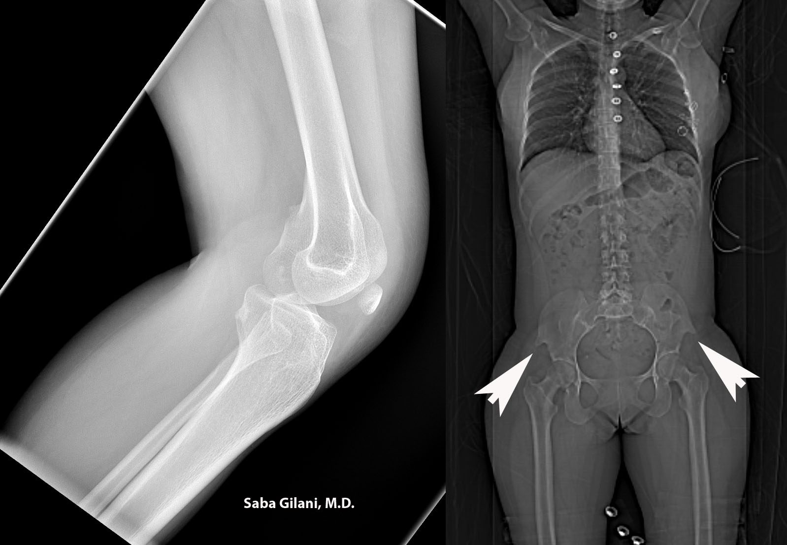
Hereditary osteo-onychodysplasia disease (HOOD), also known as Nail Patella Disease or Iliac Horn Syndrome is a genetic disorder due to an autosomal dominant mutation in the LMX1B gene. A pathognomonic finding is the presence of "iliac horns" projecting posterolaterally from the bilateral iliac bones. Other associated findings are absence or hypoplasia of the patella and radial head. Nail deformities are also common. The joint deformities can lead to osteoarthritis. About 40% of patients may also develop renal disease ranging from proteinuria to nephrotic syndrome.
Above, the lateral radiograph of the knee is from a patient who presented to the emergency room after a fall. A hypoplastic patella was incidentally noted. A review of prior imaging demonstrated bilateral iliac horns best seen on the scout image.
REFERENCES
Scott JE and Taor WS. The small patella syndrome. J Bone Joint Surg [Br] 1979;61-B:172-5.
Thompson EA, Walker T, Weens HS. Iliac horns. An osseous manifestation of hereditary arthrodysplasia associated with dystrophy of the fingernails. Radiology 1949;53:88-92.
Tuncbilek N, Karakas HM, Okten OO. Imaging of nail-patella syndrome. Hong Kong Med J 2005;11(2):116-8.
Tuesday, August 21, 2012
Lymph Node Stations for Pelvic Tumors
Most pelvic visceral tumors metastasize via lymphatics. Metastases to regional lymph nodes is consider N stage in the TNM classification system while spread to nonregional lymph nodes is considered M stage disease (distant metastases). Since upstaging influences prognosis and clinical management, knowledge of the pelvic nodal anatomy is important in evaluation of cross sectional imaging.
The following groups of pelvic lymph nodes should be considered:
1. Common iliac lymph nodes: between the aortic bifurcation and the common iliac vessel bifurcation.
REFERENCES
McMahon CJ, Rofsky NM, Pedrosa I. Lymphatic metastases from pelvic tumors: anatomic classification, characterization, and staging. Radiology 2010;254:31-46.
The following groups of pelvic lymph nodes should be considered:
1. Common iliac lymph nodes: between the aortic bifurcation and the common iliac vessel bifurcation.
- Medial, lateral, and lumbosacral subdivisions
- lumbosacral subdivision refers to lymph nodes in the lumbosacral fossa (triangular region bounded by common iliac vessels medially, psoas muscle laterally, and lower lumbar/upper sacral vertebral bodies posteriorly)
- Medial and lateral subdivisions
- obturator nodes are considered part of the medial subdivision and gather their name from proximity to the obturator internus muscle
- Anterior, lateral sacral, and presacral subdivisions
- hypogastric is a term used by some to describe the most cephalic of the internal iliac lymph nodes while others use the term to describe all internal iliac lymph nodes as a group
- Superficial subdivision nodes lie anterior to the inguinal ligament and femoral vessels
- Deep subdivision lymph nodes are enclosed within the femoral sheath
REFERENCES
McMahon CJ, Rofsky NM, Pedrosa I. Lymphatic metastases from pelvic tumors: anatomic classification, characterization, and staging. Radiology 2010;254:31-46.
Monday, August 20, 2012
Hereditary Syndromes Associated with Craniosynostoses

Dolichocephaly is a craniosynostosis involving premature closure of the sagittal suture resulting in an elongated head shape as shown above. The craniosynostoses were previously discussed here: craniosynostoses.
Hereditary syndromes that are associated with craniosynostoses are:
Crouzon syndrome: premature synostosis, maxillary hypoplasia, shallow orbits
Apert's syndrome: Coronal synostosis, midfacial hypoplasia, bilateral syndactyly, symphalangism (ankylosis of interphalangeal joints)
Pfeiffer syndrome: premature synostosis, broad thumbs and great toes, mild syndactyly
Carpenter syndrome: premature synostosis, severe developmental delay, brachydactyly, syndactyly, thumb duplication.
Acquired conditions that can result in premature synostosis include Rickets, hypophosphatasia, and mucopolysaccharidoses.
REFERENCES
Glass RBJ, Fernbach SK, Norton KI, et al. Radiographics 2004;24:507-22.
Friday, August 17, 2012
Pulmonary Patterns in Stage IV Sarcoidosis
The Siltzbach classification system defines five stages of sarcoidosis based on chest radiograph findings:
Stage 0 - No radiographic abnormality
Stage 1 - Lymphadenopathy
Stage 2 - Lymphadenopathy + pulmonary infiltrate
Stage 3 - Pulmonary infiltrate
Stage 4 - Pulmonary fibrosis
About 20% of patients with sarcoidosis will develop pulmonary fibrosis which indicates irreversible disease. Three main HRCT patterns of pulmonary fibrosis have been identified and linked to performance on pulmonary function tests:
1. Bronchial distortion in a primarily central distribution, associated with lower expiratory flow rates
2. Honeycombing in a peripheral and upper lobe distribution, associated with restriction and decreased diffusion capacity of carbon monoxide
3. Coarse, irregular linear opacities that radiate from the hila, associated with the least functional impairment.
Distinguishing between these patterns is important as it may predict treatment response to corticosteroids.
REFERENCES
Abhesera M, Valeyre D, Grenier P, et al. Sarcoidosis with pulmonary fibrosis: CT pattern and correlation with pulmonary function. AJR Am J Roentgenol 2000;174(6):1751-1757.
Criado E, Sanchez M, Ramirez J, et al. Pulmonary sarcoidosis: typical and atypical manifestations at high resolution CT with pathologic correlation. Radiographics 2010;30:1567-1586.
Thursday, August 16, 2012
Intraperitoneal Migration of an Intrauterine Device

As intrauterine devices have become a common form of contraception worldwide, their normal and abnormal imaging appearance should be recognized. The stem of a properly positioned intrauterine device (IUD) should be entirely within the endometrial cavity with the arms extending laterally at the uterine fundus. The device should never be seen in the endocervical canal.
An IUD may be expulsed from the uterine cavity or displaced within it. Perforation through the uterus and migration into the peritoneal cavity is a rare complication. An intraperitoneal IUD may cause bowel/omental adhesions, abscess formation, or perforation of intraabdominal structures. Timely surgical intervention can prevent these complications.
REFERENCES
Boortz HE, Margolis DJA, Ragavendra N, et al. Migration of intrauterine devices: radiologic findings and implications for patient care. Radiographics. 2012 March;32:335-52.
Peri N, Graham D, Levine D. Imaging of intrauterine contraceptive devices. J Ultrasound Med 2007;26(10):1389-1401.
Images courtesy of Anna Nazarenko, M.D.
Wednesday, August 15, 2012
Anomalies of the Inferior Vena Cava and Their Clinical Significance
1. Left IVC: joins the left renal vein and crosses anterior to the aorta to join the right renal vein
- can be mistaken for paraaortic adenopathy
- report of AAA rupture into the IVC
- suspect if patient has recurrent pulmonary embolism after placement of an IVC filter
- important to consider in cases of a right paratracheal mass
- can be mistaken for retrocrural adenopathy
- significant during planning of nephrectomy
- recognition during preoperative planning is important
- hemiazygous collateral pathway may be mistaken for a left mediastinal mass
- accessory hemiazygous has been reported to be mistaken for an aortic dissection
- patients may develop partial ureteral obstruction or recurrent urinary tract infections
- treatment is surgical relocation of the ureter anterior to the IVC
- patients may present with symptoms of lower extremity venous insufficiency or idiopathic deep venous thrombosis
- collateral circulation may mimic a paraspinal mass
REFERENCES
Bass JE, Redwine MD, Kramer LA, et al. Spectrum of congenital anomalies of the inferior vena cava: cross-sectional imaging findings. Radiographics. 2000 May;20:639-52.
Tuesday, August 14, 2012
The Spectrum of Esophageal Atresias

Esophageal atresia is a congenital anomaly related to incomplete formation of the esophagus with or without the presence of a tracheoesophageal (TE) fistula. While the exact cause is unknown, it is felt to be related to incomplete separation of the primitive foregut into the trachea and the esophagus. One accepted classification of esophageal atresia and TE fistulas is as follows:
A. Atresia without TE fistula
B. Atresia with proximal TE fistula
C. Atresia with distal TE fistula (most common)
D. Atresia with proximal and distal TE fistula
E. TE fistula without atresia
Esophageal atresia is usually suspected in the setting of polyhydramnios, excessive salivation, choking/cyanosis during feeding, and inability to pass a nasogastric/feeding tube to the stomach. Anteroposterior and lateral radiographs will reveal a blind-ending, air-filled proximal esophagus. Radiographs of the abdomen should also be performed to evaluate for air in the GI tract, the presence of which raises suspicion for a distal TE fistula. Fluoroscopy may be used to confirm findings in which case water soluble contrast is preferred.
The above images are from a newborn with a history of difficulty feeding. The initial radiograph reveals a dilated upper esophagus without air seen in the GI tract. A nasogastric tube was placed which coiled in the proximal esophageal stump. Via a percutaneous gastrostomy tube, contrast was introduced into the stomach which refluxed into a distal esophageal remnant without evidence for fistulous connection. Type A esophageal atresia was diagnosed.
REFERENCES
Berrocal T, Torres I, Gutierrez J, et al. Congenital anomalies of the upper gastrointestinal tract. Radiographics. 1999;19:855-72.
Monday, August 13, 2012
Metastatic Disease to the Breast
Metastatic disease to the breast is uncommon and represents less than 2% of all breast malignancies. Sources of metastases include carcinoma of the opposite breast, lymphoma, leukemia, and melanoma. In children, rhabdomyosarcoma is the most common cause while in males prostatic adenocarcinoma is the most common culprit.
Metastases to the breast differ from primary breast carcinoma in that chest wall fixation, skin/nipple retraction, spiculations, and microcalcifications are generally not seen. Metastases are more like to be multiple and bilateral than primary malignancies.
There are two mechanisms of occurrence of metastases to the breast: lymphangitic and hematogenous spread. The former occurs across the anterior thoracic wall with the contralateral breast as the source. This gives an appearance of dense subcutaneous fat with thickened trabecular pattern. As the tumor spreads via lymphatics, the glandular stroma of the breast also appears dense and irregular. This appearance is indistinguishable from that of inflammatory breast carcinoma and is most commonly seen with metastases from the contralateral breast and gastric carcinoma. A solitary discrete lesion is the most common appearance of hematogenous metastases to the breast.
REFERENCES
Feder JM, De Paredes ES, Hogge JP, et al. Unusual breast lesions: radiologic-pathologic correlation. Radiographics. 1999 October;19:S11-S26.
Lee SH, Park JM, Kook SH, et al. Metastatic tumors to the breast: mammographic and ultrasonographic findings. J Ultrasound Med. 2000;19:257-62.
Friday, August 10, 2012
Mercury Emoblism to the Lung

Nonthrombotic pulmonary emboli are rare and the imaging findings are specific to the origin of the embolus. Types of nonthrombotic emboli include septic emboli, fat emboli, amniotic fluid emboli, and air emboli. Iodinated oil emboli have been reported after transcatheter chemoembolization of hepatocellular carcinoma. Substances such as talc used in preparation of amphetamines by drug addicts can also embolize to the pulmonary arterial tree.
The above image is from a patient who attempted suicide via intravenous mercury injection. The xray image shows multiple metallic spherules throughout both lungs which is the typical imaging appearance for mercury emboli.
REFERENCES
Han D, Lee KS, Franquet T, et al. Thrombotic and nonthrombotic pulmonary arterial embolism: spectrum of imaging findings. Radiographics. 2003 November;23:1521-39.
Rossi SE, Goodman PC, Franquet T. Nonthrombotic pulmonary emboli. AJR Am J Roentgenol 2000;174:1499-1508.
Thursday, August 9, 2012
Idiopathic Dilatation of the Pulmonary Artery
Aneurysms of the main pulmonary artery are rare. About half are attributed to congenital heart disease and are usually associated with pulmonary hypertension. Acquired causes include infection (syphilis, tuberculosis, bacterial endocarditis), vasculitis (Behcet's and Hughes-Stovin syndromes), connective tissue abnormalities (Ehlers Danlos, Marfan's, cystic medial necrosis), and trauma (including malpositioned Swan-Ganz catheters causing iatrogenic pulmonary artery pseudoaneurysm). The aforementioned causes need to be excluded before idiopathic dilatation of the pulmonary artery is considered.
CECT parameters for a normal pulmonary artery are:
1. transverse diameter of main pulmonary artery < 28-29 mm, measured at the bifurcation, perpendicular to the long axis of the artery,
2. main pulmonary artery with a smaller diameter than the adjacent aorta,
3. transverse diameter of right interlobar artery < 16 mm.
Focal dilatation of the pulmonary artery exceeds these parameters. In addition to a dilated main pulmonary artery, CECT may also show enlargement of the right and left pulmonary arteries.
REFERENCES
Nair KKS and Cobanoglu AM. Idiopathic main pulmonary artery aneurysm. Ann Thorac Surg 2001;71:1688-90.
Nguyen ET, Silva CIS, Seely JM, et al. Pulmonary artery aneurysms and pseudoaneurysms in adults: findings at CT and radiography. AJR Am J Roentgenol 2007;188:W126-W134.
Ring NJ and Marshall AJ. Idiopathic dilatation of the pulmonary artery. Brit J Radiol 2002;75:532-5.
Wednesday, August 8, 2012
Significance of Intraperitoneal Free Fluid in Male Trauma Patients Without Identifiable Injuries

MDCT is routinely used in the evaluation of blunt abdominal trauma. The presence of intraperitoneal free fluid in the absence of identifiable injury presents a diagnostic challenge, especially in male patients (in female patients, free fluid, especially when seen in the pouch of Douglas, can be a normal physiologic finding).
In the late 1990s the presence of free fluid without identifiable injury necessitated exploratory laparotomy. Since then, studies have advocated for conservative management such as admitting the patient for observation rather than proceeding with surgical intervention.
In their study of 669 consecutive male trauma patients, Drasin and Anderson show that approximately 3% of patients can have the finding of isolated free fluid without clinical significance They advocate for the evaluation of factors such as size and attenuation measurements of fluid when triaging the patient and suggest that observation with repeat imaging may prevent unnecessary laparotomies.
REFERENCES
Drasin TE, Anderson SW, Asandra A, et al. MDCT evaluation of blunt abdominal trauma: clinical significance of free intraperitoneal fluid in males with absence of identifiable injury. AJR Am J Roentgenol 2008;191:1821-26.
Levin CD, Patel UJ, Wachsberg RH, et al. CT in patients with blunt abdominal trauma: clinical significance of intraperitoneal free fluid detected on a scan with otherwise normal findings. AJR Am J Roentgenol 1995;164:1381-85.
Tuesday, August 7, 2012
The Target Sign: Bowel vs Bone
The target sign is a well known entity in abdominal imaging. It refers to the CECT stratified appearance of bowel wall layers in the setting of inflammation. Specifically, there is high attenuation of the inner and outer layers of the bowel representing hyperemia of the mucosa and muscularis propria. The low attenuation of the submucosa is believed to be secondary to edema. This appearance is best seen when imaging is performed during the late arterial/early venous phase. The intensity of bowel wall enhancement is felt to correlate with the severity of disease.
The target sign is a nonspecific finding as it can be seen several entities including inflammatory bowel disease, ischemic colitis, and radiation induced colitis. While the most common CECT finding in bowel inflammation is bowel wall thickening, the presence of a target sign can lead towards the diagnosis of a benign entity since the sign is uncommon in malignancy (the exception being the occurrence of the target sign in cases of infiltrating scirrhous carcinoma of the rectum).
A less well known usage of the target sign is in reference to the appearance of peripheral nerve sheath tumors (PNSTs) on axial T2 weighted MR images where a central area of low signal intensity is surrounded by an area of high signal intensity. In this context, the target sign appearance is due to the histologic composition of the tumor: the central low signal is due to the fibrocollagen tissue in the lesion's core, while the peripheral high signal is due to the presence of myxomatous tissue.
PNSTs are classified as benign (neurofibroma, schwannoma) and malignant (neurofibrosarcoma, malignant schwannoma). The clinical distinction between benign and malignant entities is difficult due to significant overlap in their manifestation. The target sign is most commonly seen in neurofibromas which tend to have a separation of cellular and noncellular component. It is thought that when the target sign is seen in regions of a malignant PNST, the areas demonstrating the sign represent benign tissue while the areas without the target sign represent tissue that has undergone malignant transformation.
REFERENCES
Ahualli J. The target sign: bowel wall. Radiology. 2005 February;234:549-50.
Banks KP. The target sign: extremity. Radiology. 2005 March;234:899-900.
Monday, August 6, 2012
Phthisis Bulbi

Phthisis bulbi refers to an atrophic, disorganized globe with calcifications. It is an end-stage ocular condition that can be a sequela of ocular inflammation, trauma, radiation, infection, and retinal detachment. The effected eye is non-functioning.
The above images demonstrate a shrunken, calcified left globe in a patient with confirmed phthisis bulbi. The patient has had scleral banding on the right.
REFERENCES
LeBedis CA and Sakai O. Nontraumatic orbital conditions: diagnosis with CT and MR imaging in the emergent setting. Radiographics. 2008 October;28:1741-53.
Mafee MF. The eye. In: Som PM, Curtis HD, eds. Head and neck imaging. 4th ed. St. Louis, MO: Mosby, 2003; 441-527.
Friday, August 3, 2012
Spontaneous Intramural Small-Bowel Hematoma
The hemorrhage is usually in the submucosal layer of the small bowel. Intraluminal, intramesenteric, and retroperitoneal extension can occur. Hemorrhagic ascites may be present.
The CT appearance depends on the age of the hematoma. Initially, thickened small bowel loops with intramural hyperdensity will be seen. As the clot lyses, the attenuation decreases and the thickened bowel wall appears hypodense.
Treatment consists of normalizing the INR with bowel resection reserved for cases presenting with high grade bowel obstruction. Follow up imaging will demonstrate a resolution of CT findings within a few weeks.
The above clip is from a patient with a history of atrial fibrillation on warfarin who presented with a 5 day history of vague abdominal pain and was found to have a supratherapeutic INR. CECT demonstrated hemorrhagic ascites with thickened, hyperdense loops of jejunum.
REFERENCES
Abbas MA, Collins JM, Olden KW. Spontaneous intramural small-bowel hematoma: imaging findings and outcomes. AJR Am J Roentgenol. 2002 Dec;179(6):1389-94.
Boudiaf M, Soyer P, Terem C, et al. CT evaluation of small bowel obstruction. Radiographics. 2001 May;21:613-24.
Thursday, August 2, 2012
Post-traumatic Syrinx

The etiology of a syrinx can be developmental such as in the Chiari/Dandy Walker malformations or secondary to trauma, tumors, inflammation and ischemia. In cases of an unexplained syrinx, one should suspect a tumor and imaging with gadolinium may be obtained to search for a source.
The above images are from a patient who suffered a retropulsed T6 fracture. Caudal expansion of the central canal with a CSF containing structure is most consistent with a post-traumatic syrinx.
REFERENCES
Brant WE, Helms CA. Fundamentals of diagnostic radiology. Lippincott Williams & Wilkins. (2007).
Potter K and Saifuddin A. MRI of chronic spinal cord injury. Brit J Radiol 2003;76:347-352.
Wednesday, August 1, 2012
The Reversal Sign
The reversal sign refers to the inversion of the normal attenuation relationship between gray and white matter on NECT. The result is a higher attenuation of the thalami, brainstem, and cerebellum relative to the surrounding gray matter. The finding indicates diffuse cerebral edema secondary to anoxic injury and is associated with a poor prognosis.
Several theories regarding the pathogenesis of the reversal sign exist:
1. Transtentorial herniation relieves pressure on the central structures which improves tissue perfusion in the posterior fossa.
2. Hypoxia induces neovascularity in regions of normally high metabolism and capillary density.
3. Hypoxia causes petechial hemorrhages in the regions of relatively increased attenuation.
4. Increased intracranial pressure causes venous outflow obstruction and distention of deep medullary veins.
5. Elevated serum (brain) glucose levels during anoxia causes preferential damage to the cortex with relative preservation of the thalami and brainstem.
Kavanagh EC. The reversal sign. Radiology 2007;245:914-915.
Several theories regarding the pathogenesis of the reversal sign exist:
1. Transtentorial herniation relieves pressure on the central structures which improves tissue perfusion in the posterior fossa.
2. Hypoxia induces neovascularity in regions of normally high metabolism and capillary density.
3. Hypoxia causes petechial hemorrhages in the regions of relatively increased attenuation.
4. Increased intracranial pressure causes venous outflow obstruction and distention of deep medullary veins.
5. Elevated serum (brain) glucose levels during anoxia causes preferential damage to the cortex with relative preservation of the thalami and brainstem.
REFERENCES
Han BK, Towbin RB, De Courten-Meyers G, et al. Reversal sign on CT: effect on anoxic/ischemic cerebral injury in children. AJNR Am J Neuroradiol 1989;10:1191-1198.Kavanagh EC. The reversal sign. Radiology 2007;245:914-915.
Subscribe to:
Posts (Atom)