Showing posts with label General. Show all posts

Thursday, March 29, 2018

False Perpetuations: Ovarian Torsion, Doppler Ultrasound, and CT


Ryan schwope ovarian torsion ax CT
ovarian torsoin US Ryan Schwope
Contrast-enhanced CT (top) and gray-scale US (bottom) in the same patient, both modalities demonstrating right ovarian torsion. Note the enlarged and heterogenous right ovary, peripheral follicles, and ascites

  • False Perpetuation: After a normal CT for lower abdominal or pelvic pain, an ultrasound with color Doppler is necessary to "rule out ovarian torsion"
  • Ovarian/adnexal torsion is caused by complete or partial rotation of the ovarian pedicle on its long axis. This results in lymphatic and venous congestion, inturn limiting arterial inflow
    • Torsion of both the ovary and fallopian tube occurs more often than that of either structure alone
    • Occurs more frequently on the right
  • Findings on Ultrasound
    • Enlarged ovary
    • Eccentric mass (usually a cyst) serving as a lead point
    • Round/'full' ovary with a central 'ground glass' appeaerance
    • Peripheral follicles
    • The presence of (or decrease in) arterial/venous flow predicts a better outcome
      • The absense of ovarian flow suggests necrosis
    • Even if there is ovarian flow on Doppler imaging, there can still be torsion
      • One study (J Ultrasound Med 2001; 20:1083-1089) evaulated the use of Doppler in pathologic proven cases of ovarian/adnexal torsion and found:
        • No arterial or venous flow in 40% of cases
        • No venous flow, decreased arterial flow in 7% cases
        • No arterial flow, decreased venous flow in 33% cases
        • Decreased arterial and venous flow in 13% cases
        • Normal arterial and venous flow in 7% cases
      • A meta-analysis (Eur J Pediatr Surg 2015; 25:82-86) looked at different modalities in diagnosing ovarian torsion in pediatric patients. Regarding morphologic and Doppler criteria on ultrasound for the diagnosis of ovarian torsion, this study found:
        • Morphologic features: 92% sensitive and 96% specific
          • Some of the morphologic features reviewed:
            • Enlarged heterogenous ovary (compared to contralateral normal ovary)
            • Ovarian volume 12x larger than contralateral volume, or 75 mL absolute ovarian volume
            • Ovarian diameter 2.3x larger than contralteral diameter
            • Multiple peripheral cortical follicles with transudative fluid
            • Whirlpool-sign
            • Cystic mass, particularly > 5cm in diameter
        • Doppler: 55% sensitive and 87% specific
  • Thus, the diagnosis of ovarian torsion is made or excluded based on grayscale appearance, not the Doppler findings
  • Regarding CT and ovarian torsion
    • One study (Abdom Imaging 2015; 40:3206-3213) retrospectively evaluated the utility of Doppler ultrasound in the assessment of ovarian torsion following a negative contrast-enhanced CT (the ultrasound and CT were performed within a 24 hour period) found:
      • Of the 48 cases with ovarian enlargement (defined as greater than 5 cm), 11 had torsion
      • Of the 235 cases without ovarian enlargment, 0 had torsion
      • Other CT findings assessed:
        • Presense of free fluid
        • Uterine deviation
        • Fallopian tube thickening
        • Smooth wall thickening of a cystic mass
        • Ovarian fat stranding
        • Twisted Pedicle
        • Abnormal ovarian enhancement
      • The most common ultrasound finding associated with ovarian torsion was ovarian enlargement (either due to the enlarged ovary itself or a mass functioning as a lead point)
      • A completely negative CT was never associated with a Doppler ultrasound suspicious for ovarian torsion (negative predictive value of 100%)
  • There is no utility in the addition of a Doppler Ultrasound (specifically for the evaluation of ovarian torsion) following a negative contrast-enhanced CT of the abdomen and pelvis
  • Ovarian size should be used as a dominant feature in the exclusion of ovarian torsion on both CT and US
*This blog was inspired by and based on a workshop given at the Society of Abdominal Radiology 2018 annual meeting by Dr. Maitray D. Patel of Mayo Clinic Arizona

Wednesday, January 31, 2018

Dilated Cisterna Chyli: A Potential Mimicker of Lymphadenopathy

  ryan schwope
ryan schwope
Axial (top) and coronal (bottom) contrast-enhanced CT images
demonstrate a retrocrural fluid-filled tubular structure with imperceptible walls
 (black arrows), the classic imaging features of a cisterna chyli
    Ryan Schwope
    Coronal T2-weighted MRI shows the tubular cystic structure of the cisterna chili
    and it's continuity with the thoracic duct (white arrow)
  • The cistern chyli is a dilated lymphatic sac ommonly located in the right retrocrural region, at the level of L1-L2, extending 5-7 cm in CC dimension. It classically receives draining lymph from two lumbar trunks and an intestinal trunk, and continues cephalad as the thoracic duct
  • Can enhance on delayed MRI >5 min
  • Has an average size of 7.4 mm in the AP dimension, although some authors consider it dilated when ≥6 mm
  • Dilatation can be secondary to lymphatic damage from prior gastroesophageal or retroperitoneal surgery, uncompensated cirrhosis, hypoalbuminemialymphangioleiomyomatosis, elevated central venous pressure, and biliary obstruction
  • Size changes can vary depending on phase of respiration, hydration, and lower thoracic duct peristalsis
  • Important to know of this entity because it can mimic retrocrural lymphadenopathy in the oncologic setting
  • Mulitplanar reformations and MRI can help demontrsate the tubular cystic nature of the cistern chyli and its continuity with the thoracic duct 

References 


Re

Monday, April 10, 2017

False Perpetuations: Main Portal Vein Size and Portal Hypertension

Perpetuation: A main portal vein (MPV) diameter >13 mm is "consistent with portal hypertension" (pHTN)

This cutoff of 13 mm is based on weak literature (mainly from the 1980's), some of which did not include comparison values of normal patients

  • One comparative study using ultrasound found (Radiology 1982; 142: 167-172):
    • In 79 patients with pHTN
      • 36 had a MPV diameter of <13 mm 
      • 33 had a MPV diameter >/= 13 mm
      • The MPV was not visualized in 10 patients
    • In the 45 control patients
      • The MPV diameter was < 13 mm in 41 cases
      • The MPV was not visualized in 4 patients. 

More recent studies have found that there is no significant difference in MPV diameters when comparing patients without cirrhosis to patients with cirrhosis, and the normal MPV diameter is significantly larger than the 13 mm cutoff

  • A study (Eur J Gastroenterol Hepatol 2004; 16:147-155) from King's College using ultrasound (49 controls and 14 cirrhotics) found: 
    • the average MPV diameters were 9.6 cm and 10.8 cm in patients without and with cirrhosis, respectively.
  • A second study (JCAT 2008; 32: 198-203) from UCSF using CT (59 controls and 67 cirrhotics) found:
    • The average MPV diameters were 14.5 cm and 14.8 cm in patients without and with cirrhosis, respectively.
  • Using CT, the MPVs in healthy renal donor patients were measured before and after the administration of intravenous contrast, and in the axial and coronal planes (Abdom Radiol 2016; 41:1931-1936). This study found:
    • The average MPV diameter was 15.5 +/- 1.9 mm
      • This value was significantly different than 13 mm
    • Post-contrast MPVs were 0.56 mm larger compared to non-contrast
    • A positive correlation between BMI and height versus MPV diameter
In fact, the MPV size can be reduced in portal hypertension and has been described as a sign of hepatofugal MPV flow (AJR 2003; 181: 1629-1633). This study found:
  • A MPV diameter of less than 1 cm is a highly sensitive (but not very specific) for MPV flow reversal in patients with cirrhosis

Wednesday, December 9, 2015

Bisphosphonate-related Osteonecrosis of the Jaw


Ryan Schwope BROJN1
Figure 1: Axial CT of the facial bones. There is a mixed sclerotic and lytic lesion within the mandible (Figure 1, arrows) with foci of cortical interruption (Figure 2, arrowheads). 

Ryan Schwope BROJN 2
Figure 2: Sagital CT of the facial bones. There is a mixed sclerotic and lytic lesion within the mandible (Figure 1, arrows) with foci of cortical interruption (Figure 2, arrowheads).

Bisphosphonate-related osteonecrosis of the jaw (BRONJ) is associated with the use of bisphosphonates to treat severe osteoporosis, and metabolic and oncologic bone conditions, including hypercalcemia associated with malignancy. These agents inhibit osteoclasts, reducing bone resorption and osteolysis, and also possess antiangiogenic properties, reducing blood flow and necrosis. Patients with BRONJ present with pain and exposed, nonvital bone involving the maxillofacial structures. The incidence of BRONJ increases with the duration of treatment, especially with the intravenous route and concomitant steroid therapy. The osteonecrosis usually is participated by dental extraction. When spontaneous, it commonly occurs along the mylohyoid ridge. BRONJ can mimic osteomyelitis and osteoradionecrosis. A history of dental caries and the presence of periosteal elevation can help direct one towards the diagnosis of osteomyelitis. Osteoradionecrosis can be excluded if the patient has not received oropharyngeal radiation therapy. Jaw neoplasm, primary or metastatic, can potentially also mimic BRONJ. Treatment of BRONJ consists of cessation of bisphosphonate drug therapy, antibiotics for secondary infection, and surgical debridement of necrotic sequestra.

References:


Thursday, November 19, 2015

Lipoleiomyoma


Ryan Schwope
CT shows a predominantly fat attenuation, intramural uterine mass. There is rim of surrounding calcification as well as a few foci of internal soft tissue density at the superior aspect of the mass 

Ryan Schwope
   MR imaging showed the majority of the uterine mass as hyperintense on T1 weighted imaging (isointense to subcutaneous fat)
Ryan Schwope
T2 weighted image with fat saturation MRI shows the uterine mass markedly hypointense (isointense to subcutaneous fat)
Ryan Schwope
Post contrast T1 weighted MRI image with fat saturation show mld enhancement of the soft tissue component  along the superior margin of the mass; the majority of the mass is markedly hypointense (isointense to subcutaneous fat)
Uterine lipoleiomyomas are rare, benign tumors with a variable reported incidence ranging from 0.03% to 0.2%. The exact etiology of these lesions is unclear.  It is postulated lipoleiomyomas either arise from fatty metaplasia of the smooth muscle cells of leiomyomas, or from misplaced embryonic fat cells in the uterus. CT is highly specific for the diagnosis when an intrauterine mass is seen containing both macroscopic fat and soft tissue density. MR can also be confirmatory as the mass will have high T1 weighted signal which can be confirmed as fat by using a fat suppression. The role of imaging is also to differentiate lipoleiomyoma from an ovarian teratoma, a much more common entity presenting as a fat-containing pelvic mass. Lipoleiomyomas require no treatment or follow-up whereas teratomas are frequently resected. 


References


Tuesday, December 18, 2012

Imaging the Pancreas


Pancreatic adenocarcinoma carries a poor prognosis. CT is a fine modality for imaging the pancreas and determining the nonresectability of a mass. Pancreatic imaging protocols tend to be biphasic with image acquisition performed in the pancreatic parenchymal and portal venous phases. The pancreatic parenchymal phase involves a scanning delay of 40-70 seconds while the portal venous phase is imaged at a delay of 60-70 seconds. Arterial phase imaging should be reserved for patients requiring CT angiography to evaluate for tumor resectability.


REFERENCES
McNulty NJ, Francis IR, Platt JF, et al. Multi-detector row helical CT of the pancreas: effect of contrast enhanced multiphasic imaging on enhancement of the pancreas, peripancreatic vasculature, and pancreatic adenocarcinoma. Radiology 2001;220:97-102.

Friday, September 14, 2012

Cystic and Solid Lesions of the Cardiophrenic Space

The cardiophrenic space is located at the base of the mediastinum and is defined by the base of the heart, the diaphragm, and the chest wall. Normally, this space is occupied by fat although both benign and malignant lesions can be found in this location.

Cystic Lesions
  1. Pericardial cysts
    • benign
    • usually in the right cardiophrenic space
    • CT: well defined, smooth-walled cyst filled with simple fluid
    • MR: high signal intensity on T2WI
  2. Hydatid cyst
    • rare
    • may be a herniated hepatic hydatid cyst
    • CT: uni- or multilocular cyst with peripheral mural calcifications
    • MR: mother cyst (the matrix) is usually intermediate signal intensity on T1WI; daughter cysts are high signal intensity on T2WI
  3. Thymic tumors with cystic components
Solid Lesions
  1. Lymphadenopathy
    • > 8mm in short axis diameter is considered pathologic
    • lymphoma is most common cause
  2. Thymoma

REFERENCES
Pineda V, Andreu J, Caceres J, et al. Lesions of the cardiophrenic space: findings at cross-sectional imaging. Radiographics 2007;27:19-32.

Wednesday, September 12, 2012

Pepper Pot Skull

Pepper pot skull (or salt and pepper skull) refers to a granular, mottled appearance of the calvarium with tiny hyperlucent areas. Other descriptive terms for this finding include ground glass texture and loss of differentiation between the inner and outer tables of the skull. This appearance is attributed to the resorption of trabecular bone and is seen in hyperparathyroidism. While some may use the term pepper pot skull to describe the lytic skull lesions seen in multiple myeloma, these lesions typically produce larger hyperlucent areas in the calvarium and are often referred to as "punched out" lucencies.

I recommend taking a look at the reference below - a cute reminder of all of the "food signs" in radiology.

REFERENCES
Roche CJ, O'Keeffe DP, Lee WK, et al. Selections from the buffet of food signs in radiology. Radiographics 2002;22:1369-84.

Wednesday, August 22, 2012

Hereditary Osteo-onychodysplasia Disease


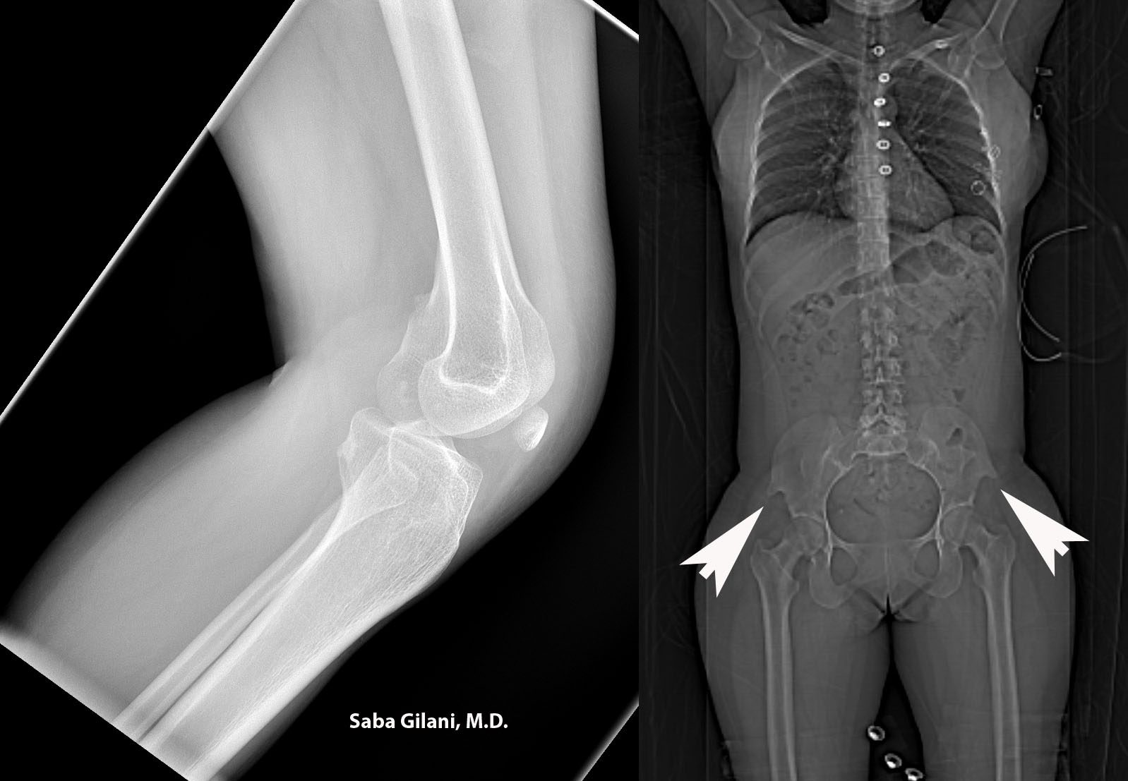
Hereditary osteo-onychodysplasia disease (HOOD), also known as Nail Patella Disease or Iliac Horn Syndrome is a genetic disorder due to an autosomal dominant mutation in the LMX1B gene. A pathognomonic finding is the presence of "iliac horns" projecting posterolaterally from the bilateral iliac bones. Other associated findings are absence or hypoplasia of the patella and radial head. Nail deformities are also common.  The joint deformities can lead to osteoarthritis. About 40% of patients may also develop renal disease ranging from proteinuria to nephrotic syndrome.

Above, the lateral radiograph of the knee is from a patient who presented to the emergency room after a fall. A hypoplastic patella was incidentally noted. A review of prior imaging demonstrated bilateral iliac horns best seen on the scout image.


REFERENCES
Scott JE and Taor WS. The small patella syndrome. J Bone Joint Surg [Br] 1979;61-B:172-5.
Thompson EA, Walker T, Weens HS. Iliac horns. An osseous manifestation of hereditary arthrodysplasia associated with dystrophy of the fingernails. Radiology 1949;53:88-92.
Tuncbilek N, Karakas HM, Okten OO. Imaging of nail-patella syndrome. Hong Kong Med J 2005;11(2):116-8.

Wednesday, August 8, 2012

Significance of Intraperitoneal Free Fluid in Male Trauma Patients Without Identifiable Injuries


MDCT is routinely used in the evaluation of blunt abdominal trauma. The presence of intraperitoneal free fluid in the absence of identifiable injury presents a diagnostic challenge, especially in male patients (in female patients, free fluid, especially when seen in the pouch of Douglas, can be a normal physiologic finding).

In the late 1990s the presence of free fluid without identifiable injury necessitated exploratory laparotomy. Since then, studies have advocated for conservative management such as admitting the patient for observation rather than proceeding with surgical intervention.

In their study of 669 consecutive male trauma patients, Drasin and Anderson show that approximately 3% of patients can have the finding of isolated free fluid without clinical significance They advocate for the evaluation of factors such as size and attenuation measurements of fluid when triaging the patient and suggest that observation with repeat imaging may prevent unnecessary laparotomies. 


REFERENCES

Drasin TE, Anderson SW, Asandra A, et al. MDCT evaluation of blunt abdominal trauma: clinical significance of free intraperitoneal fluid in males with absence of identifiable injury. AJR Am J Roentgenol 2008;191:1821-26.
Levin CD, Patel UJ, Wachsberg RH, et al. CT in patients with blunt abdominal trauma: clinical significance of intraperitoneal free fluid detected on a scan with otherwise normal findings. AJR Am J Roentgenol 1995;164:1381-85.

Sunday, July 29, 2012

New Beginnings

Please join me in welcoming Dr. Saba Gilani as the new editor of Roentgen Ray Reader. Dr. Gilani brings with her a collection of interesting cases of relevance to day-to-day practice, and, more importantly, will inject some much needed fresh energy into the blog.

Tuesday, March 6, 2012

Pencil on CT

The CT appearance of the typical pencil containing graphite and wood is shown above in cross section in a patient with a penetrating injury. The attenuation of wood is negative (~-400 HU in this case), seen best on the lung windows. The graphite has much higher attenuation (~500 in this case), but lower than that of cortical bone.

The paint seems to have higher attenuation than the wood. Two old papers from 1971 raise concerns about the lead content of paints used on pencils, but I can't find any recent data on the contents of the paint.

References

  • Pichirallo J. Lead poisoning: risks for pencil chewers? Science. 1971 Aug 6;173(3996):509-10.
  • Tenenholz T, Baxter AB, McKhann GM. Orbital assault with a pencil: evaluating vascular injury. AJR Am J Roentgenol. 1999 Jul;173(1):144.
  • Schaplowsky AF. Lead in paint on pencils. HSMHA Health Rep. 1971 Nov;86(11):961-2.

Thursday, February 23, 2012

Foreign Body


Telephone handset in rectum
Cocaine bags in colon

Foreign bodies are uncommon, but they are important and interesting. Foreign bodies may be ingested, inserted into a body cavity, or deposited into the body by a traumatic or iatrogenic injury.

Foreign body ingestions or insertions are seen in four broad categories of patients:
  • Children
  • Mentally handicapped persons. May present multiple times for unusual injuries and foreign body insertions and ingestions.
  • Adults with unusual sexual behaviors
  • "Normal" adults or children with predisposing factors or injurious situational problems. This group includes individuals who may abuse drugs or alcohol, engage in criminal activities, engage in extreme sporting activities, or may be subject to child or spousal abuse.
The rectum, vagina, urethra, nose, and ear are favorite sites for insertion of foreign objects.

References

Friday, October 14, 2011

New Proposed Criteria for Diagnosis of Nephrogenic Systemic Fibrosis

Cowper and colleagues have proposed a new system "to guide and standardize the evaluation and diagnosis of nephrogenic systemic fibrosis." In contrast to the old Cowper criteria, this is a scoring system that uses clinical and histopathological features to yield two scores: a clinical score and a histologic score. The combination is used to give the clinician an idea of how strongly nephrogenic systemic fibrosis should be suspected. Like the old system, there is no role for radiology in the diagnosis, but it's good to know how our colleagues in dermatology and pathology will be diagnosing one of the complications associated with our practice.

Major clinical criteria:
  • Patterned plaques: Red to violaceous thin, fixed skin plaques showing polygonal, reticular, or "amoeboid" morphologies.
  • Joint contractures:
  • Cobblestoning:
  • Marked induration/Peau d'orange: Unpinchable, firm, bound-down skin over the extremities. Peau d'orange refers to follicular dimpling. Must be present in the upper extremity or in the lower extremity above the knee.
Minor clinical criteria:
  • Puckering/linear banding:
  • Superficial plaque/patch: Thin, irregularly bordered hypopigmented, pink, or flesh-colored macules coalescing into patches or thin plaques. Common on the upper extremities and unusual on the trunk.
  • Dermal papules: Slightly brawny papules without epidermal changes.
  • Scleral plaques: There are new-onset white/yellow scleral plaques with dilated capillary loops in a patient younger than 45 years.
Clinical scenario
Clinical score
> 1 major criteria
4
1 major criterion
3
≥ 1 minor criteria
2
1 or no minor criteria
1
Diagnostic of another entity
0


Histologic findings:
  • Increased dermal cellularity: Increased numbers of spindled and/or epithelioid cells with few other inflammatory cells in the dermis. (Histologic score +1).
  • CD34+ cells with tram-tracking: CD34+ spindle or epithelioid cells in a reticular or parallel arrangement forming a complex network. (Histologic score +1).
  • Thick and thin collagen bundles: Both fine and ropey collagen surrounded by clefts. (Histologic score +1).
  • Preserved elastic fibers : Use elastic stain to assess elastic content. (Histologic score –1 if absent, otherwise unchanged).
  • Septal involvement : Expansion of interlobular septa by collagen, cells, and other matrix elements. Spindle cells encroach on fat lobules. (Histologic score +1).
  • Osseous metaplasia: A highly specific feature. Nonmineralized osseous tissue around elastic fibers, some of which protrude beyond the zone of ossification. (Histologic score +3).
Histologic
Clinical score
score
0
1
2
3
4
0
 
Consider alternative diagnosis
1
    Not NSF
Inconsistent
2
   
Suggestive
Consistent
3
   
Consistent
NSF
4
 
Inconsistent
   

References

Girardi M, Kay J, Elston DM, Leboit PE, Abu-Alfa A, Cowper SE. Nephrogenic systemic fibrosis: Clinicopathological definition and workup recommendations. J Am Acad Dermatol. 2011 Jul 1. [Epub ahead of print]

Tuesday, October 11, 2011

Gadolinium Contrast Agents

Because of the toxicity of free gadolinium (Gd3+), MRI contrast agents contain gadolinium bound to chelates. The stability of the gadolinium ion in the agent is, therefore, an important factor in its safety profile. Factors that can affect stability are the shape (linear vs. cyclic) and ionicity (ionic vs. non-ionic) of the chelate.

The nine gadolinium contrast agents currently available for clinical use can be classified as ionic or non-ionic and linear or cyclic. Ionic agents have negatively charged groups that tend to offer better binding of the positively charged gadolinium ion compared to the non-ionic agents.

Cyclic agents are constructed so as to encircle the gadolinium ion in a cage, which is thought to prevent its dissociation. Linear agents, on the other hand, have a flexible open chain that has a lower tendency to hang on to the the gadolinium ion.

Theoretically, of the four combinations possible (see table), the ionic cyclic agents should provide the best protection for the gadolinium ion, and the non-ionic linear agents would provide the least protection. In vivo studies, while confirming the low stability of the non-ionic linear agents, have not found a significant difference in stability between ionic and non-ionic cyclic agents.

With the revised FDA labeling, the use of Magnevist, Omniscan, and Optimark is contraindicated in patients with acute kidney injury or chronic, severe kidney disease (GFR < 30 mL/min/1.73m2).

 
Ionic
Non-ionic
Linear Ablavar (gadofosveset trisodium)
Eovist (gadoxetate disodium)
Magnevist (Gadopentetate, Gd-DTPA)
Multihance (Gadobenate, Gd-BOPTA)
Omniscan (Gadodiamide, Gd-DTPA-BMA)
OptiMark (Gadoversetamide, Gd-DTPA-BMEA)
Cyclic Dotarem (Gadoterate, Gd-DOTA) Gadavist (USA)/Gadovist (Europe, Canada) (Gadobutrol, Gd-BT-DO3A)
ProHance (Gadoteridol, Gd-HP-DO3A)


Gadolinium Release and Nephrogenic Systemic Fibrosis

A concept that is important in stability of gadolinium chelates in vivo is transmetalation. This is the process of gadolinium release through replacement by other serum cations (e.g., zinc, iron, copper, and calcium). Zinc's relatively high serum concentration (55–125 μmol/L) makes it the major player in transmetalation. The zinc replaces the gadolinium ion and is excreted in urine as zinc chelate. Measurement of zinc chelate can be used as an indicator of the amount of free gadolinium.

Copper's low serum concentration, calcium's low affinity to organic ligands, and iron's strong bond to ferritin and hemosiderin limits their availability for transmetalation with gadolinium ions.

The released gadolinium ions, in turn, bind to endogenous anions such as phosphate, citrate, hydroxide, and carbonate and deposit in tissues as insoluble compounds. It is thought that the release of cytokines in response to phagocytosis of these insoluble compounds by macrophages is responsible for the development of nephrogenic systemic fibrosis (NSF).

One such cytokine, transforming growth factor beta, is a potent fibrogenic agent that attracts circulating fibrocytes. These deposit in the dermis and other organs that contain the insoluble gadolinium deposits and mature into fibroblasts. The fibroblasts then deposit collagen in the affected organs, leading to fibrosis.

References

Morcos SK. Extracellular gadolinium contrast agents: differences in stability. Eur J Radiol. 2008 May;66(2):175-9.

Tuesday, September 27, 2011

Advanced Imaging Technology at Airports

There was an interesting discussion in this month's Radiology about airport scanners (advanced imaging technology, or AIT). Two main technologies currently exist: Backscatter X-ray (ionizing radiation) and millimeter-wave (non-ionizing radiation).

Backscatter X-ray Systems

These devices transmit x-rays to passengers (depending on the manufacturer, the x-rays are either 50 kVp or 120 kVp) and detect reflected radiation. Some of the x-rays penetrate deep into the body (see below) and some bounce off the skin. The posteroanterior and anteroposterior images are obtained mainly from the reflected photons.

Three manufacturers currently make these devices: Rapiscan (Secure 1000), American Science and Engineering (Smartcheck), and Tek84 (AIT84 Body Scanner and Castscope, the latter designed for examining casts, bandages, and artificial limbs).

Given the public concern about radiation and privacy, more thought could have been put into naming these devices. Rapiscan raises the spectre of violation, the AIT84 brings to mind George Orwell's 1984, and Castscope manages to raise associations with both castration and colonoscopy.

Public fears and unfortunate names aside, the estimated radiation from these devices is pretty low. Skin dose estimates range from 0.7 μGy to 2.5 μGy and effective dose estimates from 0.015 μSv to 0.9 μSv. By way of comparison, we get an effective dose of about 0.04 μSv/minute just by flying in an airplane.

A common misconception is that photons from backscatter devices do not penetrate deep into the body (here's one from an ACR press release). While seemingly reassuring at first consideration, the implication of this misconception is that skin doses can be many times higher than the effective dose to the entire body. This misconception is the result of confusion of dose penetration with imaging penetration, the former reflecting the behavior of the photons in the body and the latter describing the photons used for image creation (more here).

The penetration of the photons into the body does raise concern about fetal dose. However, a scan delivering an effective dose of 0.25 μSv to the mother is estimated to deliver only about 0.12 μSv to the uterus.

Millimeter-Wave Systems

Another system used in airports makes use of non-ionizing, millimeter-wave radiation. These come in active and passive varieties. Active scanners direct millimeter-wave energy at the passenger, while passive systems detect energy naturally emitted from the body and concealed objects. The data is then analyzed and reconstructed to generate a 3-dimensional holographic image (as opposed to the planar images generated by the backscatter devices).

L3 Security and Detection Systems manufactures the ProVision system currently used by the TSA.

References

  • Brenner DJ. Are x-ray backscatter scanners safe for airport passenger screening? For most individuals, probably yes, but a billion scans per year raises long-term public health concerns. Radiology. 2011 Apr;259(1):6-10.
  • Zanotti-Fregonara P, Hindié E, Brenner DJ. Radiation Risk from Airport X-ray Backscatter Scanners: Should We Fear the Microsievert? Radiology. 2011 Oct;261(1):330-1.

Wednesday, June 30, 2010

Iodinated Contrast and Thyrotoxicosis

The risk of iodine-induced thyrotoxicosis is low in euthyroid patients, ranging from 0% to about 2.5% depending on the study. The risk for hyperthyroid patients has not been defined, but in one study of elderly hyperthyroid patients 25% developed self-limited hyperthyroidism after nonionic contrast radiography. Prophylactic medications may be given, and include perchlorate with or without thiamazole, but there are no studies on the benefits of these medications.

References

  • Fricke E, Fricke H, Esdorn E, Kammeier A, Lindner O, Kleesiek K, Horstkotte D, Burchert W. Scintigraphy for risk stratification of iodine-induced thyrotoxicosis in patients receiving contrast agent for coronary angiography: a prospective study of patients with low thyrotropin. J Clin Endocrinol Metab. 2004 Dec;89(12):6092-6.
  • Martin FI, Tress BW, Colman PG, Deam DR. Iodine-induced hyperthyroidism due to nonionic contrast radiography in the elderly. Am J Med. 1993 Jul;95(1):78-82.

Saturday, November 7, 2009

Epinephrine Dose for Contrast Reactions

  • Intramuscular: This is the first-line treatment. Use 1:1,000 concentrated epinephrine injected in the lateral aspect of the thigh. Dose is 0.01 mg/kg body weight up to a total of 0.5 mg. Usual doses: 0.1–0.5 mg of 1:1,000 concentrated epinephrine.
  • Intravenous: Must be dilute, given slowly, and titrated for effect in a monitored patient. Fatal adverse reactions, though rare, can occur. Intravenous route may have to be used for a life-threatening reaction unresponsive to intramuscular injection. Usual doses: 5 mcg to 0.5 mg of 1:10,000 concentrated epinephrine.
  • Subcutaneous: Not as effective. May be no better than placebo. The 1:1,000 concentrated epinephrine is used. Usual doses: 0.1–0.5 mg of 1:1,000 concentrated epinephrine.

References

Lightfoot CB, Abraham RJ, Mammen T, Abdolell M, Kapur S, Abraham RJ. Survey of radiologists' knowledge regarding the management of severe contrast material-induced allergic reactions. Radiology. 2009 Jun;251(3):691-6.