Saturday, December 23, 2017

Myxofibrosarcoma Recurrence

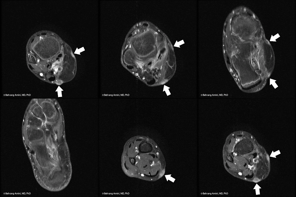
tail-like recurrence of myxofibrosarcoma (MFS)
Serial contrast-enhanced axial images of the forearm in a patient with recurrent myxofibrosarcoma (MFS). Arrows indicate slowly enlarging, tail-like recurrence along the superficial fascia in the resection bed.

Myxofibrosarcoma (MFS, previously myxoid malignant fibrous histiocytoma) is an intermediate-grade soft-tissue sarcoma with fibroblastic and myxoid components. Patients are typically in the sixth to eighth decades of life.

These lesions can range from round and well-defined (like most soft tissue sarcomas) to predominantly infiltrating, with tails extending along fascial planes. They have high T2 signal intensity related to their myxoid content and heterogeneous enhancement that can be feather-like (typically seen with myxoid neoplasms).

Despite best efforts at acquiring negative margins (made difficult by infiltrating tails), MFS has a propensity for repeated local recurrence, with rates of up to 79%. Recurrence can be tricky to detect. As seen in the image above, tail-like recurrence can be easily dismissed as being related to inflammation or trauma. Our group has found no association between the appearance of MFS at presentation (well-defined vs. tail-like) and the pattern of recurrence, so it's important to watch out for tails regardless of the initial tumor presentation.

In summary, the key to early detection of recurrent MFS is
  1. Know that you're dealing with MFS (look at the path report, and know that some pathologists may still call these "myxoid MFH").
  2. Be aware of propensity for tail-like recurrence.
  3. Make sure to compare to multiple studies dating back to post-operative baseline to increase sensitivity for detection of these slowly growing lesions.

References

Daniels C , Wang WL , Madewell JE, Wei W, Amini B. Pattern of Recurrence of Myxofibrosarcoma is not Associated with Pattern at Presentation or Rate of Delayed Diagnosis. Iran J Radiol. 2017 ;14(1):e13469.

Monday, December 11, 2017

Pleomorphic adenoma

Magnetic resonance imaging (MRI) of parotid pleomorphic adenoma. T1- and T2-weighted and post-contrast sequences

Pleomorphic adenoma (also known as benign mixed tumor) is the most common tumor of the major salivary glands, and the most common benign tumor of the parotid glands (~75% of all benign salivary gland tumors). Prior head and neck irradiation is a risk factor. They are typically solitary, slowly growing, and asymptomatic. They are typically diagnosed after palpation by the patient or incidentally on imaging studies. Surgical resection is advised due to risk of malignant transformation.

On MRI, pleomorphic adenomas, have polylobulated margins a rim of low T2 signal corresponding to a fibrous capsule. They have heterogeneous low-to-intermediate signal intensity on T1-weighted images. High T2 signal and avid, solid enhancement are considered relatively specific features, especially when present in a younger patient (< 57 years). Dynamic contrast-enhanced (DCE) MRI shows gradual enhancement. On diffusion-weighted imaging (DWI), pleomorphic adenomas tend to have very high ADC values; however, DWI is not able to differentiate between benign and malignant parotid gland tumors.

On ultrasound, pleomorphic adenomas are typically hypoechogenic. They can have mild to moderate uptake on FDG PET.

Differential considerations include:
  • Warthin tumor: 10–15% are bilateral. Can have proteinaceous cystic components with high T1 signal ranging from a few millimeters to 1–2 cm. Solid components have rapid enhancement and washout.
  • Adenoid cystic carcinoma: Small, low-grade lesions can be mistaken for pleomorphic adenomas. Variable signal intensity on T2-weighted depending on type. Low-grade tumors have high T2 signal in the solid parts. Large tumors can have cystic areas of hemorrhagic necrosis.
  • Myoepithelial adenoma: Also tend to have very high ADC values
  • Basal cell adenoma: More commonly in the superficial lobe of the parotid gland. Tend to be round and well-circumscribed tumors. Have heterogeneous enhancement on CT.
  • Carcinoma in pre-existing pleomorphic adenoma (carcinoma ex pleomorphic adenoma): Typically less well-circumscribed than benign pleomorphic adenoma. Tends to occur after 10–15 years of an existing pleomorphic adenoma, with sudden rapid growth (3–6 months) in patients in the sixth-to-eighth decades of life.
  • Lymphoma: Tend to be multiple. Can have well-defined and lobulated margins. Tend to have low T2 signal and slight enhancement.
  • Sarcoid: Can be placed in any differential, including this one.

References

Monday, December 4, 2017

Osseous Pseudoprogression after Spine Stereotactic Radiosurgery

Osseous Pseudoprogression after Spine Stereotactis Radiosurgery

One of the issues we run into when assessing response to radiation therapy is pseudoprogression: Enlargement of the area of abnormality that is not truly progression. This phenomenon is best known in brain lesions following gamma knife therapy, but has also been seen in lung lesions after stereotactic body radiotherapy (SBRT).

In the spine, we can see pseudoprogression after spine stereotactic radiosurgery (SSRS) in bone lesions (osseous pseudoprogression, or OPP), as well as in the epidural soft-tissue components of bone lesions.

The take-away messages are:
  • We have so far only seen OPP after single-fraction SSRS. This is likely due to the higher biological dose of single-fraction therapy.
  • If you see an enlarging bone lesion on MRI performed within 3-6 months after single-fraction SSRS, you can't be confident that it represents true progression, because about 1/3 of these enlarging bone lesions will represent OPP.
  • The only finding on conventional MRI that has been shown to be associated with OPP is tumor growth confined to the 80% iso-dose line and the slope of enlargement (earlier time to tumor enlargement).

References

Sunday, July 9, 2017

Hemochromatosis: Hand Manifestations

X-ray of hand in hemochromatosis
50-year-old man with subtle manifestations of hemochromatosis arthropathy. Note narrowing of the metacarpophalangeal (MCP) joints, more advanced at the index and middle fingers. Other features of hemochromatosis arthropathy are not present, specifically, there is no subchondral cyst formation, hook-like osteophytes, osteoporosis, or chondrocalcinosis and the radiocarpal articulation is unaffected.

Hemochromatosis: General
  • Autosomal recessive disorder
  • Several genes implicated
  • Body has mechanism for dealing with low Fe (increased intestinal absorption), but no effective way of dealing with excess Fe
  • Fe accumulates in organs
  • Damage via Fe2+ -> Fe3+ -> Oxidation -> Free radicals


Arthropathy
  • Present in 50% of patients when rigorous criteria are used to define arthritis
  • Males affected earlier and more severely
  • Associated with use of joint
  • Typically a chronic process
  • Acute episodes of inflammatory arthritis may occur: May be associated with recovery of CPPD crystals from joint fluid aspiration.
  • Rare syndrome of septicemia accompanied by monoarticular or oligoarticular septic arthritis caused by Yersinia species described: Prosthetic joints seem especially susceptible to this infection.


Pathophysiology
  • Hemosiderin found in superficial synovial lining cells and macrophages (phagocytosis of iron loaded synoviocytes)
  • Very little iron detected in deeper synovial layers, and only occasionally in macrophages.
  • Arthropathy generally non-inflammatory. Chronic inflammatory cell infiltrate rarely seen.
  • Minimal to no iron deposition in cartilage.
  • Chondrocalcinosis a frequent finding
  • No close spatial relationship between iron and CPPD crystals, and crystals can be found even in the absence of iron deposits.


Imaging Features
  • Overlap with those of idiopathic OA and CPPD
  • Joint space narrowing
  • Subchondral sclerosis
  • Subchondral cysts
  • Osteophyte formation (hook-like osteophytes can be seen)
  • Chondrocalcinosis in articular and non-articular cartilage
  • Osteoporosis


The deal with chondrocalcinosis
  • Fe2+ (but not Fe3+) inhibits pyrophosphatase
  • Leads to diminished hydrolyzation of inorganic pyrophosphate
  • Contributes to precipitation of inorganic pyrophosphate with calcium.

References

  • Dallos T, Sahinbegovic E, Aigner E, Axmann R, Schöniger-Hekele M, Karonitsch T, Stamm T, Farkas M, Karger T, Cavallaro A, Stölzel U, Keysser G, Datz C, Schett G, Manger B, Zwerina J. Validation of a radiographic scoring system for haemochromatosis arthropathy. Ann Rheum Dis. 2010 Dec;69(12):2145-51.
  • Frenzen K, Schäfer C, Keyßer G. Asymmetrical hemochromatosis arthropathy in a patient with a history of poliomyelitis. Rheumatol Int. 2012 Apr;32(4):1045-7.
  • van Vulpen LF, Roosendaal G, van Asbeck BS, Mastbergen SC, Lafeber FP, Schutgens RE. The detrimental effects of iron on the joint: a comparison between haemochromatosis and haemophilia. J Clin Pathol. 2015 Aug;68(8):592-600.

Sunday, July 2, 2017

Chordoma Drop Metastases

MRI of spine with chordoma drop metastasis

Chordoma drop metastases are extremely rare, with ~11 reported cases. Of these, 1 was present at the time of initial presentation and 3 were only seen at autopsy. The naturally slow growth of chordomas, lack of symptoms in several cases, and late age of onset may mask true incidence of intradural drop metastases.

The imaging features are nonspecific and tend to mirror those of the primary neoplasm.

References

Martin MP, Olson S. Intradural drop metastasis of a clival chordoma. J Clin Neurosci. 2009 Aug;16(8):1105-7.

Monday, June 26, 2017

Radiation-Associated Sarcomas

CT, bone scan (MDP), FDG PET/CT and MRI in a patient with radiation-associated sarcoma (osteosarcoma)
Patient with history of radiation therapy for head and neck cancer. Radiation field extended into the supraclavicular nodal stations. CT shows an osteoid producing soft tissue mass to the right of midline. Bone scan and PET show uptake in the lesion, as well as contralateral lymph nodes. PET shows an FDG-avid lung nodule. T1-WI post-contrast MRI with FS shows a peripherally enhancing soft tissue mass.

The incidence of radiation-associated sarcomas of bone and soft tissue is about 0.1%. They are more commonly seen in patients with breast cancer, lymphoma, head and neck malignancies, and gynecologic cancers. The distribution of primary cancers is likely related to the larger numbers of patients with these cancers and the high survival rates for these tumors.

The majority of radiation-associated tumors are soft tissue sarcomas, with bone sarcomas making up about 20-30% of cases. The majority are high grade and aggressive. The most common soft tissue sarcomas are unclassified pleomorphic sarcoma (UPS, formerly MFH), followed by angiosarcoma (particularly in breast cancer), fibrosarcoma and leiomyosarcoma (particularly in retinoblastoma). The most common bone sarcomas are osteosarcomas.

The latency between radiation and sarcoma ranges from as little as a few months to 54 years. The average is 7 to 16 years. In breast cancer, the average is 10 to 11 years (4-8 years for angiosarcomas). In childhood cancers, the average latency is between 12 to 13 years.

Risk factors include, dose (Rarely seen with low doses: <40 Gy), age at exposure, concomittant chemo exposure (particularly alkylating agents) and genetic tendency (e.g., Li-Fraumeni syndrome).

References

Maki, R. et al. Radiation-Associated Sarcomas. UpToDate

Sunday, June 18, 2017

Keloids and Hypertrophic Scars

MRI in a patient with hypertrophic scar/keloid
T1 axial post-contrast MRI of a keloid or hypertrophic scar formation along the margins of a myocutaneous flap. The enhancement can fool you into thinking that there is recurrent tumor, but the linear pattern along the flap margin is the clue that this is related to the scar.

Keloids and hypertrophic scars are fibroblastic proliferations of the dermis. Their morphologic and pathologic features overlap. They are associated with trauma, infection, and connective tissue diseases. They do not spontaneously regress and tend to recur after surgical excision, so surgery is often combined with topical corticosteroid injection or, less commonly, radiation therapy.

In contrast to hypertrophic scars, keloids tend to grow beyond the margins of the injury site, have keloid collagen, and are less likely to stain for smooth muscle actin. Keloids have a higher recurrence rate than hypertrophic scars.

Keloids are hypocellular and are composed of dense collagen. The abundance of type I collagen results in low T2 signal. Keloids occur most frequently in patients aged 15–45 years. People of African and Chinese origin have a higher predilection for keloids. The face, shoulders, forearms, and hands are most commonly affected. They tend to occur where there is increased skin tension

References

Dinauer PA, Brixey CJ, Moncur JT, Fanburg-Smith JC, Murphey MD. Pathologic and MR imaging features of benign fibrous soft-tissue tumors in adults. Radiographics. 2007 Jan-Feb;27(1):173-87.

Monday, June 12, 2017

The axillary nerve and adhesive capsulitis

MRI of anatomy of the axillary nerve and its relationship to the joint capsule.
The axillary nerve (yellow arrow) and the posterior humeral circumflex artery (red arrow) in the region of the quadrilateral space. Note proximity to the inferior capsule.

The axillary nerve is closely related to the inferior capsule of the shoulder. It passes inferior to the subscapularis muscle and travels adjacent to the capsule before entering the quadrilateral space.

The axillary nerve is associated with adhesive capsulitis in at least 2 ways.

First, the axillary nerve can be irritated in the setting of inflammation and thickening of the inferior capsule. The evidence for this is somewhat anecdotal, but makes anatomic sense. The image below is from a patient with adhesive capsulitis. Note the teres minor atrophy (green arrow) in the setting of thickening of the inferior capsule (blue arrow), and constrained fluid in the joint (orange arrow) being forced into the superior subscapularis recess (orange*). The bone lesions are from myeloma, in case you were wondering.

MRI of anatomy of the axillary nerve and its relationship to the joint capsule in adhesive capsulitis.

Second, the close proximity of the nerve to the joint capsule predisposes it to injury during arthroscopic capsule release for treatment of adhesive capsulitis. Risk of injury is decreased by placing the incision of the glenohumeral joint capsule at the glenoid insertion with the arm in the abducted and externally rotated position.

We can appreciate the extent of inflammation on other imaging modalities too. On FDG PET/CT, for example, patients with adhesive capsulitis tend to have uptake at the inferior capsule that extends into the adjacent tissues.

References

  • Sridharan R, Engle MP, Garg N, Wei W, Amini B. Focal uptake at the rotator interval or inferior capsule of shoulder on 18F-FDG PET/CT is associated with adhesive capsulitis. Skeletal Radiol. 2017 Apr;46(4):533-538.
  • Salem U, Zhang L, Jorgensen JL, Kumar R, Amini B. Adhesive capsulitis mimicking metastasis on 18F-FDG-PET/CT.Clin Nucl Med. 2015 Feb;40(2):e145-7.
  • Jerosch J, Filler TJ, Peuker ET. Which joint position puts the axillary nerve at lowest risk when performing arthroscopic capsular release in patients with adhesive capsulitis of the shoulder? Knee Surg Sports Traumatol Arthrosc. 2002 Mar;10(2):126-9.
  • E. B. G. D. Santos, P. M. E. Souza (pdf). Teres minor beyond quadrilateral space syndrome: a pictorial review. ECR 2014 conference.

Tuesday, June 6, 2017

Sacrum

I was reading an article on the role of religion in the secular Turkish state and came across this statement:
It is possible -- such is the argument of Carter Findley in his Turks in World History -- that in doing so it drew on a long Turkish cultural tradition, born in Central Asia and predating conversion to Islam, that figured a sacralisation of the state, which has vested its modern signifier, devlet, with an aura of unusual potency.

You may be wondering what the heck a congenital variant of spinal segmentation has to do with religion. From the always-excellent Online Etymology Dictionary:

Bone at the base of the spine, 1753, from Late Latin os sacrum "sacred bone," from Latin os "bone" + sacrum, neuter of sacer "sacred" (see sacred). Said to be so called because the bone was the part of animals that was offered in sacrifices. Translation of Greek hieron osteon. Greek hieros also can mean "strong," and some sources suggest the Latin is a mistranslation of Galen, who was calling it "the strong bone."

Monday, May 29, 2017

Rind-like Perirenal Soft-Tissue Masses

rind-like enhancement around the kidney in a patient with retroperitoneal fibrosis
Rind-like perirenal soft tissue masses: Retroperitoneal fibrosis

Differential diagnosis for rind-like perirenal soft-tissue masses:
  • Lymphoma (and Waldenstrom Macroglobulinemia)
    • Usually due to contiguous spread from retroperitoneal or renal lymphoma: Distinct imaging patterns: multiple masses, solitary mass, diffuse infiltrating mass, rindlike soft-tissue thickening, and direct invasion from adjacent retroperitoneal lymphadenopathy
    • Isolated perirenal lymphoma very unusual (<10% of cases): Uniformly attenuating rindlike soft-tissue mass.
    • Does not necessarily affect renal function.
  • Erdheim-Chester disease
    • Rindlike soft-tissue lesions surrounding the kidneys and ureters
    • Severe compression of renal parenchyma and ureters leads to progressive renal failure
    • Percutaneous nephrostomy made difficult because due to fibrous perinephritis.
  • Retroperitoneal Fibrosis (shown above)
    • Typically localized to infrarenal aorta and common iliac arteries
    • Isolated or related to multifocal fibrosclerosis (may include autoimmune pancreatitis, sclerosing cholangitis, scleroderma, Riedel thyroiditis, fibrotic pseudotumor of the orbit, and fibrosis involving multiple organ systems).
    • Perirenal involvement can be from extension from retroperitoneal fibrosis, without associated retroperitoneal fibrosis, or one of manifestations of multifocal fibrosclerosis
    • Perirenal involvement: Soft-tissue mass enveloping kidneys without displacing them.

References

Surabhi VR, Menias C, Prasad SR, Patel AH, Nagar A, Dalrymple NC. Neoplastic and non-neoplastic proliferative disorders of the perirenal space: cross-sectional imaging findings. Radiographics. 2008 Jul-Aug;28(4):1005-17.

Saturday, May 13, 2017

Toxic Osteoblastoma

Toxic osteoblastoma is an extremely rare variant of osteoblastoma that is associated with systemic symptoms, such as fever, anorexia, weight loss. There is also marked systemic periostitis, not only of the involved bone, but also at other skeletal sites.

Patients tend to be young children (5-7 years of age). On physical examination, there is massive swelling, warmth, and induration of the overlying skin and prominent superficial vessels overlying the lesion. Patients can also have hyperdynamic circulation and even high-output cardiac failure. Regional adenopathy can also be present.

The systemic response is thought to be due to an exaggerated immune response to the tumor. Interleukins can lead to fever and the diffuse periostitis, as well as anemia and massive limb swelling and vascular proliferation. Another possibility is toxic substances released by the tumor itself.

The lesions are highly vascular, and arteriovenous shunting within the lesion can lead to finger clubbing and diffuse periostitis and can account for hyperdynamic circulation.

Differential considerations include osteomyelitis and osteosarcoma.

References

Sunday, April 23, 2017

Nerve Root(s)


In season 3, episode 18 of Star Trek: Deep Space Nine Dr. Bashir has to deal with some deep-seated personal issues. One of these is the fact that he graduated second in his medical school class because he mistook a "pre-ganglionic fiber for a post-ganglionic nerve." Spoiler alert: He did it on purpose because he didn't want to deal with the pressure of being first.

Dr. Bashir is not alone. I see this lead to 2 errors every day in our trainees. The clinical implication is zero, because the referring physicians also don't make this distinction (two wrongs do make a right, apparently).

First, take a look at the image below:


Note that there are 2 nerve roots (dorsal and ventral) on each side (left and right). When you say a lumbar disc compresses a nerve root in the central spinal canal, you need to add an "s," because these dorsal and ventral nerve roots travels down together in the cauda equina. Next time you look at an axial T2-WI of the lumbar spine, see if you can see two distinct nerve roots on either side.

Second, note that once we're post-ganglionic, we're dealing with a nerve, not a root. So, if you're talking about a nerve root outside the foramen, you're about as anatomically correct as a Ken doll.


The same goes for the "nerve roots" of the brachial plexus and the famous Randy Travis Drinks Cold Beer mnemonic for the brachial plexus anatomy (sorry, Randy). All is not lost. Just replace Randy Travis with Nikola Tesla.

Reference

  • Basic anatomy that everyone ignores.

Sunday, April 16, 2017

Ventriculus Terminalis

MRI of ventriculus terminalis (also known as the terminal ventricle and the fifth ventricle)
The ventriculus terminalis (also known as the terminal ventricle and the fifth ventricle) is a rarely identified cerebrospinal fluid cavity within the conus medullaris. The ventriculus terminalis does not communicate with the subarachnoid space or the central canal of the spinal cord, and may actually be an embryonal remnant of the primitive central canal, leading some to refer to it a sinus terminalis instead.

They are occasionally associated with caudal regression of the spinal cord, Chiari type I malformation, lipomyelomeningoceles, and lumbosacral "lipomas." Some authors believe that all of us have some sort of cystic CSF space at the conus medullaris, but that it's simply larger [and detectable on imaging] in some people and tends to regress (but not completely resolve) over time.

The characteristic imaging features are more commonly seen in children: Cystic lesion of the conus medullaris without spinal cord signal abnormality. In adults, ventriculus terminalis is more likely to have septations and be associated with spinal cord edema, kyphotic deformity and spinal arteriovenous malformations.

Rarely, ventriculus terminalis can enlarge in the presence of meningeal hemorrhage or deformities of the vertebral canal. An enlarged or symptomatic ventriculus terminalis can be treated by cyst fenestration with or without shunting to the subarachnoid space, pleural cavity, or peritoneal cavity.

References

Monday, April 10, 2017

False Perpetuations: Main Portal Vein Size and Portal Hypertension

Perpetuation: A main portal vein (MPV) diameter >13 mm is "consistent with portal hypertension" (pHTN)

This cutoff of 13 mm is based on weak literature (mainly from the 1980's), some of which did not include comparison values of normal patients

  • One comparative study using ultrasound found (Radiology 1982; 142: 167-172):
    • In 79 patients with pHTN
      • 36 had a MPV diameter of <13 mm 
      • 33 had a MPV diameter >/= 13 mm
      • The MPV was not visualized in 10 patients
    • In the 45 control patients
      • The MPV diameter was < 13 mm in 41 cases
      • The MPV was not visualized in 4 patients. 

More recent studies have found that there is no significant difference in MPV diameters when comparing patients without cirrhosis to patients with cirrhosis, and the normal MPV diameter is significantly larger than the 13 mm cutoff

  • A study (Eur J Gastroenterol Hepatol 2004; 16:147-155) from King's College using ultrasound (49 controls and 14 cirrhotics) found: 
    • the average MPV diameters were 9.6 cm and 10.8 cm in patients without and with cirrhosis, respectively.
  • A second study (JCAT 2008; 32: 198-203) from UCSF using CT (59 controls and 67 cirrhotics) found:
    • The average MPV diameters were 14.5 cm and 14.8 cm in patients without and with cirrhosis, respectively.
  • Using CT, the MPVs in healthy renal donor patients were measured before and after the administration of intravenous contrast, and in the axial and coronal planes (Abdom Radiol 2016; 41:1931-1936). This study found:
    • The average MPV diameter was 15.5 +/- 1.9 mm
      • This value was significantly different than 13 mm
    • Post-contrast MPVs were 0.56 mm larger compared to non-contrast
    • A positive correlation between BMI and height versus MPV diameter
In fact, the MPV size can be reduced in portal hypertension and has been described as a sign of hepatofugal MPV flow (AJR 2003; 181: 1629-1633). This study found:
  • A MPV diameter of less than 1 cm is a highly sensitive (but not very specific) for MPV flow reversal in patients with cirrhosis

Sunday, March 26, 2017

Ulnar Dimelia

Ulnar dimelia is a rare congenital disorder characterized by duplication of the ulna, absence of the radius, and polydactyly. Patients can also have arterial anomalies, such as absence of the radial artery, duplication of the ulnar artery, and abnormal arterial arches in the hand. Nerve anomalies may also be present and include shortening of the radial nerve and duplication of ulnar nerve (with or without connections to the median nerve)

Radiopaedia has some great images of type I ulnar dimelia

Distinguishing features of the two types of ulnar dimelia
Feature Type I Type II
Index finger 1 2
Lunate 1 2
Trapezoid 1 2

References

  • Afshar A. Ulnar dimelia without duplicated arterial anatomy. J Bone Joint Surg Br. 2010 Feb;92(2):293-6. doi: 10.1302/0301-620X.92B2.23057.
  • Tomaszewski R, Bulandra A. Ulnar dimelia-diagnosis and management of a rare congenital anomaly of the upper limb. J Orthop. 2015 Feb 18;12(Suppl 1):S121-4.

Sunday, March 19, 2017

Chondroblastoma

X-ray (radiograph), CT, and MRI of chondroblastoma of the femur.

General

  • Terminology: “giant cell variant” (1927) → epiphyseal chondromatous giant cell tumor → calcifying giant cell tumor → chondroblastoma
  • 1% to 2% of all primary bone tumors
  • 9% of all benign bone tumors
  • Mean age of 15-18 years
  • M >>F
  • Mean duration of symptoms 8.7 months
  • Trivia: Most common benign neoplasm of the patella

Imaging Features

  • Epiphysis/apophysis +/- metaphyseal/diaphyseal involvement
  • Metaphyseal/diaphyseal occurrence without epiphyseal/apophyseal involvement exceptionally rare
  • Proximal tibia >> proximal femur > distal femur > proximal humerus
  • Well-defined, sclerotic margins on radiographs
  • Can involve the cortex, resulting in expansion, thinning, or disruption.
  • Stippled matrix calcification seen in minority of cases
  • Periosteal reaction seen in majority of cases
  • Extensive peri-lesional edema on MRI is common
  • Homogeneously hypointense on T1
  • Variable on T2: can be diffusely hypointense, or have small cystic areas of increased T2 signal or fluid-fluid levels
  • Heterogeneous and moderate enhancement in solid portions. Less commonly, homogeneous and marked enhancement

Differential Diagnosis

Management/Prognosis

  • Curettage or resection
  • RFA (small lesions, small series, not common)
  • Local recurrence rate: 5.0% after curettage
  • Local recurrence rate: 0% after resection
  • Recurrence most frequent in the proximal humerus
  • Malignant transformation and benign pulmonary metastases extremely rare

References

Sunday, March 12, 2017

Fat-containing bone lesions

FDG PET and MRI of a patient with sarcoidosis and fat-containing bone lesions.

The presence of fat within a bone lesion is almost always reassuring, although rare exceptions exist. The following differential diagnosis is based on a combination of published papers and my own anecdotal experience:
  • Hemangioma:
  • Intra-osseous lipoma and lipoma variants, including fibrolipoma, angiolipoma and myelolipoma.
  • Enchondroma:
  • Liposclerosing myxofibrous tumor (LSMFT): Nearly all occur in the intertrochanteric region. Now felt to represent a variant of fibrous dysplasia.
  • Osteoporosis: Can give the appearance of lucent bone lesions on CT. These won't have defined margins, and measurement of internal attenuation will reveal the fatty nature of the lesion.
  • Bone infarction: Trivial, but included for the sake of completeness
  • Paget disease of bone:
  • Focal red marrow rest: Ill-defined, intermediate T1 signal. May contain subtle areas of internal fat.
  • Lymphoma: Not truly a fat-containing lesion, but can entrap fat as the tumor infiltrates marrow.
  • Sarcoid: The case above shows a patient with sarcoid and nodal, hepatic, and osseous involvement. Can have fuzzy margins ("brush border").
  • Treated metastasis: One of the ways metastases respond to therapy is by developing internal fat. Myeloma lesions can even entirely "disappear" due to fatty replacement.
  • Indolent metastases: Medullary thyroid cancer, adenoid cystic carcinoma.
  • Intra-osseous hibernoma: Rare.
  • Solid variant of aneurysmal bone cyst:
  • Nonossifying fibroma:
  • Erdheim-Chester disease:
  • Malignancy arising from a fat-containing lesion: For example, osteosarcoma arising from bone infarction or Paget.

References

Sunday, March 5, 2017

Subperiosteal hemorrhage in neurofibromatosis type 1


Neurofibromatosis type 1 (NF-1), in addition to a neuroectodermal disorder, is accompanied by mesodermal dysplasia that is accompanied by skeletal changes. The typical osseous findings include bowing of the legs, increase in length of long bones, pseudarthrosis, subperiosteal cyst formation, local bony erosions from adjacent lesions, and intramedullary neurofibromas. Except for the last two, which are due to direct involvement by neurofibromas, the remainder are due to dysplastic changes in bones.

A lesser known osseous presentation in bone is the propensity for subperiosteal hemorrhage and hematoma formation. The cause is unknown, but may be related to:
  • Vascular abnormalities: For example, diffuse flat hemangiomas or plexiform dilated veins, which have been described in patients with hypertrophy of the extremities
  • Dysplastic periosteum: The thinking is that mesodermal dysplasia manifests as an abnormally loose periosteum with poor callus response. This would predispose the patient to the formation and propagation of large subperiosteal hematomas.
  • Direct involvement by neurofibromas: Subperiosteal infiltration by neurofibromatous tissues may loosen the periosteum and allow for massive hemorrhage following minor trauma.

References

Sunday, February 26, 2017

The Lamina Dura


The lamina dura is the bony lining of the socket (alveolus) of a tooth. The periodontal ligaments extend from the lamina dura to the cementum of the tooth, an keep the tooth in place. The lamina dura is cribriform plate produced by the periodontal ligament and fibers of the periodontal ligament are embedded within it.

While loss of the lamina dura (arrow in image above) is sometimes said to be pathognomonic for hyperparathyroidism, it can be seen in a wide range of conditions:
  • Hyperparathyroidism: The case above is from a patient with primary hyperparathyroidism.
  • Osteomalacia
  • Osteoporosis
  • Paget disease
  • Leukemia
  • Myelomatosis
  • Cushing disease


The lamina dura can be thickened in bisphosphonate-related osteonecrosis of jaw (BRONJ)

References

Sunday, February 19, 2017

Systemic Mastocytosis


Systemic mastocytosis (SM) refers to mast cell infiltration in extra-cutaneous tissues. The symptoms of systemic mastocytosis are due to degranulation of mast cells and/or accumulation of mast cells in target organs.

Degranulation of mast cells

Symptoms can be caused by secretion of the following factors:
  • Histamine: Pruritus, urticaria, hypotension, gastric hypersecretion, bronchoconstriction.
  • Heparin: Local anticoagulation, osteoporosis
  • Leukotrienes: Bronchoconstriction
  • Prostaglandins: Bronchoconstriction, flushing
  • Platelet-activating factor:
  • Proteases:
  • Tumor necrosis factor:

Accumulation of mast cells in organs

Accumulation of mast cells in organs can cause organ dysfunction. The so-called B findings refer to organ involvement without organ dysfunction. C findings refer organ involvement with organ dysfunction. The example above shows hepatic involvement with cirrhosis (white arrow) and ascites (yellow arrow) and nodal involvement with bulky adenopathy (red arrow). We also have involvement with diffuse sclerosis. Interestingly, the non-radiology literature stresses the more common osteoporosis, with scarce mention of the sclerosis that tends to dominate the radiology literature.

Diagnosis systemic mastocytosis

The diagnosis of SM requires either, 1 major and 1 minor OR 3 minor criteria. Warning: Boring for radiologists

The one major criterion is: Multifocal, dense infiltrates of mast cells (≥15 mast cells in aggregates) in sections of bone marrow and/or other extra-cutaneous organ(s).

Minor criteria are:
  • Bone marrow or other extra-cutaneous organs: >25% of mast cells in the infiltrate are spindle-shaped or have atypical morphology, or of all mast cells in bone marrow aspirate smears, >25% are immature or atypical.
  • Activating point mutation at codon 816 of KIT in bone marrow, blood, or another extra-cutaneous organ.
  • Mast cells in bone marrow, blood, or other extracutaneous organs express CD2 and/or CD25 in addition to normal mast cell markers.
  • Serum total tryptase persistently > 20 mg/mL (unless associated w clonal myeloid disorder).

Subtypes

  • Indolent (ISM): No C findings
  • Smoldering (SSM): 2+ B findings, no C findings
  • Aggressive (ASM): C findings, no MCL features*
  • Mast cell leukemia (MCL): BMBx diffuse infiltration by atypical, immature mast cells. Aspirate smears ≥20% mast cells.
  • SM with associated hematologic neoplasm (SM-AHN): SM + MDS, MPN, AML, lymphoma, other

References

Akin C, Gotlib J. Systemic mastocytosis: Determining the subtype of disease. UpToDate

Sunday, February 12, 2017

The Cervical Split: A Pseudofracture


A horizontal line projecting over a cervical vertebral body on lateral radiographs can simulate a fracture or a butterfly vertebral body. This pseudofracture, the so-called cervical split, can result from the lucency between contiguous uncovertebral osteophytes, or, as in the case above, cervical scoliosis resulting in projection of the facet joint over the vertebral body.

A cervical split due to uncovertebral joint osteophyte formation is said to be always accompanied by disc space narrowing.

References

Thursday, February 2, 2017

Chronic Subperiosteal Iliac Hematoma


Subperiosteal iliac hematoma is caused by traumatic avulsion of the periosteum in children and young adults. The loose attachment of periosteum in young patients allows it to be displaced by hematoma in trauma. In the chronic phase, it is typically incidentally discovered by radiologists.

In the chronic phase, it presents as a lens-shaped ossified process on the internal aspect of the iliac wing with a ghost cortex (dotted line in the image above). It may or may not have the well-defined central lesion we have in this case.

References

Guillin R, Moser T, Koob M, Khoury V, Chapuis M, Ropars M, Cardinal E. Subperiosteal hematoma of the iliac bone: imaging features of acute and chronic stages with emphasis on pathophysiology. Skeletal Radiol. 2012 Jun;41(6):667-75.