Wednesday, November 30, 2011

The Yune Soft Tissue Index

The Yune soft tissue index is used to assess the amount of soft tissue at the tips of the distal phalanges. This can help in early detection of acral soft-tissue atrophy (for example, in patients with scleroderma).

You take the amount of soft tissue at the tip of the finger (A) and compare it to the width of the base of the distal phalanx (B). If A is larger than B/4, then the amount of soft tissue is normal. If A is less than B/5, then the amount of soft tissue is abnormal.

The case above in a patient with scleroderma seemed borderline by eye. Let's see if the Yune soft tissue can help.
A = 2.9 mm
B = 13.4 mm

2.9 [?] (13.4/4)
2.9 < 3.35
∴ Not normal

2.9 [?] (13.4/5)
2.9 > 2.68
∴ Not abnormal

∴ Borderline.

References

Freyschmidt J, Brossmann J, Wiens J, Sternberg A. The Hand - Distal Phalanges. In Freyschmidt's Köhler and Zimmer: Borderlands of normal and early pathologic findings in skeletal radiography. Fifth revised edition. Thieme (2003). P 81.

Tuesday, November 29, 2011

Histiocytic Disorders

A new classification scheme for histiocytic disorders has been promulgated by the World Health Organization's Committee on Histiocytic/Reticulum Cell Proliferations and the Reclassification Working Group of the Histiocyte Society.

The new classification scheme is based on the biological behavior of the disease and the lineage of the predominant cell type.

A very cursory review of immunology is helpful before proceeding to the classification. A histiocyte is a cell of the mononuclear phagocyte system (formerly the reticuloendothelial system). The histiocytes we'll be concerned with are monocytes, macrophages, and dendritic cells. Precursor cells in the marrow give rise to circulating monocytes, which differentiate into the tissue-resident phagocytes, macrophages and dendritic cells under the proper cytokine milieu. Langerhans cells are a class of dendritic cells that typically reside in the epidermis.

Under the new classification scheme, histiocytic disorders are classified based on biologic behavior into those with malignant behavior and those with varied biological behavior, and based on predominant cell type into monocyte-, dendritic cell-, or macrophage-related disorders. The term varied biological behavior is meant to convey the spectrum of behavior in this class, which can range in severity from self-limited to lethal.

Langerhans cell histiocytosis, the most well known of the histiocytic disorders by radiologists, is classified as a dendritic cell-related histioctyic disorder of varied biological behavior. Terms such as eosinophilic granuloma, Hand-Schüller-Christian disease, and Letterer-Siwe disease are now (> 10 years) considered obsolete or unnecessary, and should be avoided unless you're trying to be difficult during case conference.

Erdheim-Chester disease, an extremely rare disorder that somehow pops up during radiology residency and on boards, is classified as part of Juvenile xanthogranuloma and related disorders.

The outline below summarizes the current classification scheme:

Disorders of Varied Biological Behavior
  • Dendritic cell-related
    • Langerhans cell histiocytosis
    • Secondary dendritic cell processes
    • Juvenile xanthogranuloma and related disorders
      • Erdheim-Chester disease
      • Solitary histiocytomas with juvenile xanthogranuloma phenotype
    • Solitary histiocytomas of various dendritic cell phenotypes
  • Macrophage-related
    • Hemophagocytic syndromes
    • Primary hemophagocytic lymphohistiocytosis (familial , sporadic)
    • Secondary hemophagocytic syndromes (infection-associated, malignancy-associated, other)
    • Rosai-Dorfman disease (sinus histiocytosis with massive adenopathy)
    • Solitary histiocytoma with macrophage phenotype
Malignant Disorders
  • Monocyte-related
    • Leukemias (Monocytic leukemia M5A and B, acute myelomonocytic leukemia M4, chronic myelomonocytic leukemia)
    • Extramedullary monocytic tumor or sarcoma
  • Dendritic cell-related histiocytic sarcoma (localized or disseminated) based on phenotype
    • Langerhans cells
    • Follicular dendritic cell
    • Interdigitating dendritic cell
    • Others
  • Macrophage-related histiocytic sarcoma (localized or disseminated)

References

  • Chow A, Brown BD, Merad M. Studying the mononuclear phagocyte system in the molecular age. Nat Rev Immunol. 2011 Oct 25;11(11):788-98.
  • Favara BE, Feller AC, Pauli M, Jaffe ES, Weiss LM, Arico M, Bucsky P, Egeler RM, Elinder G, Gadner H, Gresik M, Henter JI, Imashuku S, Janka-Schaub G, Jaffe R, Ladisch S, Nezelof C, Pritchard J. Contemporary classification of histiocytic disorders. The WHO Committee On Histiocytic/Reticulum Cell Proliferations. Reclassification Working Group of the Histiocyte Society. Med Pediatr Oncol. 1997 Sep;29(3):157-66.
  • Lieberman PH, Jones CR, Steinman RM, Erlandson RA, Smith J, Gee T, Huvos A, Garin-Chesa P, Filippa DA, Urmacher C, Gangi MD, Sperber M. Langerhans cell (eosinophilic) granulomatosis. A clinicopathologic study encompassing 50 years. Am J Surg Pathol. 1996 May;20(5):519-52.

Monday, November 28, 2011

Bifid Ribs

A bifid rib, also known as a forked or bifurcated rib, is a relatively common anatomic variant where the sternal end of a rib divides into upper and lower divisions. It is thought that the lower division represents the intended development of the rib, and the upper division represents the anomalous development.

Bifid ribs are more common in males, slightly more common on the right, and occur most frequently in the third and foruth ribs, followed by the fifth, sixth, and second ribs. Both divisions have their own costal cartilage, which may fuse before joining with the sternum.

The space between the upper division and the rib above (white *) is usually narrowed, while the lower intercostal space (red *) is wider than normal.

Familiarity with the underlying muscular, vascular, and neural anatomy may be important for planning surgical and percutaneous procedures.
  • The intercostal muscles traverse the space between the upper and lower divisions.
  • Intercostal nerves run their normal course along the inferior margin of the parent rib and then usually along the inferior margin of the lower division. At least one case of a pair of intercostal nerves traveling along both the upper and lower margins of the parent rib has been reported.
  • I assume that the intercostal arteries take the same course as the nerves, but can't find any supporting evidence.
Bifid ribs are usually isolated, unilateral, and asymptomatic. They may, however, present as a palpable abnormality on physical examination or an asymmetric opacity on chest radiography.

Bifid ribs can also be seen in association with other conditions, most famously with basal cell nevus syndrome, where they may be multiple and bilateral.

References

  • Osawa T, Sasaki T, Matsumoto Y, Tsukamoto A, Onodera M, Nara E, Chen JK, Fujimura A, Nozaka Y. Bifid ribs observed in the third and the fourth ribs. Kaibogaku Zasshi. 1998 Dec;73(6):633-5.
  • Song WC, Kim SH, Park DK, Koh KS. Bifid rib: anatomical considerations in three cases. Yonsei Med J. 2009 Apr 30;50(2):300-3.

Sunday, November 27, 2011

Imaging Features of Fanconi Anemia

Fanconi anemia is a genetically and phenotypically heterogeneous recessive disorder of progressive pancytopenia, various congenital malformations, and predisposition to hematologic and solid malignancies.

Many of the congenital anomalies can be detected on imaging studies. These include:
  • Growth disturbances: Intrauterine growth retardation, short stature, delayed ossification.
  • Central nervous system: Hydrocephalus, single ventricle, absent septum pellucidum/corpus callosum, vascular malformations, moyamoya, Chiari malformations/absent septum pellucidum/corpus callosum.
  • Skull: Microcephaly, craniosynostosis, micrognathia, frontal bossing, small or absent external auditory canal, absent tympanic membrane, microtia, fused ossicles.
  • Spine: Spina bifida, Klippel-Feil anomaly, vertebral body anomalies, sacral agenesis or hypoplasia, kyphosis, scoliosis.
  • Radial ray anomalies: Thenar hypoplasia; dislocation of the radial head; radioulnar synostosis; absence or hypoplasia of the radius, scaphoid, trapezium, and/or thumb; floating thumb; bifid thumb; digitalized thumb; and abnormal thumb placement.
  • Extremities (other): Brachydactyly, arachnodactyly clubfoot, dysplastic or absent ulna, humeral abnormalities, absent clavicles, Sprengel deformity, congenital hip dysplasia/dislocation, Legg-Calve-Perthes disease, leg length discrepancy, soft-tissue syndactylism of the toes, metatarsus varus, medial deviation of the toes, hammer toes.
  • Gastrointestinal: Esophageal atresia, tracheoesophageal fistula, duodenal atresia, duodenal web, malrotation, foregut duplication cyst, anal atresia. Biliary atresia, annular pancreas.
  • Renal anomalies: Renal aplasia, horseshoe kidney, low-lying kidney(s), renal ectopy, hydronephrosis, hydroureter, urethral stenosis, reflux.
  • Genital anomalies: Hypogenitalism, cryptorchidism, hypospadias, bicornate uterus, aplasia or hypoplasia of vagina and uterus, atresia of vagina, hypoplasic uterus, hypoplastic/absent ovary.
  • Cardiopulmonary: Patent ductus arteriosis, ventricular septal defect, pulmonic or aortic stenosis, coarcation of the aorta, double aortic arch, cardiomyopathy, tetralogy of Fallot, pulmonary atresia.
  • Osteoporosis:

References

  • Auerbach AD. Fanconi anemia and its diagnosis. Mutat Res. 2009 Jul 31;668(1-2):4-10.
  • Juhl JH, Wesenberg RL, Gwinn JL. Roentgenographic findings in Fanconi's anemia. Radiology. 1967 Oct;89(4):646-53.
  • De Kerviler E, Guermazi A, Zagdanski AM, Gluckman E, Frija J. The clinical and radiological features of Fanconi's anaemia. Clin Radiol. 2000 May;55(5):340-5.

Saturday, November 26, 2011

The Dot-in-Circle Sign

The dot-in-circle sign refers to the appearance of mycetoma on T2-weighted and post-contrast images. It consists of a tiny hypointense focus within a high-intensity spherical lesion. Numerous lesions are typically seen, separated by a low-signal intensity matrix. It is thought that the small central hypointense focus represents the fungal ball or granule and the surrounding hyperintense area represents the inflammatory granuloma. The intervening tissue of low signal intensity represents the fibrous matrix.

The mycetomas can be found in the soft tissues or bone and can represent infection by fungi (eumycetoma) or aerobic actinomycetes (actinomycetomas). Recall from medical school that actinomycetes have microscopic characteristics similar to those of fungi, and were formerly classified as fungi.

Patients present with a history of a firm, painless nodule that follows an indolent but progressive course. The initial infection may communicate with the skin with discharge of fungal granules. Alternatively, the infection may fester internally and even lead to osteomyelitis and significant destruction and deformity. Definitive diagnosis may be provided by biopsy, or be elusive in cases of fastidious organisms.

The case above is a STIR image of the foot, revealing multiple round hyperintense lesions with central hypointense foci along the dorsum of the foot. The patient had a 10-year history a slowly growing foot mass following trauma and presented to us after biopsy (unknown) and unspecified treatment and at an outside facility. Biopsy at our facility revealed no evidence of fungal infection but was positive for bacteria. This complicated picture was suspected to be due to actinomycetoma.

Geography trivia: Mycetomas are more commonly found in the dry topics. The entity of Madura foot (mycetoma of the foot) was first described in the Madura district of Southern India in 1846. The infection usually starts with penetrating injury to the foot (e.g., thorn prick) with inoculation of organisms that are normal inhabitants of the soil.

References

Friday, November 25, 2011

Ischiofemoral Impingement

Normally the femur can rotate without contacting the ischium or proximal hamstring tendons. Abnormal contact between the ischium and femur is referred to as ischiofemoral impingement and can cause hip pain. The abnormal contact can be due to congenital narrowing of the space between the ischial tuberosity and lesser trochanter or be the result of abnormal positioning following hip arthroplasty.

Radiographs may reveal sclerosis and cystic change at the lesser trochanter and ischium. On MRI, one can see crowding of the fibers of the quadratus femoris muscle belly as it passes between the ischium or hamstring tendons and the posteromedial femur. Edema can also be seen, centered in the muscle belly at the site of maximal impingement.

The main differential consideration is quadratus femoris strain, which presents with edema along the distal myotendinous junction near the posteromedial aspect of the proximal femur.

Two spaces have been defined for the purposes of assessing the free rotation of the femur without contacting the ischium or proximal hamstring tendons. The ischiofemoral space is "the smallest distance between the lateral cortex of the ischial tuberosity and medial cortex of the lesser trochanter." The quadratus femoris space is "the smallest space between the superolateral surface of the hamstring tendons and the posteromedial surface of the iliopsoas tendon or lesser trochanter" (this delimits the space for passage of the quadratus femoris muscle). Unfortunately, these measurements depend on the degree of hip rotation during imaging, and the validity of exact numbers remains unclear.

References

  • Kassarjian A, Tomas X, Cerezal L, Canga A, Llopis E. MRI of the quadratus femoris muscle: anatomic considerations and pathologic lesions. AJR Am J Roentgenol. 2011 Jul;197(1):170-4.
  • Patti JW, Ouellette H, Bredella MA, Torriani M. Impingement of lesser trochanter on ischium as a potential cause for hip pain. Skeletal Radiol. 2008 Oct;37(10):939-41.
  • Torriani M, Souto SC, Thomas BJ, Ouellette H, Bredella MA. Ischiofemoral impingement syndrome: an entity with hip pain and abnormalities of the quadratus femoris muscle. AJR Am J Roentgenol. 2009 Jul;193(1):186-90.

Thursday, November 24, 2011

Parietal Thinning

Biparietal thinning (previously known as involutionskrankheit, malum senile biparietale, senile atrophy, biparietal thinness, and biparietal osteodystrophy) is thought to represent a manifestation of osteoporosis in the skull. Histologically it represents loss of the external table and compensatory remodeling of the diploe. It occurs in about 0.5% of the population, more commonly in women and in older people.

Radiographically, it manifests as a sharp lucency in the parietal region on lateral skull radiographs and flattening or grooving in the parietal regions on frontal views.

References

  • Bruyn GW. Biparietal osteodystrophy. Clin Neurol Neurosurg. 1978;80(3):125-48.
  • Cederlund CG, Andrén L, Olivecrona H. Progressive bilateral thinning of the parietal bones. Skeletal Radiol. 1982;8(1):29-33.
  • Epstein BS. The concurrence of parietal thinness with postmenopausal, senile, or idiopathic osteoporosis. Radiology. 1953 Jan;60(1):29-35. No abstract available.
  • Phillips RC. Cranial anomaly, pathology, or normal variant? Thin parietal bones in ancient Egyptian human remains. (January 1, 2007). Dissertations available from ProQuest. Paper AAI3292064.

Wednesday, November 23, 2011

Ankylosis

Ankylosis is rigidity across a joint and can be classified as extra-articular and intra-articular based on the level of fusion. Ankylosis can occur following trauma and electrical and thermal injury.

Extra-articular ankylosis is due to bridging heterotopic ossification. The underlying joint is preserved. The elbow, shoulder and hip are most commonly affected. Interestingly, these joints can be affected regardless of the site of injury.

The earliest radiographic manifestation of extra-articular ankylosis is periarticular calcification that may progress to heterotopic ossification. Extra-articular ankylosis usually responds to physical therapy and surgical excision.

Intra-articular ankylosis (shown above) occurs within the joint and most commonly occurs at the interphalangeal joints. Intra-articular ankylosis results in serious functional and cosmetic problems and is not as easily treated as the extra-articular type. Treatment options include arthrodesis, osteotomy, arthroplasty, or amputation.

References

  • Balen PF, Helms CA. Bony ankylosis following thermal and electrical injury. Skeletal Radiol. 2001 Jul;30(7):393-7.
  • Tomak Y, Piskin A, Gulman B, Tomak L. Treatment of U-shaped bone ankylosis of the knee with the Ilizarov method. A case report. J Bone Joint Surg Am. 2005 May;87(5):1104-7.

Tuesday, November 22, 2011

Ulnar Attachment of the Annular Ligament

A small fossa can be seen in the proximal ulna at the attachment of the annular ligament. The size of this fossa ranges from a small notch, as seen above, to a wider saucer. It should not be mistaken for a pathological process.

References

Keats TE and Anderson MW. Atlas of Normal Roentgen Variants That May Simulate Disease. 8th edition, page 556; Mosby (2004).

Monday, November 21, 2011

Superior Sublabral Recess

The superior sublabral recess is a synovial recess between the superior labrum and the glenoid rim created by the attachment of the biceps tendon on the supraglenoid tubercie. Because of this recess, the labrum does not attach to the glenoid rim at the 12 o'clock position.

The size of this recess is variable, and has been classified into various types by different authors. One scheme divides the sublabral recess into 3 types. In a type 1 attachment, the labral-bicipital complex is firmly attached to the glenoid rim and an arthroscopic probe cannot be inserted between the labrum and the glenoid. A type 2 attachment has a small sulcus between the labrum and the glenoid rim, while a type III attachment has a deep sulcus between the labrum and the glenoid rim that allows a probe to be inserted between the two.

The superior sublabral recess can be continuous with the sublabral foramen.

References

Sunday, November 20, 2011

POEMS Syndrome

POEMS syndrome is a paraneoplastic syndrome related to a plasma cell dyscrasia that is also known as Crow-Fukase syndrome, Takatsuki syndrome. The acronyms POEMS (Polyneuropathy, Organomegaly, Endocrinopathy, M protein and Skin changes) and PEP (Plasma cell dyscrasia, Endocrinopathy and Polyneuropathy), capture some—but not all—of the associated manifestations. Other manifestations include sclerotic bone lesions, Castleman disease, papilledema, thrombocytosis, erythrocytosis, pleural effusions, edema, and ascites.

Given this potential variability in presentation, Dispenzieri et al. proposed major and minor criteria for the diagnosis of POEMS syndrome. The major criteria are polyneuropathy and monoclonal plasmaproliferative disorder.

Minor criteria include sclerotic bone lesions, Castleman disease, Organomegaly (splenomegaly, hepatomegaly, or lymphadenopathy), Edema (edema, pleural effusion, or ascites), endocrinopathy (adrenal, thyroid, pituitary, gonadal, parathyroid, pancreatic), skin changes (hyperpigmentation, hypertrichosis, plethora, hemangiomata, white nails), papilledema.

They proposed that two major criteria and at least one minor criterion differentiate POEMS syndrome from neuropathy associated with monoclonal gammopathy of undetermined significance, myeloma, and Waldenström disease.

The vast majority of POEMS syndrome patients have radiographic evidence of bone lesions at presentation. Slightly less than half of these lesions are purely sclerotic (well-defined or fluffy), approximately half are mixed sclerotic and lytic, and a small number (2%) are purely lytic bone lesions, which tend to have scelrotic margins giving them a unique ring-like appearance. More than half of patients with bone lesions had more than one lesion.

Resnick has described a pattern of bony proliferation that is pathognomonic for POEMS syndrome: irregular and spiculated bone contours at areas of tendinous and ligamentous attachment, posterior elements of the spine (facet joints, laminae, transverse processes, and costovertebral articulations).

Differential considerations for the sclerotic bone lesions include: Differential considerations for the proliferative spine lesions include:
  • Diffuse idiopathic skeletal hyperostosis (DISH): Also has flowing anterior ossifications.
  • Seronegative spondyloarthropathy: Look for syndesmophytes, sacroiliac joint ankylosis.
  • Fluorosis: Also has increased bone density.
  • Hypoparathyroidism:
  • X-linked hypophosphatemia:
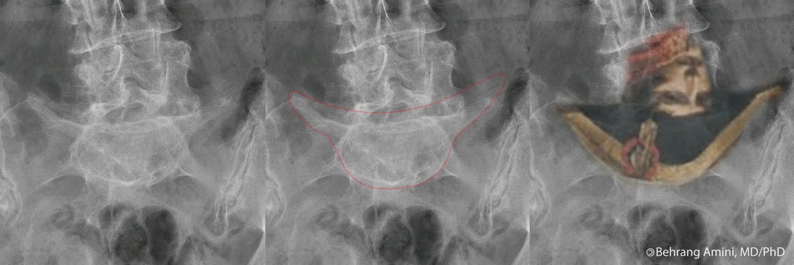
The images above are from a patient with POEMS syndrome. We see sclerotic lesions in the humerus, pelvis, and proximal femur, some of which have the typical ring-like appearance (e.g., the left intertrochanteric region). Looking through our 20 or so cases of POEMS here, I wasn't able to find any with the pathognomonic proliferative changes described by Resnick, so a look at the original paper is worthwhile to get familiar with this appearance.

Special thanks to Dr. James Dimaala for the case.

References

  • Chong ST, Beasley HS, Daffner RH. POEMS syndrome: radiographic appearance with MRI correlation. Skeletal Radiol. 2006 Sep;35(9):690-5.
  • Dispenzieri A, Kyle RA, Lacy MQ, Rajkumar SV, Therneau TM, Larson DR, Greipp PR, Witzig TE, Basu R, Suarez GA, Fonseca R, Lust JA, Gertz MA. POEMS syndrome: definitions and long-term outcome. Blood. 2003 Apr 1;101(7):2496-506.
  • Owens CL, Weir EG, Ali SZ. Cytopathologic findings in "POEMS" syndrome associated with Castleman disease. Diagn Cytopathol. 2007 Aug;35(8):512-5.
  • Resnick D, Greenway GD, Bardwick PA, Zvaifler NJ, Gill GN, Newman DR. Plasma-cell dyscrasia with polyneuropathy, organomegaly, endocrinopathy, M-protein, and skin changes: the POEMS syndrome. Distinctive radiographic abnormalities. Radiology. 1981 Jul;140(1):17-22.

Saturday, November 19, 2011

Myxoinflammatory Fibroblastic Sarcoma

Myxoinflammatory fibroblastic sarcoma is a low-grade sarcoma with myxoid stroma and a mixed acute and chronic inflammatory infiltrate. Three main types of neoplastic cells are seen: spindled cells, large polygonal and bizarre ganglion-like cells, and variably sized multivacuolated lipoblast-like cells. All three types also have features of fibroblasts. The lesion predominantly affects the extremities, usually the hands and feet.

MRI findings are variable. The lesions can be well-defined or poorly defined. The signal characteristics are also variable. They can be low signal intensity on T1-weighted images and high signal intensity on T2-weighted images, mimicking a cyst on unenhanced imaging. Post-contrast imaging reveals these to be solid lesions with avid enhancement ranging from mildly heterogeneous to homogeneous.

Differential considerations include much more common entities, such as ganglion cyst (in cases of predominantly myxoid lesions), giant cell tumor of the tendon sheath (in cases of well-defined heterogeneous lesions near tendon sheaths), and tenosynovitis (in cases of poorly defined lesions near tendon sheaths), nodular fasciitis (for lesions occurring in the subcutaneous tissues), and other soft tissue sarcomas.

The case above is from a 50-year-old woman who palpated a nodule in her right knee. The images reveal a heterogeneous, poorly defined lesion in the subcutaneous tissues abutting the vastus medialis muscle. The lesion is predominantly T1- and T2- hypointense with central areas of T2 hyperintensity and a peripheral T2-hyperintense rim. Post-contrast imaging was not performed.

References

  • Kindblom LG and Meis-Kindblom JM. Myxoinflammatory fibroblastic sarcoma. in Pathology and Genetics of Tumours of Soft Tissue and Bone. Fletcher CDM, Unni KK, Mertens F (eds). IARCPress Lyon, 2002. pp 96-97.
  • Narváez JA, Martinez S, Dodd LG, Brigman BE. Acral myxoinflammatory fibroblastic sarcomas: MRI findings in four cases. AJR Am J Roentgenol. 2007 May;188(5):1302-5.
  • Tateishi U, Hasegawa T, Onaya H, Satake M, Arai Y, Moriyama N. Myxoinflammatory Fibroblastic Sarcoma: MR Appearance and Pathologic Correlation. AJR Am J Roentgenol. 2005 Jun;184(6):1749-53.

Friday, November 18, 2011

Implantable Spinal Fusion Stimulator

The implantable spinal fusion stimulators by Biomet consist of a direct current generator (pink arrow) in a titanium shell with a focal coating of platinum (serves as the anode) connected to insulated nonmagnetic steel leads (white arrows) that in turn connect to bare wire cathodes (blue arrows). The cathodes get embedded in pieces of bone graft by the surgeon and are thought to stimulate fusion of the allograft. The SpF-XL IIb Spinal Fusion Stimulator model is shown in the image above.

The generator is powered by a lithium iodine battery that is capable of delivering 20-μA of current for about 6 months. After that, the device is non-functional.

The device can be safely used in magnets with static field strengths of 1.5 T or less. The cathodes should ideally be placed at least 1 cm from nerve roots (to reduce the possibility of nerve excitation during MR imaging). It would also be nice if the stimulator is placed as far as possible from the spinal canal and bone graft to decrease the effect of artifact.

In addition,
  • Radiographs should be obtained prior to MRI to verify that the leads are intact. Broken leads can lead to excessive heating during imaging. If radiographs cannot make this determination, the risk of heating should be discussed with the patient prior to the study.
  • MRI should be performed in magnets with static fields of 1.5 T or less.
  • Spin-echo, fast spin-echo, and gradient echo pulse sequences are permissible.
  • Echo planar techniques, conditions that produce exposures to high levels of RF energy (exceeding a whole-body averaged specific absorption rate of 1.0 W/kg), exposure to gradient fields that exceed 20 T/sec, or unconventional MR sequences should be avoided.
  • Patients should be continuously observed during the study and instructed to report any unusual sensations including any feelings of warming, burning, or neuromuscular excitation or stimulation.

References

  • Biomet. MRI Safety Information - SpF®-XL IIb.
  • Schwardt JD, Jankowski GB. Preformed extendable mesh cathode for implantable bone growth stimulator. US Patent 6,112,122
  • Shellock FG, Hatfield M, Simon BJ, Block S, Wamboldt J, Starewicz PM, Punchard WF. Implantable spinal fusion stimulator: assessment of MR safety and artifacts. J Magn Reson Imaging. 2000 Aug;12(2):214-23.
  • Tejano NA, Puno R, Ignacio JM. The use of implantable direct current stimulation in multilevel spinal fusion without instrumentation. A prospective clinical and radiographic evaluation with long-term follow-up. Spine (Phila Pa 1976). 1996 Aug 15;21(16):1904-8.

Thursday, November 17, 2011

The J Sign

The intact inferior glenohumeral ligament normally extends from the glenoid labrum to the anatomic neck of the humerus, forming a U shape on oblique coronal images. Avulsion of the right inferior glenohumeral ligament from the neck of the humerus results in its humeral end falling inferiorly forming a J shape. This J-shaped appearance of the torn right inferior glenohumeral ligament in cases of humeral avulsion of the glenohumeral ligament (HAGL) is referred to as the J sign. On the left side, the J is reversed.

Special thanks to Dr. David Wells for the case.

References

Carlson CL. The "J" sign. Radiology. 2004 Sep;232(3):725-6.

Wednesday, November 16, 2011

Brachymesophalangia

Brachymesophalangia is the most common hereditary anomaly of the middle phalanges. It is more common in the small finger (brachymesophalangia-5 or brachymesophalangia-V) and is seen in up to 20% of the Japanese population and about 1% of the European population. About 2/3 of patients with Down syndrome have brachymesophalangia.

As seen in the image above, the short, often trapezoidal middle phalanx can result in angular deviation of the distal phalanx (clinodactyly).

In 1951, Bell classified brachydactyly into types A-E.
Type Description
A1 Brachymesophalangia II-IV with brachybasophalangia I
A2 Brachymesophalangia II with absent epiphyses, delta phalanx, and radial clinodactyly
A3 Brachymesophalangia V with radial clinodactyly (case shown above)
A4 Brachymesophalangia II-V with bifid distal phalanx of thumb and dystelephalangia V
B Brachymesophalangia with brachytelephalangia II-V (or absent distal phalanges)
C Brachymesophalangia II, III, and V with hyperphalangia of index and middle finger proximal phalanges
D Brachytelephalangia I with broadening
E Brachymetacarpia III-V

Brachybasophalangia: Short proximal phalanges
Brachymetacarpia: Short metacarpals
Brachytelephalangia: Short distal phalanges
Dystelephalangia: Deformed terminal phalanx (Kirner deformity)

References

  • Bell J. On brachydactyly and symphalangism. Treasury of human inheritance. Vol 5, part 1. London: Cambridge University Press, 1951.
  • Freyschmidt J, Brossmann J, Wiens J, Sternberg A. Chapter 2 - Upper Extremity. In Freyschmidt's Köhler and Zimmer: Borderlands of normal and early pathologic findings in skeletal radiography. Fifth revised edition. Thieme (2003). Pp 85-86.

Tuesday, November 15, 2011

Medial Meniscus Root Tears

Circumferential collagen fibers in menisci absorb compressive forces, resisting longitudinal loading (hoop stress) and meniscal extrusion. The normal hoop tension for the meniscus is reduced by radial tears that extend to the capsular margin of the meniscus. Functionally speaking, the loss of hoop tension of a meniscus is equivalent to total meniscectomy and leads to early degenerative change.

Root tears are more commonly seen in the posterior horn of the medial meniscus. Complete radial tear of the medial meniscus root leads first to meniscal extrusion and subsequently to the development of osteoarthritis. The image above shows a complete radial tear of the posterior root of the medial meniscus on the coronal image (pink arrow) with a ghost meniscus on the sagittal image (white arrow held by Casper) and meniscal extrusion (blue arrow). Bone marrow T2 hyperintensity is seen in the medial femoral condyle and the medial tibial plateau, with loss of articular cartilage at the medial compartment and a small subchondral fracture at the medial tibial plateau.

References

Lerer DB, Umans HR, Hu MX, Jones MH. The role of meniscal root pathology and radial meniscal tear in medial meniscal extrusion. Skeletal Radiol. 2004 Oct;33(10):569-74.

Monday, November 14, 2011

Accessory Epiphysis (Pseudoepiphysis)

A pseudoepiphysis is an accessory epiphysis that does not significantly contribute to the longitudinal growth of a tubular bone. One or more can occur in the same patient as normal variants, but they have also been associated with Down syndrome and hypothyroidism (~80% of patients with Down syndrome have pseudoepiphyses).

When found incidentally in patients without chromosomal and metabolic abnormalities, they they are most common in the distal thumb metacarpal followed by the proximal index finger metacarpal. In patients with Down syndrome, they are more common in the proximal index finger metacarpal followed by the distal thumb metacarpal (i.e., the order of the two most common sites is reversed).

References

  • Freyschmidt J, Brossmann J, Wiens J, Sternberg A. The Hand-General Aspects. In Freyschmidt's Köhler and Zimmer: Borderlands of normal and early pathologic findings in skeletal radiography. Fifth revised edition. Thieme (2003). Pp 23-24.
  • Kozin SH and Waters PM. Fractures and dislocations of the hand and carpus in children. In Rockwood and Wilkins' Fractures in Children. Seventh edition. Wolters Kluwer (2010). P 227.
  • Ogden JA, Ganey TM, Light TR, Belsole RJ, Greene TL. Ossification and pseudoepiphysis formation in the "nonepiphyseal" end of bones of the hands and feet. Skeletal Radiol. 1994 Jan;23(1):3-13.

Sunday, November 13, 2011

Os Carpi Centrale

The os carpi centrale (os centrale) is an uncommon accessory carpal bone that is located dorsally between the first and second carpal rows and articulates with the scaphoid, capitate, and trapezoid, but not with the trapezium. It can have the appearance of a segment cut out of the scaphoid that has ossified independently of it. The os carpi centrale is usually round and dense, and lacks an internal trabecular architecture. It can be bilateral and duplicated.

When the os carpi centrale is unossified, it is radiographically occult and can present as a large gap between the distal scaphoid and a groove in the capitate. On the other extreme, the os carpi centrale can fuse with the adjacent scaphoid, capitate, or trapezoid

A similar bone is found in the writs of of the Orangutan, but the os centrale in these primates articulates with the trapezium.

Because of its location, it may be confused with a scaphoid fracture. Patients are usually asymptomatic; however, some may present with painful clicking or pain from osteonecrosis.

The main differential considerations are bipartite scaphoid, scaphoid fracture, and an unfused scaphoid ossification center. Dysplasia epiphysealis hemimelica and congenital hypothyroidism can also be considered but the appearance of these conditions is usually distinct.

References

  • Abascal F, Cerezal L, del Piñal F, García-Valtuille R, García-Valtuille A, Canga A, Torcida J. Unilateral osteonecrosis in a patient with bilateral os centrale carpi: imaging findings. Skeletal Radiol. 2001 Nov;30(11):643-7.
  • Freyschmidt J, Brossmann J, Wiens J, Sternberg A. Carpus-Scaphoid. In Freyschmidt's Köhler and Zimmer: Borderlands of normal and early pathologic findings in skeletal radiography. Fifth revised edition. Thieme (2003). Pp 155-157.
  • Turner W. Some Variations in the Bones of the Human Carpus. J Anat Physiol. 1883 Jan;17(Pt 2):244-9.

Saturday, November 12, 2011

Wrisberg Rip

Wrisberg rip is a term coined by the folks at RadSource for a longitudinal tear in the posterior horn of the lateral meniscus that extends laterally from the Wrisberg ligament attachment. They feel that the tear is the result of traction from the ligament of Wrisberg in cases of anterior cruciate ligament tears.

The Wrisberg rip should be differentiated from the normal appearance of the so-called Wrisberg pseudo-tear: A vertical/oblique signal intensity region at the junction of the a meniscofemoral ligament (most commonly the ligament of Wrisberg) with the posterior horn of the lateral meniscus. When seen on only one sagittal slice, the pseudotear is thought to be caused by a volume averaging of the ligament connection with the meniscus. It can also be seen on multiple slices depending on the angle of insertion of the meniscofemoral ligament.

The case above shows a Wrisberg rip. The sagittal images A-C are from lateral to medial. The pink and white arrows indicate the posterior horn of the lateral meniscus on either side of a tear extending laterally from the attachment of the ligament of Wrisberg (blue arrow). The axial images (D, E) show the same thing from a different perspective. Panel F shows the anterior cruciate ligament tear that is seen in association with this meniscal tear pattern.

References

  • de Abreu MR, Chung CB, Trudell D, Resnick D. Meniscofemoral ligaments: patterns of tears and pseudotears of the menisci using cadaveric and clinical material. Skeletal Radiol. 2007 Aug;36(8):729-35.
  • Vahey TN, Bennett HT, Arrington LE, Shelbourne KD, Ng J. MR imaging of the knee: pseudotear of the lateral meniscus caused by the meniscofemoral ligament. AJR Am J Roentgenol. 1990 Jun;154(6):1237-9.

Friday, November 11, 2011

Low-Lying Peroneus Brevis Muscle Belly

A low-lying peroneus brevis muscle belly is one that has muscle tissue extending beyond the retromalleolar groove and the distal tip of the fibula. Between ~10%-25% of patients with a peroneus longus or brevis tendon tear have a low-lying peroneus brevis muscle belly when examined by MRI, suggesting that this variant can predispose the muscle and tendon to tears. It is thought that increased muscle mass in the retromalleolar groove causes overcrowding and results in peroneal tendon injury. A similar mechanism is thought to underlie the association of the presence of a peroneus quartus muscle to peroneal tendon tears.

The main differential consideration for the appearance of a low-lying peroneus brevis muscle belly is an accessory muscle, such as the peroneus quartus.

References

  • Freccero DM, Berkowitz MJ. The relationship between tears of the peroneus brevis tendon and the distal extent of its muscle belly: an MRI study. Foot Ankle Int. 2006 Apr;27(4):236-9.
  • Geller J, Lin S, Cordas D, Vieira P. Relationship of a low-lying muscle belly to tears of the peroneus brevis tendon. Am J Orthop (Belle Mead NJ). 2003 Nov;32(11):541-4.
  • Khoury NJ, el-Khoury GY, Saltzman CL, Kathol MH. Peroneus longus and brevis tendon tears: MR imaging evaluation. Radiology. 1996 Sep;200(3):833-41.
  • Rosenberg ZS, Beltran J, Cheung YY, Colon E, Herraiz F. MR features of longitudinal tears of the peroneus brevis tendon. AJR Am J Roentgenol. 1997 Jan;168(1):141-7.

Thursday, November 10, 2011

Fredericson Grading of Tibial Stress Reaction

Fredericson system is a 5-level grading scheme for tibial stress reactions developed by studying symptomatic runners. In symptomatic patients, it correlates with the time for return to full impact activity.

The MRI findings have to be correlated to symptoms, as a follow-up study showed that almost half of asymptomatic college distance runners had signs of tibial stress reaction on MRI. In addition, these changes did not predict future tibial stress reactions or stress fractures.
Grade MRI Findings Time to full-impact activity*
0 Normal MRI findings.  
1 Increased periosteal T2 signal. Normal marrow signal intensity on all images (shown here) 2 to 3 weeks
2 Grade 1 plus bone marrow signal changes on T2-weighted images 4 to 6 weeks
3 Grade 2 plus bone marrow signal changes on T1-weighted images (shown above) 6 to 9 weeks
4 Grade 3 plus a clearly visible fracture plane  
*Full impact activity: Ablility to return to running on grass or a soft dirt track

References

  • Bergman AG, Fredericson M, Ho C, Matheson GO. Asymptomatic tibial stress reactions: MRI detection and clinical follow-up in distance runners. AJR Am J Roentgenol. 2004 Sep;183(3):635-8.
  • Fredericson M, Bergman AG, Hoffman KL, Dillingham MS. Tibial stress reaction in runners. Correlation of clinical symptoms and scintigraphy with a new magnetic resonance imaging grading system. Am J Sports Med. 1995 Jul-Aug;23(4):472-81.

Wednesday, November 9, 2011

Aggregoma

Deposition of monoclonal light chains in B-cell dyscrasias can be systemic or localized, and fibrillar and non-fibrillar, and can be categorized under the broad definition of monoclonal immunoglobulin deposition diseases: "monoclonal expansion of a B-cell and plasma-cell population producing an excess immunoglobulin polypeptide with structural characteristics predisposing to tissue deposition in either the fibrillar or nonfibrillar state."

Systemic forms of amyloid (fibrillar) deposition include light chain (AL) and heavy chain (AH) amyloidosis, while systemic forms of non-amyloid (non-fibrillar) deposition include light chain deposition disease (LCDD), heavy chain deposition disease (HCDD) and light–heavy chain deposition disease (LHCDD).

The localized form of amyloid deposition is known as an amyloidoma. By analogy, aggregomas are localized tumoral masses of monoclonal light chain deposition, consisting of nonfibillar and Congo-red-negative proteins.

Imaging findings have not been described, but are presumably similar to those of amyloidomas.

References

  • Rostagno A, Frizzera G, Ylagan L, Kumar A, Ghiso J, Gallo G. Tumoral non-amyloidotic monoclonal immunoglobulin light chain deposits ('aggregoma'): presenting feature of B-cell dyscrasia in three cases with immunohistochemical and biochemical analyses. Br J Haematol. 2002 Oct;119(1):62-9.
  • Buxbaum J, Gallo G. Nonamyloidotic monoclonal immunoglobulin deposition disease. Light-chain, heavy-chain, and light- and heavy-chain deposition diseases. Hematol Oncol Clin North Am. 1999 Dec;13(6):1235-48.

Tuesday, November 8, 2011

Meniscal Cysts

Meniscal cysts are seen in about 5%-10% of knee MRIs. Meniscal cysts are more commonly parameniscal (perimeniscal), but can also be intermeniscal or both. The majority, though not all, of meniscal cysts are associated with meniscal tears. Conversely, parameniscal cysts are seen in less than 10% of cases of meniscal tears.

Meniscal cysts are thought to arise from dissection of synovial fluid through meniscal tears, and entrapment through a one-way valve mechanism.

On MRI, they are more commonly seen in the medial compartment. Earlier studies, however, reported a ration of lateral to medial cysts of up to 7:1. The discrepancy is thought to be related to the higher sensitivity of MRI for the detection of meniscal cysts compared to physical examination (which is better at detecting lateral cysts due to the smaller amount of fatty soft tissue on the lateral aspect of the knee), conventional knee arthrography, and arthroscopy (due to the difficult surgical approach to the posterior horn of the medial meniscus, where most medial cysts are located).

Parameniscal cysts typically present as loculated fluid signal with a connection to the adjacent meniscus. Intrameniscal cysts present as increased signal within an enlarged meniscus with an expanded contour. The T2 signal of intrameniscal cysts may not be as high as that of parameniscal cysts.

When located medially, parameniscal cysts are usually adjacent to the posterior horn. The majority also extend anteriorly adjacent to the body. Cysts can also extend centrally and end up posterior to or surrounding the posterior cruciate ligament, simulating a ganglion.

When located laterally, parameniscal cysts are usually adjacent to the anterior horn, with half of the cysts extending posteriorly adjacent to the body. The image above shows a cyst adjacent to the body of the lateral meniscus in communication with a horizontal tear.

The majority of medial parameniscal cysts, as well as parameniscal cysts overlying the body or posterior horn of the lateral meniscus are associated with meniscal tears. By comparison, only about 2/3 of cysts adjacent to or extending to the anterior horn of the lateral meniscus are associated with meniscal tears. It is not clear why these cysts are less likely to have an underlying meniscal tear.

References

  • Anderson JJ, Connor GF, Helms CA. New observations on meniscal cysts. Skeletal Radiol. 2010 Dec;39(12):1187-91.
  • Bergin D, Hochberg H, Zoga AC, Qazi N, Parker L, Morrison WB. Indirect soft-tissue and osseous signs on knee MRI of surgically proven meniscal tears. AJR Am J Roentgenol. 2008 Jul;191(1):86-92.
  • Campbell SE, Sanders TG, Morrison WB. MR imaging of meniscal cysts: incidence, location, and clinical significance. AJR Am J Roentgenol. 2001 Aug;177(2):409-13.
  • De Smet AA, Graf BK, del Rio AM. Association of parameniscal cysts with underlying meniscal tears as identified on MRI and arthroscopy. AJR Am J Roentgenol. 2011 Feb;196(2):W180-6.

Monday, November 7, 2011

Clear Cell Variant of Chondrosarcoma

Clear cell chondrosarcoma is a low-grade malignancy that behaves less aggressively than conventional chondrosarcoma. It accounts for about 2% of all cases of chondrosarcoma, is more commonly seen in men, and usually affects patients in their 20s to 40s.

It tends to affect the epiphyses of long tubular bones, most commonly the femur and humerus. In such cases, patients can present with joint pain and effusion and limited range of motion.

Radiographs typically reveal a predominantly lytic lesion with a narrow zone of transition sometimes with a sclerotic margin. The lesion may be expansile, have central calcifications and ossifications, and demonstrate endosteal scalloping. Periosteal reaction and soft tissue extension are rare. The imaging appearance can mimic chondroblastoma, especially when seen in younger patients: While clear cell chondrosarcoma has homogeneous intermediate signal intensity on T1-weighted images and heterogeneous high signal intensity on T2-weighted images, chondroblastoma usually has low signal intensity on T1- and T2-weighted images.

Also of interest: cartilage lesions by age and location.

References

Greenspan A, Jundt G, Remagen W. Cartilage (Chondrogenic) Lesions. In Differential Diagnosis of Orthopaedic Oncology, 2nd Edition. 2007 Lippincott Williams & Wilkins; pp 222-224.

Sunday, November 6, 2011

Inverted Napoleon's Hat Sign

The inverted Napoleon's hat sign refers to the appearance of the L5 vertebra on frontal radiographs in patients with anterolisthesis of L5 on S1. It is formed by overlap of L5 on the sacrum on the frontal radiograph, with the vertebral body forming the dome of Napoleon's hat and the transverse processes forming tapered brim of the hat.

References

Talangbayan LE. The inverted Napoleon's hat sign. Radiology. 2007 May;243(2):603-4.

Saturday, November 5, 2011

Metal Artifact Reduction Sequence (MARS)

The metal artifact reduction sequence (MARS) is a modified spin-echo pulse sequence that uses view angle tilting (application of the slice-select gradient at the same time as the frequency encoding gradient). Additional modifications are also made to optimize imaging.

All together, the MARS technique involves:
  • View angle tilting: Used to remvoe geometric distortion. The slice-select gradient is applied at the same time as the frequency encoding gradient (i.e., during signal readout), giving the spins the same narrow frequency band and removing distortion due to magnetic field inhomogeneity. This comes at the cost of introducing blurring in the frequency encode direction.
  • Increasing the slice-select gradient strength: This makes the imaging gradients as large as possible relative to the susceptibility-induced gradients produced by metal. Increasing the slice-select gradient strength also results in a decrease in slice thickness.
  • Increasing the frequency encoding gradient strength: This makes the imaging gradients as large as possible relative to the susceptibility-induced gradients produced by the metal implants. Also decreases geometric distortion in the image. Increasing the frequency encoding gradient strength, however, requires a larger bandwidth, which reduces the signal-to-noise ratio (30% to 50% lower, depending on the field-of-view).

References

  • Cho ZH, Kim DJ, Kim YK. Total inhomogeneity correction including chemical shifts and susceptibility by view angle tilting. Med Phys. 1988 Jan-Feb;15(1):7-11.
  • Lee MJ, Janzen DL, Munk PL, MacKay A, Xiang QS, McGowen A. Quantitative assessment of an MR technique for reducing metal artifact: application to spin-echo imaging in a phantom. Skeletal Radiol. 2001 Jul;30(7):398-401.
  • Olsen RV, Munk PL, Lee MJ, Janzen DL, MacKay AL, Xiang QS, Masri B. Metal artifact reduction sequence: early clinical applications. Radiographics. 2000 May-Jun;20(3):699-712.

Friday, November 4, 2011

Semimembranosus-Tibial Collateral Ligament Bursa

The semimembranosus-tibial collateral ligament bursa is a U-shaped structure that wraps around the anterior expansion of the semimembnanosus tendon. It is positioned between the semimembranosus-gastrocnemius (cupcake in the diagram) and middle collateral ligament bursae superiorly and the pes anserinus bursa (Canada goose in the diagram) inferiorly. The semimembranosus-tibial collateral ligament bursa does not communicate the adjacent bursae or with the knee joint.

The U shape of the bursa (blue) is formed by deep and superficial arms. The deep arm (dotted line) is located between the semimembranosus tendon and the medial tibial condyle. The superficial arm (solid line) is located between the semimembranosus tendon and the medial collateral ligament (tibial collateral ligament). The two arms join along the anterosuperior margin of the semimembranosus tendon ("elbow").

Special thanks to Dr. Luke Yoon for the case.

References

Thursday, November 3, 2011

Lipomatosis of Nerve

Lipomatosis of nerve, formerly known as fibrolipomatous hamartoma, is the anomalous growth of fibrofatty tissue within a nerve, resulting in fusiform enlargement with or without associated nerve-territory oriented osseous overgrowth and proliferation of subcutaneous fat (1/3-2/3 of cases). Histological changes in the nerves are identical to those seen in cases of macrodystrophia lipomatosa, and the world health organization lists macrodystrophia lipomatosa as a synonym for lipomatosis of nerve. Other synonyms include: lipofibroma, fibrolipomatosis, intraneural lipoma, perineural lipoma, median nerve lipoma, and neural fibrolipoma.

While commonly first noted at birth or in early childhood, patients may not present for treatment until adulthood. Women and girls are more likely to have associated macrodactyly. Patients usually present with a gradually enlarging mass, with or without associated with motor or sensory deficits.

Lipomatosis of nerve most commonly affects the median nerve and its digital branches, followed by the ulnar nerves; however, it can affect any other nerve, including cranial nerves and the brachial plexus.

MRI findings are usually pathognomonic: Evenly distributed fat tissue splaying nerve fascicles, resulting in fusiform enlargement of the nerve. No significant edema is typically present within the nerve. The amount of fat varies and can be barely detectable.

In the face of pathognomonic imaging findings, biopsy of a major peripheral nerve in not needed, as it can result in neurologic deficit. Lipomatosis of nerve also cannot be surgically excised for the same reason; however, in the case of disease limited to cutaneous nerves the functional loss will be negligible. Decompression is helpful is tight spaces such as the median nerve in the carpal tunnel.

The images above show a case of lipomatosis of the median nerve, just deep to the plamaris longus tendon. The patient was treated with carpal tunnel release without significant improvement in symptoms.

References

  • Nielsen GP. Lipomatosis of nerve. in Pathology and Genetics of Tumours of Soft Tissue and Bone. Fletcher CDM, Unni KK, Mertens F (eds). IARCPress Lyon, 2002. pp 24-25.
  • Wong BZ, Amrami KK, Wenger DE, Dyck PJ, Scheithauer BW, Spinner RJ. Lipomatosis of the sciatic nerve: typical and atypical MRI features. Skeletal Radiol. 2006 Mar;35(3):180-4.

Wednesday, November 2, 2011

The Structure of Articular Cartilage

Articular cartilage consists of relatively small numbers of chondrocytes in a matrix of proteoglycans compressed by and trapped within a collagen network. The negatively charged side chains of proteoglycans create an osmotic gradient across the surface of cartilage and draw water into the tissue (the water content of cartilage is between 70%–80%). The collagen fibers provide tensile resistance to the swelling caused by the water and anchor the cartilage to the underling bone.
Articular cartilage is organized as a multilayered structure made up of four zones. The most superficial zone contains ellipsoid chondrocytes and collagen fibers oriented parallel to the articular surface. Deep to this is a transitional zone made up of collagen fibers arranged in a more random fashion. The next layer is the radial zone, made up of large collagen fibers and cells arranged in columns. The radial zone has the highest proteoglycan content and the lowest water content. Finally, there is the calcified zone, which separates the articular cartilage from the subchondral bone. The tidemark is the boundary between the radial and calcified zones.

References

  • Goodwin DW. MRI appearance of normal articular cartilage. Magn Reson Imaging Clin N Am. 2011 May;19(2):215-27.
  • Wong M, Carter DR. Articular cartilage functional histomorphology and mechanobiology: a research perspective. Bone. 2003 Jul;33(1):1-13.

Tuesday, November 1, 2011

GraftJacket Tendon Repair

The GraftJacket® matrix is an acellular human dermal matrix made by Wright Medical Technology that is processed to remove the epidermis and dermal cells, leaving an acellular scaffold of collagen, elastin, chondroitin sulfate, proteoglycans, hyaluronic acid, laminin, tenacin, and fibroblast growth factor. The acellular nature is thought to make it less immunogenic. The scaffold is supposed to provide strength as well as an intact extracellular framework with preserved vascular channels to support rapid revascularization and cellular repopulation. The system can be used to augment or replace irreparable portions of the rotator cuff tendon.

In the shoulder, the GraftJacket is used to span the cuff tear in younger, more active patients who have irreparable rotator cuff tears. Ideally, patients are motivated, intelligent, and younger and have disabling pain but an intact biceps tendon and well-maintained active motion with a functioning subscapularis muscle. The biceps tendon is used as an anterior anchor point for the graft.

References

  • Barber FA, Burns JP, Deutsch A, Labbé MR, Litchfield RB. A Prospective, Randomized Evaluation of Acellular Human Dermal Matrix Augmentation for Arthroscopic Rotator Cuff Repair. Arthroscopy. 2011 Oct 5.
  • Bond JL, Dopirak RM, Higgins J, Burns J, Snyder SJ. Arthroscopic replacement of massive, irreparable rotator cuff tears using a GraftJacket allograft: technique and preliminary results. Arthroscopy. 2008 Apr;24(4):403-409.e1.
  • Wong I, Burns J, Snyder S. Arthroscopic GraftJacket repair of rotator cuff tears. J Shoulder Elbow Surg. 2010 Mar;19(2 Suppl):104-9.