Showing posts with label Variants. Show all posts

Sunday, April 16, 2017

Ventriculus Terminalis

MRI of ventriculus terminalis (also known as the terminal ventricle and the fifth ventricle)
The ventriculus terminalis (also known as the terminal ventricle and the fifth ventricle) is a rarely identified cerebrospinal fluid cavity within the conus medullaris. The ventriculus terminalis does not communicate with the subarachnoid space or the central canal of the spinal cord, and may actually be an embryonal remnant of the primitive central canal, leading some to refer to it a sinus terminalis instead.

They are occasionally associated with caudal regression of the spinal cord, Chiari type I malformation, lipomyelomeningoceles, and lumbosacral "lipomas." Some authors believe that all of us have some sort of cystic CSF space at the conus medullaris, but that it's simply larger [and detectable on imaging] in some people and tends to regress (but not completely resolve) over time.

The characteristic imaging features are more commonly seen in children: Cystic lesion of the conus medullaris without spinal cord signal abnormality. In adults, ventriculus terminalis is more likely to have septations and be associated with spinal cord edema, kyphotic deformity and spinal arteriovenous malformations.

Rarely, ventriculus terminalis can enlarge in the presence of meningeal hemorrhage or deformities of the vertebral canal. An enlarged or symptomatic ventriculus terminalis can be treated by cyst fenestration with or without shunting to the subarachnoid space, pleural cavity, or peritoneal cavity.

References

Monday, April 10, 2017

False Perpetuations: Main Portal Vein Size and Portal Hypertension

Perpetuation: A main portal vein (MPV) diameter >13 mm is "consistent with portal hypertension" (pHTN)

This cutoff of 13 mm is based on weak literature (mainly from the 1980's), some of which did not include comparison values of normal patients

  • One comparative study using ultrasound found (Radiology 1982; 142: 167-172):
    • In 79 patients with pHTN
      • 36 had a MPV diameter of <13 mm 
      • 33 had a MPV diameter >/= 13 mm
      • The MPV was not visualized in 10 patients
    • In the 45 control patients
      • The MPV diameter was < 13 mm in 41 cases
      • The MPV was not visualized in 4 patients. 

More recent studies have found that there is no significant difference in MPV diameters when comparing patients without cirrhosis to patients with cirrhosis, and the normal MPV diameter is significantly larger than the 13 mm cutoff

  • A study (Eur J Gastroenterol Hepatol 2004; 16:147-155) from King's College using ultrasound (49 controls and 14 cirrhotics) found: 
    • the average MPV diameters were 9.6 cm and 10.8 cm in patients without and with cirrhosis, respectively.
  • A second study (JCAT 2008; 32: 198-203) from UCSF using CT (59 controls and 67 cirrhotics) found:
    • The average MPV diameters were 14.5 cm and 14.8 cm in patients without and with cirrhosis, respectively.
  • Using CT, the MPVs in healthy renal donor patients were measured before and after the administration of intravenous contrast, and in the axial and coronal planes (Abdom Radiol 2016; 41:1931-1936). This study found:
    • The average MPV diameter was 15.5 +/- 1.9 mm
      • This value was significantly different than 13 mm
    • Post-contrast MPVs were 0.56 mm larger compared to non-contrast
    • A positive correlation between BMI and height versus MPV diameter
In fact, the MPV size can be reduced in portal hypertension and has been described as a sign of hepatofugal MPV flow (AJR 2003; 181: 1629-1633). This study found:
  • A MPV diameter of less than 1 cm is a highly sensitive (but not very specific) for MPV flow reversal in patients with cirrhosis

Sunday, February 12, 2017

The Cervical Split: A Pseudofracture


A horizontal line projecting over a cervical vertebral body on lateral radiographs can simulate a fracture or a butterfly vertebral body. This pseudofracture, the so-called cervical split, can result from the lucency between contiguous uncovertebral osteophytes, or, as in the case above, cervical scoliosis resulting in projection of the facet joint over the vertebral body.

A cervical split due to uncovertebral joint osteophyte formation is said to be always accompanied by disc space narrowing.

References

Monday, December 24, 2012

Pyelonephritis Mimic


The above CECT images are from a patient who presented to the ER with a recurrent history of left flank pain. A cursory look shows regions of decreased parenchymal enhancement in the left kidney with inflammatory changes of the left perinephric fat which may favor a diagnosis of pyelonephritis. However, there is subtle retroperitoneal adenopathy (left paraaortic and surrounding the left renal artery). Biopsy revealed primary Ewing's sarcoma of the left kidney.

Friday, November 16, 2012

Meniscal Flounce Revisited


Previously discussed here, meniscal flounce is a normal variant characterized by a single fold along the free edge of a meniscus. By definition it exists in the absence of a meniscal tear. While usually seen in the medial meniscus, the above images demonstrate the finding on the lateral meniscus. 


REFERENCES
Park JS, Kyung NR, Yoon KH. Meniscal flounce on knee MRI: correlation with meniscal locations after positional changes. AJR Am J Roentgenol 2006;187:364-70.
Yu JS, Cosgarea AJ, Kaeding CC. Meniscal flounce MR imaging. Radiology 1997;203:513-5.

Wednesday, November 14, 2012

Enlarged Parietal Foramina


Enlarged parietal foramina are considered a benign normal variant in calvarial ossification. They are associated with anomalies of cerebral venous development, variations in occipital cortical infolding, scalp defects, and cleft palate. Although sometimes palpable, these defects are usually discovered incidentally. For patients with prominent foramina, cerebral vascular imaging may be considered for work up of aforementioned anomalies.


REFERENCES
Fink AM and Maixner W. Enlarged parietal foramina: MR imaging features in the fetus and neonate. AJNR 2006;27:1379-81.
Reddy AT, Hedlund GL, Percy AK. Enlarged parietal foramina: association with cerebral venous and cortical anomalies. Neurology 2000;54(5):1175-8.

Tuesday, October 23, 2012

Portal Vein Varix


Varix of the portal vein is a rare congenital anomaly of unknown origin and refers to focal dilatation of the portal vein. It is sometimes improperly referred to as an aneurysm of the portal vein. The varix is usually asymptomatic and tends to be an incidental finding.

The above images are from a patient who presented with right sided flank pain and was presumed to have renal colic. NCECT demonstrates a portal vein varix with luminal high density consistent with thrombosis.


REFERENCES
Lee WK, Chang SD, Duddalwar VA, et al. Imaging assessment of congenital and acquired abnormalities of the portal venous system. Radiographics 2011;31:905-26. 

Thursday, August 23, 2012

Normal Variants in the Pediatric Cervical Spine

Interpreting cervical spine x-rays in the pediatric population can be a challenge due to normal anatomic variants. At age 8-10 a child's cervical spine reaches adult proportions. Normal variants to be considered in the younger populations include:

1. The atlantodens interval (ADI) may be up to 5mm in the pediatric patient whereas the upper limit of normal is 3mm in an adult.
2. Pseudo-Jefferson fracture: up to 6mm displacement of the lateral masses of atlas on the axis on the open mouth view is normal for children up to age 7.
3. Pseudo-subluxation of C2 on C3 (and to a lesser extent C3 on C4): normal mobility of upper cervical spine (due to ligamentous laxity) can cause up to a 4mm anterior displacement of C2 on C3. The Swischuk line - a line drawn through the posterior arch of C2 should be within 2mm of the spinolaminar line drawn at C1-C3 - can be used to determine whether this finding is normal or due to possible hangman's fracture. A discrepancy of > 2mm can indicate a fracture.
4. Anterior "wedging" of up to 3mm is a normal finding. 
5. Ossification centers and unfused apophyses may mimic fractures.
6. Absence of cervical spine lordosis may be seen up to age 16.

REFERENCES
Curtin P and McElwain J. Assessment of the "nearly normal" cervical spine radiograph: C2-C3 pseudosubluxation in an adult with whiplash injury. Emerg Med J 2005;22:907-8.
Lustrin ES, Karakas SP, Ortiz AO, et al. Pediatric cervical spine: normal anatomy, variants, and trauma. Radiographics 2003;23:539-60.

Wednesday, August 22, 2012

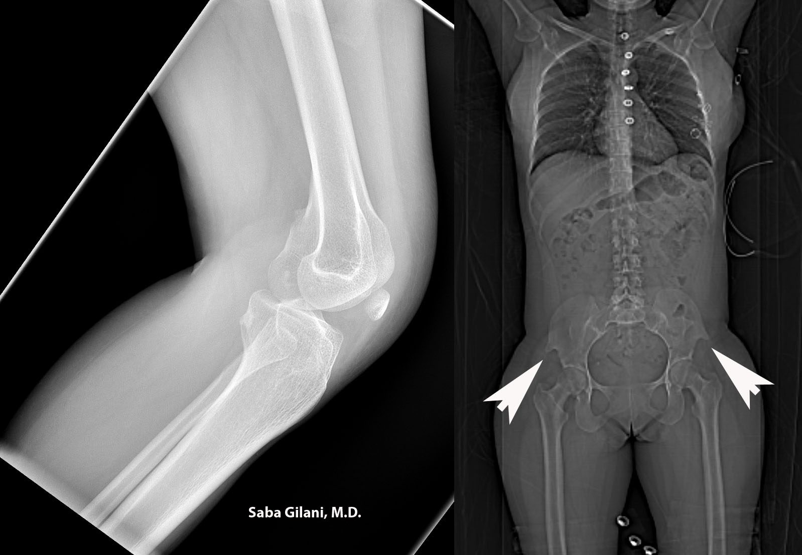
Hereditary Osteo-onychodysplasia Disease


Hereditary osteo-onychodysplasia disease (HOOD), also known as Nail Patella Disease or Iliac Horn Syndrome is a genetic disorder due to an autosomal dominant mutation in the LMX1B gene. A pathognomonic finding is the presence of "iliac horns" projecting posterolaterally from the bilateral iliac bones. Other associated findings are absence or hypoplasia of the patella and radial head. Nail deformities are also common.  The joint deformities can lead to osteoarthritis. About 40% of patients may also develop renal disease ranging from proteinuria to nephrotic syndrome.

Above, the lateral radiograph of the knee is from a patient who presented to the emergency room after a fall. A hypoplastic patella was incidentally noted. A review of prior imaging demonstrated bilateral iliac horns best seen on the scout image.


REFERENCES
Scott JE and Taor WS. The small patella syndrome. J Bone Joint Surg [Br] 1979;61-B:172-5.
Thompson EA, Walker T, Weens HS. Iliac horns. An osseous manifestation of hereditary arthrodysplasia associated with dystrophy of the fingernails. Radiology 1949;53:88-92.
Tuncbilek N, Karakas HM, Okten OO. Imaging of nail-patella syndrome. Hong Kong Med J 2005;11(2):116-8.

Wednesday, August 15, 2012

Anomalies of the Inferior Vena Cava and Their Clinical Significance


1. Left IVC: joins the left renal vein and crosses anterior to the aorta to join the right renal vein
  • can be mistaken for paraaortic adenopathy
  • report of AAA rupture into the IVC
2. Double IVC: the left IVC ends at the level of the left renal vein which crosses anterior to the aorta to join the confluence of the right IVC/renal vein.
  • suspect if patient has recurrent pulmonary embolism after placement of an IVC filter
3. Azygous continuation of the IVC: above the renal veins the IVC passes posterior to the diaphragmatic crus and continues as the azygous vein which joins the superior vena cava in the right paratracheal region. The hepatic segment of the IVC empties directly into the right atrium.
  • important to consider in cases of a right paratracheal mass 
  • can be mistaken for retrocrural adenopathy
4. Circumaortic left renal vein: two left renal veins are present; the superior renal vein receives the left adrenal vein while the inferior renal vein receives the left gonadal vein.
  • significant during planning of nephrectomy
5. Retroaortic left renal vein: a single left renal vein passes posterior to the aorta
  • recognition during preoperative planning is important
6. Double IVC with retroaaortic right renal vein and hemiazygous continuation of the IVC: confluence of the right renal vein and right IVC crosses posterior to the aorta to join the left IVC and continue cephlad as the hemiazygous vein. Several collateral pathways for the hemiazygous vein exist in the thorax - hemiazygous crossing posterior to the aorta to join the azygous vein, hemiazygous joining the cardinal vein of the heart via a persistent left SVC, and accessory hemiazygous continuation to the brachocephalic vein
  • hemiazygous collateral pathway may be mistaken for a left mediastinal mass
  • accessory hemiazygous has been reported to be mistaken for an aortic dissection
7. Circumcaval ureter: always occurring on the right, the proximal ureter courses posterior to the IVC and emerges to the right of the aorta and continues anterior to the right iliac vessels.
  • patients may develop partial ureteral obstruction or recurrent urinary tract infections
  • treatment is surgical relocation of the ureter anterior to the IVC
8. Absent infrarenal IVC with preservation of the suprarenal segment: external and internal iliac veins converge as lumbar veins which continue cephlad as the paravertebral collateral veins to join the azygous and hemiazygous. The confluence of the renal veins forms a normal suprarenal IVC.
  • patients may present with symptoms of lower extremity venous insufficiency or idiopathic deep venous thrombosis
  • collateral circulation may mimic a paraspinal mass


REFERENCES

Bass JE, Redwine MD, Kramer LA, et al. Spectrum of congenital anomalies of the inferior vena cava: cross-sectional imaging findings. Radiographics. 2000 May;20:639-52.

Tuesday, August 14, 2012

The Spectrum of Esophageal Atresias


Esophageal atresia is a congenital anomaly related to incomplete formation of the esophagus with or without the presence of a tracheoesophageal (TE) fistula. While the exact cause is unknown, it is felt to be related to incomplete separation of the primitive foregut into the trachea and the esophagus. One accepted classification of esophageal atresia and TE fistulas is as follows:

A. Atresia without TE fistula
B. Atresia with proximal TE fistula
C. Atresia with distal TE fistula (most common)
D. Atresia with proximal and distal TE fistula
E. TE fistula without atresia

Esophageal atresia is usually suspected in the setting of polyhydramnios, excessive salivation, choking/cyanosis during feeding, and inability to pass a nasogastric/feeding tube to the stomach. Anteroposterior and lateral radiographs will reveal a blind-ending, air-filled proximal esophagus. Radiographs of the abdomen should also be performed to evaluate for air in the GI tract, the presence of which raises suspicion for a distal TE fistula. Fluoroscopy may be used to confirm findings in which case water soluble contrast is preferred.

The above images are from a newborn with a history of difficulty feeding. The initial radiograph reveals a dilated upper esophagus without air seen in the GI tract. A nasogastric tube was placed which coiled in the proximal esophageal stump. Via a percutaneous gastrostomy tube, contrast was introduced into the stomach which refluxed into a distal esophageal remnant without evidence for fistulous connection. Type A esophageal atresia was diagnosed.


REFERENCES

Berrocal T, Torres I, Gutierrez J, et al. Congenital anomalies of the upper gastrointestinal tract. Radiographics. 1999;19:855-72.

Saturday, March 17, 2012

Os Vesalianum Pedis

[image needed]
The os vesalianum pedis (to be differentiated from the os vesalianum manius in the hand) is an uncommon accessory bone that is seen in less than 0.5% of the population. It is located within the peroneus brevis tendon adjacent to the tip of a well-developed tuberosity of the fifth metatarsal and articulates with the cuboid. It is rarely symptomatic.

Differential considerations include:
  • Avulsion fracture: Oriented in the transverse plane.
  • Normal ossification center: The normal ossification center of the tuberosity of the fifth metatarsal is oriented parallel to the metatarsal shaft.
  • Iselin disease: Traction apophysitis at the base of the fifth metatarsal. Similar to Osgood Schlatter disease, ossicles can break free, resulting in a fragmented appearance.

References

  • Gillespie H. Osteochondroses and apophyseal injuries of the foot in the young athlete. Curr Sports Med Rep. 2010 Sep-Oct;9(5):265-8.
  • Mellado JM, Ramos A, Salvadó E, Camins A, Danús M, Saurí A. Accessory ossicles and sesamoid bones of the ankle and foot: imaging findings, clinical significance and differential diagnosis. Eur Radiol. 2003 Dec;13 Suppl 4:L164-77. Review.

Monday, March 12, 2012

Diffusely Increased FDG Activity in the Thyroid

The thyroid may occasionally have diffusely increased uptake on FDG-PET scans obtained for malignancy elsewhere. Between 1%-3% of patients undergoing a PET scan will have such uptake. This finding can be seen as a normal variant or associated with chronic lymphocytic (Hashimoto) thyroiditis, with or without the presence of hypothyroidism and can be seen even in patients receiving thyroid hormone replacement therapy.

Focal uptake in the thyroid should raise concern for carcinoma or metastasis.

References

Friday, March 9, 2012

Lateral Recess of the Sphenoid Sinus

Pneumatization of the sphenoid sinus can extend laterally, creating a lateral recess. Pneumatization can then further extend into the pterygoid process, the great wing of the sphenoid bone (as seen abvoe) or both. This can be clinically relevant, as surgical access to lesions in this lateral recess can be challenging.

Coronal images reveal projection of the sphenoid sinus sidewall beyond the foramen rotundum (r). The lateral recess extends into the greater wing of the sphenoid bone above the vidian canal (v) and below the foramen rotundum (r), separating the two. There may be normal side-to-side asymmetry of the vidian-rotundum distance.

The main differential consideration in an opacified sinus is an expansile or erosive process in the sphenoid sinus (e.g., mucoceles or polyps). The most specific evidence of such a process is gross sinus wall erosion and flattening or erosion of the rims of vidian canal or the foramen rotundum.

The patient above has bilateral lateral recesses, with the one on the right extending to the sphenoid wing. The patient also has multiple facial fractures.

References

  • Giannetti AV, Guimarães RE, Santiago AP, Perpétuo FO, Machado MA. A tomographic study of the skull base in primary spontaneous cerebrospinal fluid leaks. Neuroradiology. 2011 Jul 8.
  • Lewin JS, Curtin HD, Eelkema E, Obuchowski N. Benign expansile lesions of the sphenoid sinus: differentiation from normal asymmetry of the lateral recesses. AJNR Am J Neuroradiol. 1999 Mar;20(3):461-6.
  • Tami TA. Surgical management of lesions of the sphenoid lateral recess. Am J Rhinol. 2006 Jul-Aug;20(4):412-6.

Thursday, March 1, 2012

Notch on the Lateral Aspect of the Radial Metaphysis

A notch can be seen as a normal variant on the lateral aspect of the radial metaphysis. This notch will eventually fill in by overgrowth of the epiphysis of the radial head.

References

McCarthy SM, Ogden JA. Radiology of postnatal skeletal development. VI. Elbow joint, proximal radius, and ulna. Skeletal Radiol. 1982;9(1):17-26.

Tuesday, January 24, 2012

Portal Vein Pulsatility

The normal flow in the portal venous system is typically continuously hepatopetal, with minimal if any pulsatility in rhythm with the cardiac cycle. Marked portal venous pulsatility can be classified as continuous pulsatile or reversed pulsatile flow.

The continuous pulsatile pattern is continuously hepatopetal, but with marked pulsatility. This pattern can be seen in patients with congestive heart failure, but can also be seen as a normal finding in thin subjects, where there is an inverse correlation of pulsatility to body mass.

The second pattern, reversed pulsatile flow is characterized by marked pulsatility and reversal of flow direction related to the cardiac cycle. There is a strong correlation of reversed pulsatile flow to high (> 20 mmHg) right atrial pressures in patients with chronic heart failure. However, reversed pulsatile flow can also be seen with tricuspid insufficiency, liver disease (cirrhosis, Budd–Chiari syndrome, hepatic outflow obstruction (constrictive pericarditis, mediastinal hematoma, pericardial mass or effusion), and shunts (portal vein–hepatic vein fistula, portocaval shunt). High abdominal pressures during deep inspiration can also cause transient reversal of flow. This is more commonly seen in patients with severe right heart failure or liver disease, but can also be seen in patients without these conditions.

Therefore, pulsatile portal venous flow by itself should not be construed as a sign of cardiac abnormality, especially if there is no reversal of the portal venous flow. The image above, however, shows a reversed pulsatile flow pattern and is from a patient with congestive heart failure.

Special thanks to Dr. Hansel Otero for the case.

References

Wednesday, January 18, 2012

Flexor Digitorum Accessorius Longus Muscle

The flexor digitorum accessorius longus muscle (white arrows) is an accessory muscle of the medial compartment of the ankle that is seen in less than 10% of the population. It is more commonly seen in males and is frequenly bilateral.

The muscle arises from variety of structures in the posterior compartment of the calf distal to the soleal line. Its tendon passes posterior to the flexor hallucis longus muscle (pink arrows) and the medial malleolus and inserts into the quadratus plantae muscle (blue arrow) or the flexor digitorum longus tendon. The presence of a flexor digitorum accessorius longus muscle has been associated with tenosynovitis of the flexor hallucis longus tendon.

The relationship of the tendon to the neurovascular structures (yellow arrows) of the ankle is also important, as compression of these structures can lead to tarsal tunnel syndrome. The flexor digitorum accessorius longus tendon is located posterior and superficial to the tibial nerve as it courses deep to the flexor retinaculum through the tarsal tunnel.

On axial MR images, the muscle is seen within the tarsal tunnel, typically superficial to the neurovascular bundle. At this point, the appearance may be similar to the peroneocalcaneus internus muscle. The flexor digitorum accessorius longus muscle, however, may contain fleshy fibers in the tarsal tunnel, which may help in differentiating the two. In addition, unlike the flexor digitorum accessorius longus muscle, the peroneocalcaneus internus muscle insert onto the calcaneus.

References

Saturday, December 31, 2011

Kump's Hump

Kump's hump, also known as kump's bump, is named after the radiologist Warren Kump, who described an undulation of the anteromedial aspect of the distal tibial physis.

Kump's hump is the site of first closure of the physis and should not be mistake for a fracture. On MRI, the normal loss of cartilaginous signal intensity of the physis begins at Kump's hump.

Kump's hump can also present a pitfall on MRI. The undulation at this location can simulate physeal closure on T1-weighted images, a phenomenon that is due to partial volume averaging of the physis with the adjacent epiphysis and metaphysis.

References

  • Chung T, Jaramillo D. Normal maturing distal tibia and fibula: changes with age at MR imaging. Radiology. 1995 Jan;194(1):227-32.
  • Keats TE and Anderson MW. Atlas of Normal Roentgen Variants That May Simulate Disease. 8th edition, page 812; Mosby (2004).
  • Kump WL. Vertical fractures of the distal tibial epiphysis. Am J Roentgenol Radium Ther Nucl Med. 1966 Jul;97(3):676-81.

Thursday, December 22, 2011

Ossification of the Sacrotuberous Ligaments

Ossification of the sacrotuberous ligaments has a caudocranial direction of growth. On frontal radiographs, pencil-like opacities project over the obturator foramina and extend cephalad and medially towards the sacrum. On cross-sectional imaging, the ossifications have a ventrodorsal flattened appearance and extend cephalad from the ischial tuberosities medially and posteriorly.

It has been suggested that ossification of the sacrotuberous ligaments is a good indicator of diffuse idiopathic skeletal hyperostosis (DISH), although others have contested this association. For what it's worth, our patient, a 50-year-old man, had no manifestations of DISH in the spine. Ossification of the sacrotuberous ligaments has also been associated with pudendal nerve entrapment.

Atherosclerotic calcifications can mimic the appearance of ossified sacrotuberous ligaments, but recognition of the course and tubular nature of these calcifications should be sufficient to avoid confusion.

References

  • Arora J, Mehta V, Suri RK, Rath G. Unilateral partial ossification of sacrotuberous ligament: anatomico-radiological evaluation and clinical implications. Rom J Morphol Embryol. 2009;50(3):505-8.
  • Prescher A, Bohndorf K. Anatomical and radiological observations concerning ossification of the sacrotuberous ligament: is there a relation to spinal diffuse idiopathic skeletal hyperostosis (DISH)? Skeletal Radiol. 1993 Nov;22(8):581-5.
  • Robert R, Prat-Pradal D, Labat JJ, Bensignor M, Raoul S, Rebai R, Leborgne J. Anatomic basis of chronic perineal pain: role of the pudendal nerve. Surg Radiol Anat. 1998;20(2):93-8.

Friday, December 16, 2011

Annular Pancreas

Annular pancreas is the second most common congenital pancreatic anomaly (after pancreas divisum), occurring in 1 in 2000 people. Its embryogenesis is unclear, but the end result is pancreatic tissue surrounding the second portion of the duodenum. This may be partial or complete.

Annular pancreas can be extramural or intramural. In the extramural type, the ventral pancreatic duct encircles the duodenum to join the main pancreatic duct. In the intramural type, the pancreatic tissue is intermingled with the duodenal wall muscle and small pancreatic ducts drain directly into the duodenum.

Patients with extramural annular pancreas can present with high gastrointestinal obstruction, sometimes with pancreatitis. Patients with intramural annular pancreas can present with symptoms of duodenal ulceration.

Contrast fluoroscopy can suggest the diagnosis by revealing narrowing at the level of the major papilla. ERCP or MRCP can show the duct of Wirsung encircling the duodenum in the patient with extramural annular pancreas. Cross sectional imaging will show pancreatic tissue around the second portion of the duodenum, as shown above in two patients with annular pancreas.

References