The double duct sign refers to the concomitant dilatation of the common bile duct and pancreatic duct seen on MRCP (although the same can be visualized on CT, US, and ERCP). Visualization of the double duct sign typically suggests the presence of a malignancy - either and ampullary or pancreatic head carcinoma. Other malignancies with this appearance include cholangiocarcinoma of the distal common bile duct, lymphoma, metastases, and in rare cases, Kaposi's sarcoma. Primary retroperitoneal fibrosis has also been reported to cause simultaneous dilatation of both bile ducts.
However, some studies have shown that the double duct sign is not as specific a predictor of pancreatic malignancy as initially thought to be since benign entities can present similarly. The most common benign causes are chronic pancreatitis and ampullary stenosis. Plumley et al., have suggested that the specific bile duct morphology on ERCP is a better predictor of benign versus malignant causes of ductal dilatation: abrupt stenosis of a bile duct with irregular margins is more likely to indicate malignancy while gradual ductal stenosis, especially when associated with smooth margins, is more indicative of benign lesions. Other predictors of benign disease include glandular calcifications and ectasia of the branching bile ducts (seen in chronic pancreatitis).
REFERENCES
Ahualli J. The Double Duct Sign. Radiology 2007;244:314-5.
Edge MD, Hoteit M, Patel AP. Clinical significance of main pancreatic duct dilation on computed tomography: single and double duct dilation. World J Gastroenterol 2007;31(11):1701-5.
Plumley TF, Rohrmann CA, Freeny PC, et al. Double duct sign: reassessed significance in ERCP. Am J Roentgenol 1982;138:31-5.
Wednesday, August 29, 2012

Tuesday, August 28, 2012
Internal Derangement of the Temporomandibular Joint

Internal Derangement of the Temporomandibular Joint (TMJ), more commonly known as dislocation of the TMJ, occurs due to an abnormal positional and functional relationship between the articular disc and the articulating surface. The images above are from a patient with anterior dislocations of the bilateral TMJs.
While this abnormality can usually be detected on plain films or bone NECT, MRI is the best modality to evaluate the TMJ especially in cases of recurrent derangement. On MRI, the low signal intensity articular disc will be displaced relative to the mandibular condyle. T1WI can show specific disc abnormalities including perforation, fibrosis or adhesions. The presence of a joint effusion can be detected as high signal on T2WI. The presence of synovitis can be detected as enhancement of the disc on T1WI after administration of contrast.
REFERENCES
Sommer OJ, Aigner F, Rudisch A, et al. Cross-sectional and functional imaging of the temporomandibular joint: radiology, pathology, and basic biomechanics of the jaw. Radiographics 2003;23:e14.
Tomas X, Pomes J, Berenguer J, et al. MR imaging of temporomandibular joint dysfunction: a pictorial review. Radiographics 2006;26:765-81.
Monday, August 27, 2012
Intramedullary Osteosclerosis
A nonhereditary condition that effects the long bones of the lower extremities (tibia > femur, fibula). On imaging studies it presents with endosteal sclerosis extending into the medullary canal, possibly obliterating the canal. There is no associated periosteal reaction or soft tissue swelling. Bone scan will show intense radiopharmaceutical uptake.This condition is more common in women and usually presents as pain that is exacerbated by activity.
Differential diagnosis:
1. Ribbing disease - may look exactly the same. The main difference is that Ribbing disease tends to have a familial inheritance pattern (thought to be autosomal recessive).
2. Stress fracture - usually a focal sclerosis and periosteal reaction will be present.
3. Sclerotic metastases - patchy, discontinuous presentation, population tends to be older.
4. Progressive diaphyseal displasia (Camurati-Engelmann disease) - presents in young boys, has skull and vertebral body involvement, disease begins in the diaphysis of long bones and may extend to the metaphysis, has periosteal and endosteal sclerosis.
5. Melorheostosis (Leri-Weill disease) - a sclerosing bone dysplasia which presents unilaterally in a long bone of the upper or lower extremity and may have associated soft tissue calcifications.
REFERENCES
Balkissoon ARA and Hayes CW. Intramedullary osteosclerosis. Radiology 1999;212:708-10.
Chanchairujira K, Chung CB, Lai YM, et al. Intramedullary osteosclerosis: imaging features in nine patients. Radiology 2001;220:225-30.
Differential diagnosis:
1. Ribbing disease - may look exactly the same. The main difference is that Ribbing disease tends to have a familial inheritance pattern (thought to be autosomal recessive).
2. Stress fracture - usually a focal sclerosis and periosteal reaction will be present.
3. Sclerotic metastases - patchy, discontinuous presentation, population tends to be older.
4. Progressive diaphyseal displasia (Camurati-Engelmann disease) - presents in young boys, has skull and vertebral body involvement, disease begins in the diaphysis of long bones and may extend to the metaphysis, has periosteal and endosteal sclerosis.
5. Melorheostosis (Leri-Weill disease) - a sclerosing bone dysplasia which presents unilaterally in a long bone of the upper or lower extremity and may have associated soft tissue calcifications.
REFERENCES
Balkissoon ARA and Hayes CW. Intramedullary osteosclerosis. Radiology 1999;212:708-10.
Chanchairujira K, Chung CB, Lai YM, et al. Intramedullary osteosclerosis: imaging features in nine patients. Radiology 2001;220:225-30.
Friday, August 24, 2012
Name the Device

Deep Brain Stimulator: electrodes implanted in the thalamus for treatment of Parkinson's disease.
REFERENCES
Hunter TB, Yoshino MT, Dzioba RB, et al. Medical devices of the head, neck, and spine. Radiographics 2004;24:257-285.
Thursday, August 23, 2012
Normal Variants in the Pediatric Cervical Spine
Interpreting cervical spine x-rays in the pediatric population can be a challenge due to normal anatomic variants. At age 8-10 a child's cervical spine reaches adult proportions. Normal variants to be considered in the younger populations include:
1. The atlantodens interval (ADI) may be up to 5mm in the pediatric patient whereas the upper limit of normal is 3mm in an adult.
2. Pseudo-Jefferson fracture: up to 6mm displacement of the lateral masses of atlas on the axis on the open mouth view is normal for children up to age 7.
3. Pseudo-subluxation of C2 on C3 (and to a lesser extent C3 on C4): normal mobility of upper cervical spine (due to ligamentous laxity) can cause up to a 4mm anterior displacement of C2 on C3. The Swischuk line - a line drawn through the posterior arch of C2 should be within 2mm of the spinolaminar line drawn at C1-C3 - can be used to determine whether this finding is normal or due to possible hangman's fracture. A discrepancy of > 2mm can indicate a fracture.
4. Anterior "wedging" of up to 3mm is a normal finding.
5. Ossification centers and unfused apophyses may mimic fractures.
6. Absence of cervical spine lordosis may be seen up to age 16.
REFERENCES
Curtin P and McElwain J. Assessment of the "nearly normal" cervical spine radiograph: C2-C3 pseudosubluxation in an adult with whiplash injury. Emerg Med J 2005;22:907-8.
Lustrin ES, Karakas SP, Ortiz AO, et al. Pediatric cervical spine: normal anatomy, variants, and trauma. Radiographics 2003;23:539-60.
1. The atlantodens interval (ADI) may be up to 5mm in the pediatric patient whereas the upper limit of normal is 3mm in an adult.
2. Pseudo-Jefferson fracture: up to 6mm displacement of the lateral masses of atlas on the axis on the open mouth view is normal for children up to age 7.
3. Pseudo-subluxation of C2 on C3 (and to a lesser extent C3 on C4): normal mobility of upper cervical spine (due to ligamentous laxity) can cause up to a 4mm anterior displacement of C2 on C3. The Swischuk line - a line drawn through the posterior arch of C2 should be within 2mm of the spinolaminar line drawn at C1-C3 - can be used to determine whether this finding is normal or due to possible hangman's fracture. A discrepancy of > 2mm can indicate a fracture.
4. Anterior "wedging" of up to 3mm is a normal finding.
5. Ossification centers and unfused apophyses may mimic fractures.
6. Absence of cervical spine lordosis may be seen up to age 16.
REFERENCES
Curtin P and McElwain J. Assessment of the "nearly normal" cervical spine radiograph: C2-C3 pseudosubluxation in an adult with whiplash injury. Emerg Med J 2005;22:907-8.
Lustrin ES, Karakas SP, Ortiz AO, et al. Pediatric cervical spine: normal anatomy, variants, and trauma. Radiographics 2003;23:539-60.
Wednesday, August 22, 2012
Hereditary Osteo-onychodysplasia Disease
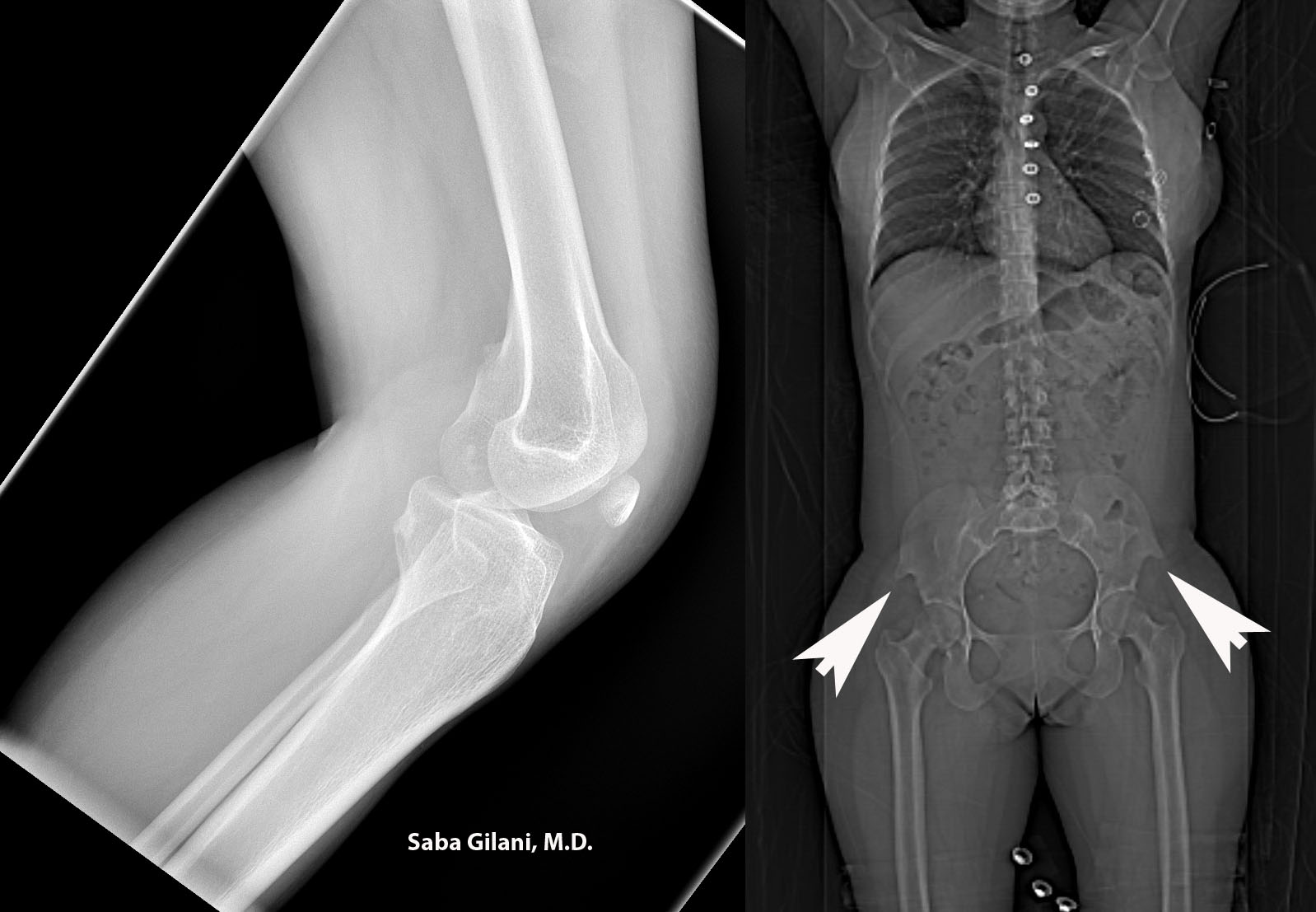
Hereditary osteo-onychodysplasia disease (HOOD), also known as Nail Patella Disease or Iliac Horn Syndrome is a genetic disorder due to an autosomal dominant mutation in the LMX1B gene. A pathognomonic finding is the presence of "iliac horns" projecting posterolaterally from the bilateral iliac bones. Other associated findings are absence or hypoplasia of the patella and radial head. Nail deformities are also common. The joint deformities can lead to osteoarthritis. About 40% of patients may also develop renal disease ranging from proteinuria to nephrotic syndrome.
Above, the lateral radiograph of the knee is from a patient who presented to the emergency room after a fall. A hypoplastic patella was incidentally noted. A review of prior imaging demonstrated bilateral iliac horns best seen on the scout image.
REFERENCES
Scott JE and Taor WS. The small patella syndrome. J Bone Joint Surg [Br] 1979;61-B:172-5.
Thompson EA, Walker T, Weens HS. Iliac horns. An osseous manifestation of hereditary arthrodysplasia associated with dystrophy of the fingernails. Radiology 1949;53:88-92.
Tuncbilek N, Karakas HM, Okten OO. Imaging of nail-patella syndrome. Hong Kong Med J 2005;11(2):116-8.
Tuesday, August 21, 2012
Lymph Node Stations for Pelvic Tumors
Most pelvic visceral tumors metastasize via lymphatics. Metastases to regional lymph nodes is consider N stage in the TNM classification system while spread to nonregional lymph nodes is considered M stage disease (distant metastases). Since upstaging influences prognosis and clinical management, knowledge of the pelvic nodal anatomy is important in evaluation of cross sectional imaging.
The following groups of pelvic lymph nodes should be considered:
1. Common iliac lymph nodes: between the aortic bifurcation and the common iliac vessel bifurcation.
REFERENCES
McMahon CJ, Rofsky NM, Pedrosa I. Lymphatic metastases from pelvic tumors: anatomic classification, characterization, and staging. Radiology 2010;254:31-46.
The following groups of pelvic lymph nodes should be considered:
1. Common iliac lymph nodes: between the aortic bifurcation and the common iliac vessel bifurcation.
- Medial, lateral, and lumbosacral subdivisions
- lumbosacral subdivision refers to lymph nodes in the lumbosacral fossa (triangular region bounded by common iliac vessels medially, psoas muscle laterally, and lower lumbar/upper sacral vertebral bodies posteriorly)
- Medial and lateral subdivisions
- obturator nodes are considered part of the medial subdivision and gather their name from proximity to the obturator internus muscle
- Anterior, lateral sacral, and presacral subdivisions
- hypogastric is a term used by some to describe the most cephalic of the internal iliac lymph nodes while others use the term to describe all internal iliac lymph nodes as a group
- Superficial subdivision nodes lie anterior to the inguinal ligament and femoral vessels
- Deep subdivision lymph nodes are enclosed within the femoral sheath
REFERENCES
McMahon CJ, Rofsky NM, Pedrosa I. Lymphatic metastases from pelvic tumors: anatomic classification, characterization, and staging. Radiology 2010;254:31-46.
Subscribe to:
Posts (Atom)