skip to main |
skip to sidebar
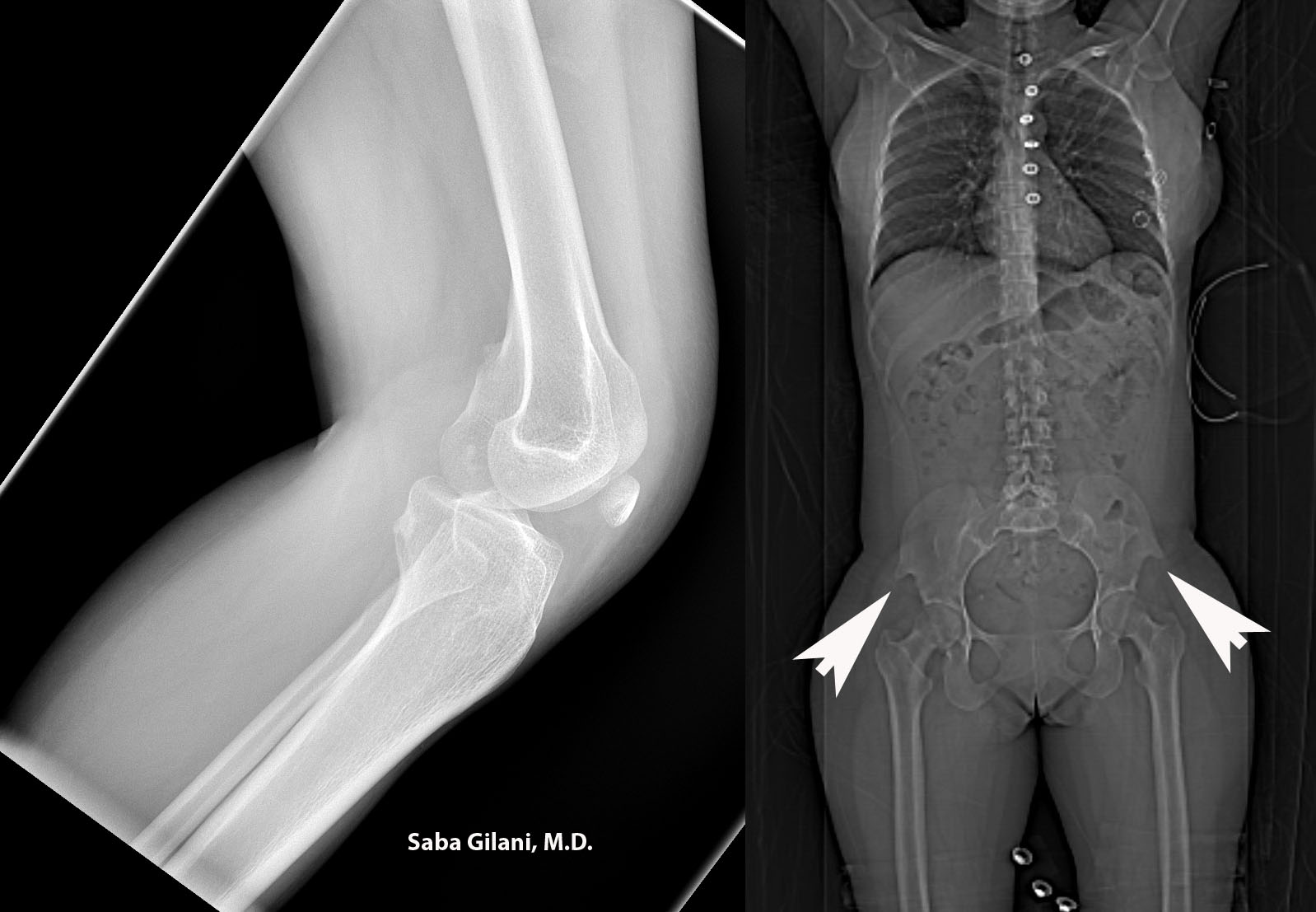
Hereditary osteo-onychodysplasia disease (HOOD), also known as Nail Patella Disease or Iliac Horn Syndrome is a genetic disorder due to an autosomal dominant mutation in the LMX1B gene. A pathognomonic finding is the presence of "iliac horns" projecting posterolaterally from the bilateral iliac bones. Other associated findings are absence or hypoplasia of the patella and radial head. Nail deformities are also common. The joint deformities can lead to osteoarthritis. About 40% of patients may also develop renal disease ranging from proteinuria to nephrotic syndrome.
Above, the lateral radiograph of the knee is from a patient who presented to the emergency room after a fall. A hypoplastic patella was incidentally noted. A review of prior imaging demonstrated bilateral iliac horns best seen on the scout image.
REFERENCES
Scott JE and Taor WS. The small patella syndrome. J Bone Joint Surg [Br] 1979;61-B:172-5.
Thompson EA, Walker T, Weens HS. Iliac horns. An osseous manifestation of hereditary arthrodysplasia associated with dystrophy of the fingernails. Radiology 1949;53:88-92.
Tuncbilek N, Karakas HM, Okten OO. Imaging of nail-patella syndrome. Hong Kong Med J 2005;11(2):116-8.
Most pelvic visceral tumors metastasize via lymphatics. Metastases to regional lymph nodes is consider N stage in the TNM classification system while spread to nonregional lymph nodes is considered M stage disease (distant metastases). Since upstaging influences prognosis and clinical management, knowledge of the pelvic nodal anatomy is important in evaluation of cross sectional imaging.
The following groups of pelvic lymph nodes should be considered:
1. Common iliac lymph nodes: between the aortic bifurcation and the common iliac vessel bifurcation.
- Medial, lateral, and lumbosacral subdivisions
- lumbosacral subdivision refers to lymph nodes in the lumbosacral fossa (triangular region bounded by common iliac vessels medially, psoas muscle laterally, and lower lumbar/upper sacral vertebral bodies posteriorly)
2. External iliac lymph nodes: between the common iliac vessel bifurcation and the inguinal ligament
- Medial and lateral subdivisions
- obturator nodes are considered part of the medial subdivision and gather their name from proximity to the obturator internus muscle
3. Internal iliac lymph nodes: surround the branches of the internal iliac vessels
- Anterior, lateral sacral, and presacral subdivisions
- hypogastric is a term used by some to describe the most cephalic of the internal iliac lymph nodes while others use the term to describe all internal iliac lymph nodes as a group
4. Inguinal lymph nodes: inferior to the inguinal ligament
- Superficial subdivision nodes lie anterior to the inguinal ligament and femoral vessels
- Deep subdivision lymph nodes are enclosed within the femoral sheath
5. Perivisceral nodes: include perirectal, periprostatic, perivesicular nodes
REFERENCES
McMahon CJ, Rofsky NM, Pedrosa I. Lymphatic metastases from pelvic tumors: anatomic classification, characterization, and staging. Radiology 2010;254:31-46.
Dolichocephaly is a craniosynostosis involving premature closure of the sagittal suture resulting in an elongated head shape as shown above. The craniosynostoses were previously discussed here: craniosynostoses.
Hereditary syndromes that are associated with craniosynostoses are:
Crouzon syndrome: premature synostosis, maxillary hypoplasia, shallow orbits
Apert's syndrome: Coronal synostosis, midfacial hypoplasia, bilateral syndactyly, symphalangism (ankylosis of interphalangeal joints)
Pfeiffer syndrome: premature synostosis, broad thumbs and great toes, mild syndactyly
Carpenter syndrome: premature synostosis, severe developmental delay, brachydactyly, syndactyly, thumb duplication.
Acquired conditions that can result in premature synostosis include Rickets, hypophosphatasia, and mucopolysaccharidoses.
REFERENCES
Glass RBJ, Fernbach SK, Norton KI, et al. Radiographics 2004;24:507-22.
The Siltzbach classification system defines five stages of sarcoidosis based on chest radiograph findings:
Stage 0 - No radiographic abnormality
Stage 1 - Lymphadenopathy
Stage 2 - Lymphadenopathy + pulmonary infiltrate
Stage 3 - Pulmonary infiltrate
Stage 4 - Pulmonary fibrosis
About 20% of patients with sarcoidosis will develop pulmonary fibrosis which indicates irreversible disease. Three main HRCT patterns of pulmonary fibrosis have been identified and linked to performance on pulmonary function tests:
1. Bronchial distortion in a primarily central distribution, associated with lower expiratory flow rates
2. Honeycombing in a peripheral and upper lobe distribution, associated with restriction and decreased diffusion capacity of carbon monoxide
3. Coarse, irregular linear opacities that radiate from the hila, associated with the least functional impairment.
Distinguishing between these patterns is important as it may predict treatment response to corticosteroids.
REFERENCES
Abhesera M, Valeyre D, Grenier P, et al. Sarcoidosis with pulmonary fibrosis: CT pattern and correlation with pulmonary function. AJR Am J Roentgenol 2000;174(6):1751-1757.
Criado E, Sanchez M, Ramirez J, et al. Pulmonary sarcoidosis: typical and atypical manifestations at high resolution CT with pathologic correlation. Radiographics 2010;30:1567-1586.
As intrauterine devices have become a common form of contraception worldwide, their normal and abnormal imaging appearance should be recognized. The stem of a properly positioned intrauterine device (IUD) should be entirely within the endometrial cavity with the arms extending laterally at the uterine fundus. The device should never be seen in the endocervical canal.
An IUD may be expulsed from the uterine cavity or displaced within it. Perforation through the uterus and migration into the peritoneal cavity is a rare complication. An intraperitoneal IUD may cause bowel/omental adhesions, abscess formation, or perforation of intraabdominal structures. Timely surgical intervention can prevent these complications.
REFERENCES
Boortz HE, Margolis DJA, Ragavendra N, et al. Migration of intrauterine devices: radiologic findings and implications for patient care. Radiographics. 2012 March;32:335-52.
Peri N, Graham D, Levine D. Imaging of intrauterine contraceptive devices. J Ultrasound Med 2007;26(10):1389-1401.
Images courtesy of Anna Nazarenko, M.D.
1. Left IVC: joins the left renal vein and crosses anterior to the aorta to join the right renal vein
- can be mistaken for paraaortic adenopathy
- report of AAA rupture into the IVC
2. Double IVC: the left IVC ends at the level of the left renal vein which crosses anterior to the aorta to join the confluence of the right IVC/renal vein.
- suspect if patient has recurrent pulmonary embolism after placement of an IVC filter
3. Azygous continuation of the IVC: above the renal veins the IVC passes posterior to the diaphragmatic crus and continues as the azygous vein which joins the superior vena cava in the right paratracheal region. The hepatic segment of the IVC empties directly into the right atrium.
- important to consider in cases of a right paratracheal mass
- can be mistaken for retrocrural adenopathy
4. Circumaortic left renal vein: two left renal veins are present; the superior renal vein receives the left adrenal vein while the inferior renal vein receives the left gonadal vein.
- significant during planning of nephrectomy
5. Retroaortic left renal vein: a single left renal vein passes posterior to the aorta
- recognition during preoperative planning is important
6. Double IVC with retroaaortic right renal vein and hemiazygous continuation of the IVC: confluence of the right renal vein and right IVC crosses posterior to the aorta to join the left IVC and continue cephlad as the hemiazygous vein. Several collateral pathways for the hemiazygous vein exist in the thorax - hemiazygous crossing posterior to the aorta to join the azygous vein, hemiazygous joining the cardinal vein of the heart via a persistent left SVC, and accessory hemiazygous continuation to the brachocephalic vein
- hemiazygous collateral pathway may be mistaken for a left mediastinal mass
- accessory hemiazygous has been reported to be mistaken for an aortic dissection
7. Circumcaval ureter: always occurring on the right, the proximal ureter courses posterior to the IVC and emerges to the right of the aorta and continues anterior to the right iliac vessels.
- patients may develop partial ureteral obstruction or recurrent urinary tract infections
- treatment is surgical relocation of the ureter anterior to the IVC
8. Absent infrarenal IVC with preservation of the suprarenal segment: external and internal iliac veins converge as lumbar veins which continue cephlad as the paravertebral collateral veins to join the azygous and hemiazygous. The confluence of the renal veins forms a normal suprarenal IVC.
- patients may present with symptoms of lower extremity venous insufficiency or idiopathic deep venous thrombosis
- collateral circulation may mimic a paraspinal mass
REFERENCES
Bass JE, Redwine MD, Kramer LA, et al. Spectrum of congenital anomalies of the inferior vena cava: cross-sectional imaging findings. Radiographics. 2000 May;20:639-52.
Esophageal atresia is a congenital anomaly related to incomplete formation of the esophagus with or without the presence of a tracheoesophageal (TE) fistula. While the exact cause is unknown, it is felt to be related to incomplete separation of the primitive foregut into the trachea and the esophagus. One accepted classification of esophageal atresia and TE fistulas is as follows:
A. Atresia without TE fistula
B. Atresia with proximal TE fistula
C. Atresia with distal TE fistula (most common)
D. Atresia with proximal and distal TE fistula
E. TE fistula without atresia
Esophageal atresia is usually suspected in the setting of polyhydramnios, excessive salivation, choking/cyanosis during feeding, and inability to pass a nasogastric/feeding tube to the stomach. Anteroposterior and lateral radiographs will reveal a blind-ending, air-filled proximal esophagus. Radiographs of the abdomen should also be performed to evaluate for air in the GI tract, the presence of which raises suspicion for a distal TE fistula. Fluoroscopy may be used to confirm findings in which case water soluble contrast is preferred.
The above images are from a newborn with a history of difficulty feeding. The initial radiograph reveals a dilated upper esophagus without air seen in the GI tract. A nasogastric tube was placed which coiled in the proximal esophageal stump. Via a percutaneous gastrostomy tube, contrast was introduced into the stomach which refluxed into a distal esophageal remnant without evidence for fistulous connection. Type A esophageal atresia was diagnosed.
REFERENCES
Berrocal T, Torres I, Gutierrez J, et al. Congenital anomalies of the upper gastrointestinal tract. Radiographics. 1999;19:855-72.