Monday, June 26, 2017

Radiation-Associated Sarcomas

CT, bone scan (MDP), FDG PET/CT and MRI in a patient with radiation-associated sarcoma (osteosarcoma)
Patient with history of radiation therapy for head and neck cancer. Radiation field extended into the supraclavicular nodal stations. CT shows an osteoid producing soft tissue mass to the right of midline. Bone scan and PET show uptake in the lesion, as well as contralateral lymph nodes. PET shows an FDG-avid lung nodule. T1-WI post-contrast MRI with FS shows a peripherally enhancing soft tissue mass.

The incidence of radiation-associated sarcomas of bone and soft tissue is about 0.1%. They are more commonly seen in patients with breast cancer, lymphoma, head and neck malignancies, and gynecologic cancers. The distribution of primary cancers is likely related to the larger numbers of patients with these cancers and the high survival rates for these tumors.

The majority of radiation-associated tumors are soft tissue sarcomas, with bone sarcomas making up about 20-30% of cases. The majority are high grade and aggressive. The most common soft tissue sarcomas are unclassified pleomorphic sarcoma (UPS, formerly MFH), followed by angiosarcoma (particularly in breast cancer), fibrosarcoma and leiomyosarcoma (particularly in retinoblastoma). The most common bone sarcomas are osteosarcomas.

The latency between radiation and sarcoma ranges from as little as a few months to 54 years. The average is 7 to 16 years. In breast cancer, the average is 10 to 11 years (4-8 years for angiosarcomas). In childhood cancers, the average latency is between 12 to 13 years.

Risk factors include, dose (Rarely seen with low doses: <40 Gy), age at exposure, concomittant chemo exposure (particularly alkylating agents) and genetic tendency (e.g., Li-Fraumeni syndrome).
References

Maki, R. et al. Radiation-Associated Sarcomas. UpToDate

Sunday, June 18, 2017

Keloids and Hypertrophic Scars

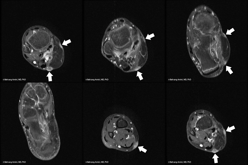
MRI in a patient with hypertrophic scar/keloid
T1 axial post-contrast MRI of a keloid or hypertrophic scar formation along the margins of a myocutaneous flap. The enhancement can fool you into thinking that there is recurrent tumor, but the linear pattern along the flap margin is the clue that this is related to the scar.

Keloids and hypertrophic scars are fibroblastic proliferations of the dermis. Their morphologic and pathologic features overlap. They are associated with trauma, infection, and connective tissue diseases. They do not spontaneously regress and tend to recur after surgical excision, so surgery is often combined with topical corticosteroid injection or, less commonly, radiation therapy.

In contrast to hypertrophic scars, keloids tend to grow beyond the margins of the injury site, have keloid collagen, and are less likely to stain for smooth muscle actin. Keloids have a higher recurrence rate than hypertrophic scars.

Keloids are hypocellular and are composed of dense collagen. The abundance of type I collagen results in low T2 signal. Keloids occur most frequently in patients aged 15–45 years. People of African and Chinese origin have a higher predilection for keloids. The face, shoulders, forearms, and hands are most commonly affected. They tend to occur where there is increased skin tension

References

Dinauer PA, Brixey CJ, Moncur JT, Fanburg-Smith JC, Murphey MD. Pathologic and MR imaging features of benign fibrous soft-tissue tumors in adults. Radiographics. 2007 Jan-Feb;27(1):173-87.

Monday, June 12, 2017

The axillary nerve and adhesive capsulitis

MRI of anatomy of the axillary nerve and its relationship to the joint capsule.
The axillary nerve (yellow arrow) and the posterior humeral circumflex artery (red arrow) in the region of the quadrilateral space. Note proximity to the inferior capsule.

The axillary nerve is closely related to the inferior capsule of the shoulder. It passes inferior to the subscapularis muscle and travels adjacent to the capsule before entering the quadrilateral space.

The axillary nerve is associated with adhesive capsulitis in at least 2 ways.

First, the axillary nerve can be irritated in the setting of inflammation and thickening of the inferior capsule. The evidence for this is somewhat anecdotal, but makes anatomic sense. The image below is from a patient with adhesive capsulitis. Note the teres minor atrophy (green arrow) in the setting of thickening of the inferior capsule (blue arrow), and constrained fluid in the joint (orange arrow) being forced into the superior subscapularis recess (orange*). The bone lesions are from myeloma, in case you were wondering.

MRI of anatomy of the axillary nerve and its relationship to the joint capsule in adhesive capsulitis.

Second, the close proximity of the nerve to the joint capsule predisposes it to injury during arthroscopic capsule release for treatment of adhesive capsulitis. Risk of injury is decreased by placing the incision of the glenohumeral joint capsule at the glenoid insertion with the arm in the abducted and externally rotated position.

We can appreciate the extent of inflammation on other imaging modalities too. On FDG PET/CT, for example, patients with adhesive capsulitis tend to have uptake at the inferior capsule that extends into the adjacent tissues.

References

  • Sridharan R, Engle MP, Garg N, Wei W, Amini B. Focal uptake at the rotator interval or inferior capsule of shoulder on 18F-FDG PET/CT is associated with adhesive capsulitis. Skeletal Radiol. 2017 Apr;46(4):533-538.
  • Salem U, Zhang L, Jorgensen JL, Kumar R, Amini B. Adhesive capsulitis mimicking metastasis on 18F-FDG-PET/CT.Clin Nucl Med. 2015 Feb;40(2):e145-7.
  • Jerosch J, Filler TJ, Peuker ET. Which joint position puts the axillary nerve at lowest risk when performing arthroscopic capsular release in patients with adhesive capsulitis of the shoulder? Knee Surg Sports Traumatol Arthrosc. 2002 Mar;10(2):126-9.
  • E. B. G. D. Santos, P. M. E. Souza (pdf). Teres minor beyond quadrilateral space syndrome: a pictorial review. ECR 2014 conference.

Tuesday, June 6, 2017

Sacrum

I was reading an article on the role of religion in the secular Turkish state and came across this statement:
It is possible -- such is the argument of Carter Findley in his Turks in World History -- that in doing so it drew on a long Turkish cultural tradition, born in Central Asia and predating conversion to Islam, that figured a sacralisation of the state, which has vested its modern signifier, devlet, with an aura of unusual potency.

You may be wondering what the heck a congenital variant of spinal segmentation has to do with religion. From the always-excellent Online Etymology Dictionary:

Bone at the base of the spine, 1753, from Late Latin os sacrum "sacred bone," from Latin os "bone" + sacrum, neuter of sacer "sacred" (see sacred). Said to be so called because the bone was the part of animals that was offered in sacrifices. Translation of Greek hieron osteon. Greek hieros also can mean "strong," and some sources suggest the Latin is a mistranslation of Galen, who was calling it "the strong bone."