 |
| CT shows a predominantly fat
attenuation, intramural uterine mass. There is rim of surrounding
calcification as well as a few foci of internal soft tissue density at the
superior aspect of the mass |
 |
| MR imaging showed the majority of the uterine mass as hyperintense on T1 weighted imaging (isointense to subcutaneous
fat) |
 |
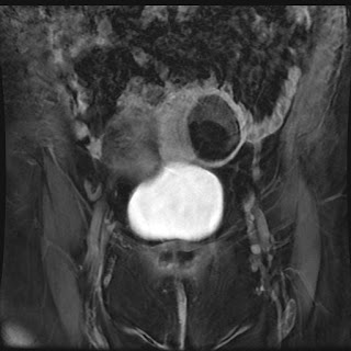
| T2 weighted image with fat saturation MRI shows the uterine mass markedly hypointense (isointense to subcutaneous fat) |
 |
| Post contrast T1 weighted MRI image with fat saturation show mld enhancement of the soft tissue component along the superior margin of the mass; the majority of the mass is markedly hypointense (isointense to subcutaneous fat) |
Uterine
lipoleiomyomas are rare, benign tumors with a variable reported incidence
ranging from 0.03% to 0.2%. The exact etiology of these lesions is
unclear. It is postulated lipoleiomyomas
either arise from fatty metaplasia of the smooth muscle cells of leiomyomas, or
from misplaced embryonic fat cells in the uterus. CT is highly specific for
the diagnosis when an intrauterine mass is seen containing both macroscopic fat
and soft tissue density. MR can also be confirmatory as the mass will have high
T1 weighted signal which can be confirmed as fat by using a fat suppression. The role of imaging is also to differentiate lipoleiomyoma from an ovarian
teratoma, a much more common entity presenting as a fat-containing pelvic mass.
Lipoleiomyomas require no treatment or follow-up whereas teratomas are
frequently resected.
References
- Preito A,Crespo C, Pardo A, Docal I, Calzada J, Alonso P. Uterine lipoleiomyomas: US andCT findings. Abdom Imaging 2000;25:655-7
- MaebayashiT, Imai K, Takekawa Y, et al. Radiologic features of uterine lipoleiomyoma. JComput Assist Tomogr 2003;27:162-5
- AvritscherR, Iyer RB, Ro J, Whitman G. Lipoleiomyoma of the uterus. AJR Am J Roentgenol2001;177:856
- Dodd GD III,Budzik RF. Lipomatous uterine tumors: diagnosis by ultrasound, CT, and MR. JComput Assist Tomogr 1990;14:629-32


