- Grade I (Circumscribed astrocytomas): Relatively benign biology and indolent clinical course. Include pilocytic astrocytoma, pleomorphic xanthoastrocytoma, subependymal giant cell astrocytoma.
- Grade II (Diffuse astrocytomas): Shown above. Cell types include fibrillary, gemistocytic, and protoplasmic astrocytomas. 1/3 occur in the frontal lobe and 1.3 occur in the temporal lobe. Cystic change and calcification may occur. Less likely to show enhancement.
- Grade III (Anaplastic astrocytomas): Usually evolve from lower grade astrocytomas. Ill-defined borders and extensive surrounding edema. More likely to show enhancement. Should not have cystic change.
- Grade IV (Glioblastoma multiforme): Most common variety of astrocytoma. Occur de novo in the elderly and are thought to arise from lower grades in younger patients. Characterized by necrosis.
Thursday, March 31, 2011
Low-Grade Astrocytoma
Astrocytomas are the most common primary, solitary, supratentorial brain lesion in adults (all these qualifiers are meant to hide the fact that brain lesions are most commonly metastases). The World Health Organization classifies them into 4 grades.
Wednesday, March 30, 2011
Pituitary Stalk Lesions
- Metastases:
- Sarcoid/Tuberculosis/Lymphoma:
- Langerhans cell histiocytosis: May have absence of the posterior pituitary bright spot.
- Pituicytoma: Also known as choristoma.
- Germinoma:
Tuesday, March 29, 2011
Orbital Cavernous Hemangioma
Orbital cavernous hemangiomas are type 3 low-flow arteriovenous malformations. They typically affect women in their 40s, who most commonly present with painless proptosis. Orbital cavernous hemangiomas may be intra- or extra-conal. They should be differentiated from orbital capillary hemangiomas, which are hamartomatous proliferations of vascular endothelial cells and occur in infancy.
On CT, orbital cavernous hemangiomas are homogeneous lesions with variable attenuation depending on the presence of microcalcifications (coarse macrocalcifications are not typical). Larger lesions can cause remodeling of adjacent bone. Avid enhancement is typical. On MRI, the lesions are homogeneous and isointense to muscle. A hypointense pseudocapsule may also be seen. T2-weighted images reveal a hyperintense lesions with or without internal septations. On dynamic MRI, the lesions demonstrate small areas of enhancement initially with progressive enhancement on later phases.
Conventional angiography reveals contrast puddles extending into the late venous phase. Due to their low-flow characteristics, the lesions are not visible on routine MRA sequences.
References
Xian J, Zhang Z, Wang Z, Li J, Yang B, Chen Q, Chang Q, He L. Evaluation of MR imaging findings differentiating cavernous haemangiomas from schwannomas in the orbit. Eur Radiol. 2010 Sep;20(9):2221-8.Monday, March 28, 2011
Superficial Gastric Erosions
Superficial gastric erosions affect only the epithelial lining of the stomach. In contrast to ulcers, they do not penetrate the muscularis mucosa.
They can be demonstrated on air-contrast upper gastrointestinal studies as tiny flecks of barium (erosion) surrounded by a radiolucent halo (edematous mucosa).
They can be idiopathic or seen in association with:
They can be demonstrated on air-contrast upper gastrointestinal studies as tiny flecks of barium (erosion) surrounded by a radiolucent halo (edematous mucosa).
They can be idiopathic or seen in association with:
- Alcohol abuse:
- Medications: NSAIDs
- Crohn disease: Aphthoid ulcers
- Viral gastritis: Herpes, cytomegalovirus
- Candidiasis:
References
Eisenberg RL. Chapter 15: Superficial gastric erosions. in Gastrointestinal Radiology: A Pattern Approach (4th ed). Lippincott Williams & Wilkins (2003). Pages 202 - 205.Sunday, March 27, 2011
Orbital Dermoid/Epidermoid
Dermoids of the orbital region can be classified as superficial subcutaneous (also known as simple or exophytic) or deep (also known as complicated or endophytic). Superficial dermoids present in infancy and are not associated with proptosis. Deep dermoids are usually seen in adults and are associated with proptosis. They can cause scalloping of adjacent bone and even bone defects, allowing them to extend into adjacent paranasal sinuses, into the temporal fossa, intracnanially, or through
the orbital fissures.
Radiographs and CT may show a soft tissue mass causing scalloping of bone. CT may also show the fat content, although the attenuation may be higher in mixed lesions. MRI is the best modality for demonstrating intracranial extension. MRI findings are variable. T1-weighted images will typically show a strongly hyperintense lesion if fat predominates, but will be intermediate intensity otherwise. On T2-weighted images, the lesions are typically isointense or mildly hypointense. Post-contrast images reveal thin rim-enhancement.
The image above shows a case of pathologically proven periorbital dermoid. The MRI characteristics aren't those typically described for these lesions. There is a well-defined cystic lesion within the subcutaneous tissue of the left lateral orbital wall adjacent to the left frontozygomatic suture. No intracranial or intraorbital extension was seen, but there was bony remodeling. The lesion has a thin rim that demonstrates minimal enhancement. DWI and ADC images didn't reveal restricted diffusion.
References
Nugent RA, Lapointe JS, Rootman J, Robertson WD, Graeb DA. Orbital dermoids: features on CT. Radiology. 1987 Nov;165(2):475-8.Saturday, March 26, 2011
Cupola Effect
The cupola effect refers to oral barium forming a dome over a pedunculated esophageal tumor and flowing into the space between the tumor and the esophageal wall.
The cupola effect is one of the features of spindle cell carcinoma (carcinosarcoma) of the esophagus. Indeed, when a polypoid, pedunculated and bulky esophageal mass with a cupola effect has scalloped edges and expands the lumen of the esophagus, spindle cell carcinoma should be the primary diagnosis.
References
- McCort JJ. Esophageal carcinosarcoma and pesudosarcoma. Radiology. 1972 Mar;102(3):519-24.
- Olmsted WW, Lichtenstein JE, Hyams VJ. Polypoid epithelial malignancies of the esophagus. AJR Am J Roentgenol. 1983 May;140(5):921-5.
Friday, March 25, 2011
Malgaigne Fracture
A Malgaigne fracture is a vertical shear injury on one side of the pelvis with disruption of both the anterior and posterior pelvic rings. It consists of a fracture through the ilium and ipsilateral pubic rami, or a dislocation of the sacroiliac joint and the pubic symphysis, or any combination of these. The acetabulum is the lateral fragment and is unstable.
The case above shows fractures through the left inferior and superior pubic rami and widening of the ipsilateral sacroiliac joint. The widening of the sacroiliac joint is most likely positional, but illustrates the point.
References
Hunter TB, Peltier LF, Lund PJ. Radiologic history exhibit. Musculoskeletal eponyms: who are those guys? Radiographics. 2000 May-Jun;20(3):819-36.Thursday, March 24, 2011
Acute Transverse Myelopathy
Acute transverse myelopathy (myelitis) is a monophasic, acute inflammatory process involving both the dorsal and ventral halves of the spinal cord. Patients present with paraplegia, sensory impairment at a specific spinal level, and sphincter dysfunction.
Acute transverse myelopathy can be idiopathic or be caused by:
- Multiple scleorosis:
- Viral infection:
- Acute disseminated encephalomyelitis:
- Collagen vascular disorders: Systemic lupus erythematosis
- Vascular disorders:
- Paraneoplastic syndromes:
Radiology
- Choi KH, Lee KS, Chung SO, Park JM, Kim YJ, Kim HS, Shinn KS. Idiopathic transverse myelitis: MR characteristics. AJNR Am J Neuroradiol. 1996 Jun-Jul;17(6):1151-60.
- DeSanto J, Ross JS. Spine infection/inflammation. Radiol Clin North Am. 2011 Jan;49(1):105-27.
Wednesday, March 23, 2011
Nasolacrimal Duct Dacrocystocele
A nasolacrimal duct dacrocystocele (also known as a dacryocele, amniotocele, and nasolacrimal mucocele) is an uncommon medial canthal mass in children. There may rarely be a connection to a cyst in the nasal cavity. It is called dacryocystitis when infected.
CT shows a low-attenuation thin-walled cyst along the medial canthus associated with smooth enlargement of the nasolacrimal duct. The cyst is so large that it superiorly displaces inferior turbinate (arrow). In some cases, it can extend further and deviate the nasal septum. Contrast enhanced images (not shown) will demonstrate minimal if any thin peripheral enhancement. It is of variable signal intensity on T1-weighted images depending on protein content and is usually T2-hyperintense.
Fat stranding and other usual inflammatory changes can be seen with infection.
Differential considerations include:
- Dermoid/Epidermoid: Usually affects the lateral canthus. Will have fat content with thin rim-enhancement, with or without calcification.
- Acquired dacrocystocele: Usually in adults. This is caused by a stenosis of the nasolacrimal duct from inflammation, cancer, trauma (e.g., LeFort 2 fracture, sinus surgery).
- Nasoorbital cephalocele: Only a consideration if you have a single slice. Scrolling up will reveal that the cyst is contiguous with intracranial contents through a bony defect.
References
- Castillo M, Merten DF, Weissler MC. Bilateral nasolacrimal duct mucocele, a rare cause of respiratory distress: CT findings in two newborns. AJNR Am J Neuroradiol. 1993 Jul-Aug;14(4):1011-3.
- Meyer JR, Quint DJ, Holmes JM, Wiatrak BJ. Infected congenital mucocele of the nasolacrimal duct. AJNR Am J Neuroradiol. 1993 Jul-Aug;14(4):1008-10.
- Rand PK, Ball WS Jr, Kulwin DR. Congenital nasolacrimal mucoceles: CT evaluation. Radiology. 1989 Dec;173(3):691-4.
Tuesday, March 22, 2011
Multiple-System Atrophy
Multiple-system atrophy is a neurodegenerative disorder that involves the basal ganglia, olivopontocerebellar system, and the autonomic system to varying degrees. Differentiation from idiopathic Parkinson disease is often difficult, especially early in the disease. Different subtypes have been defined based on the dominant symptoms:
- MSA-p (striatonigral degeneration): Parkinsonism predominates, but with less evidence of rigidity and resistance to levodopa. The basal ganglia, particularly the putamen, are affected. MRI may show putaminal atrophy and hyperintensity (T2-hyperintense rim at the putaminal edge).
- MSA-c (olivopontocerebellar atrophy): Atrophy and increased signal intensity are seen within the pons, cerebellum, and middle cerebellar peduncles. On MRI, the olivary nuclei and medulla are small. There is atrophy of the pons and the cerebellar hemispheres with relative sparing of the vermis. The transverse pontine fibers, brachium pontis, middle cerebellar peduncles, and bulbar olives may be T2-hyperintense. The putamen, globus pallidus, and substantia nigra may be T2-hypointense. The hot cross bun sign may be seen on T2-weighted images as a T2-hyperintense cross in the pons due to loss of myelinated transverse pontocerebellar fibers and neurons in the pontine raphe.
- MSA-a (Shy-Drager syndrome): Autonomic failure is dominant, with orthostatic hypotension, urinary incontinence, inability to sweat, and extrapyramidal and cerebellar disturbances. As with MSA-p, there is T2-hyperintensity with the putamen.
References
- Gallucci M, Limbucci N, Catalucci A, Caulo M. Neurodegenerative diseases. Radiol Clin North Am. 2008 Jul;46(4):799-817, vii.
- Schrag A, Kingsley D, Phatouros C, Mathias CJ, Lees AJ, Daniel SE, Quinn NP. Clinical usefulness of magnetic resonance imaging in multiple system atrophy. J Neurol Neurosurg Psychiatry. 1998 Jul;65(1):65-71.
- Shrivastava A. The hot cross bun sign. Radiology. 2007 Nov;245(2):606-7
Monday, March 21, 2011
Globe Lesions in Children: Quick and Dirty
- Retinopathy of prematurity (ROP): Abnormal vascular development due to prolonged oxygen therapy leads to hemorrhage. Usually bilateral. Increased attenuation in a small globe associated with retinal detachment.
- Persistent hyperplastic primary vitreous (PHPV): Persistence of embryonic vitreous vascualrity leads to hemorrhage. Usually unilateral. Increased attenuation in a small globe associated with cataracts and retinal detachment. Hyperintense on T1 and T2.
- Retinoblastoma (Rb): Most common globe tumor in children, with almost all cases presenting before age 5. Majority unilateral. Most often has calcium. Hyperintense on T1 and hypointense on T2. Avid enhancement.
- Retinal astrocytoma: Look for signs of tuberous sclerosis or neurofibromatosis type 1. Most often has calcium.
- Coats disease: Subretinal exudates with V-shaped retinal detachment in boys. Contrary to the oversimplification of the chart, the globe is actually smaller than in patients with retinoblastoma. No tumor mass. Enhancement of the detached leaves of the retina. Hyperintense on T1 and T2.
- Toxocariasis: Due to hypersensitivity to the larva. Patients have a history of contact with dogs or cats. Can calcify if chronic. Traction detachment of the retina. Isointense on T1 and hyperintense on T2.
References
- Smirniotopoulos JG, Bargallo N, Mafee MF. Differential diagnosis of leukokoria: radiologic-pathologic correlation. Radiographics. 1994 Sep;14(5):1059-79.
- Galluzzi P, Venturi C, Cerase A, Vallone IM, Bracco S, Bardelli AM, Hadjistilianou T, Gennari P, Monti L, Filosomi G. Coats disease: smaller volume of the affected globe. Radiology. 2001 Oct;221(1):64-9.
Sunday, March 20, 2011
Cortical Nephrocalcinosis
- Acute cortical necrosis: Most common cause. Can be due to severe acute hypotension, drugs (amphoteracin), or toxins (antifreeze).
- Chronic glomerulonephritis: Second most common cause. Can be due to diabetes, lupus, or membranous glomerulonephritis.
- Chronic transplant rejection:
- Oxalosis: Due to altered bile metabolism from small bowel pathology (e.g., Crohn disease) or small bowel resection.
- Alport syndrome: Associated with hereditary nerve deafness. Rare cause of cortical nephrocalcinosis.
- Sickle cell anemia: Rare cause of cortical nephrocalcinosis.
References
Dyer RB, Chen MY, Zagoria RJ. Abnormal calcifications in the urinary tract. Radiographics. 1998 Nov-Dec;18(6):1405-24.Saturday, March 19, 2011
Undifferentiated (Embryonal) Sarcoma of the Liver
Undifferentiated (embryonal) sarcoma of the liver is the fourth or fifth most common liver tumor in children. It may or may not share the same histiogenesis as mesenchymal hamartoma, with the two representing the ends of a spectrum.
CT and MR findings can resemble those of a cystic lesion, possibly related to the abundant myxoid stroma, leading to misdiagnosis as a hydatid cyst or amebic abscess. US is useful in demonstrating the solid nature of the tumor.
Suggestive findings include a large hepatic lesion with a seemingly cystic appearance on CT and MR images and a largely solid appearance on US images. The tumors demonstrate predominantly low T1 and high T2 signal. Areas of hemorrhage appear as T1 hyperintensity and increased attenuation on CT. Post-contrast images reveal enhancing internal nodules.
On ultrasound, our case demonstrates a large, predominantly solid lesion within the liver with small cystic spaces and no significant vascularity. CT shows a large, predominantly low-attenuation lesion within the right lobe of the liver, with several internal septa and areas of increased. No definite enhancement was seen. On MRI, the lesion is predominantly T1-hypointense and T2-hyperintense with nodular enhancement superiorly.
The differential diagnosis for a cystic lesion in the liver (based on its appearance on CT and MR) includes biliary cystadenoma and cystadenocarcinoma, cystic degeneration of hepatocellular carcinoma, infection (abscess, bacterial and parasitic), metastatic disease, and resolving hematoma.
References
- Buetow PC, Buck JL, Pantongrag-Brown L, Marshall WH, Ros PR, Levine MS, Goodman ZD. Undifferentiated (embryonal) sarcoma of the liver: pathologic basis of imaging findings in 28 cases. Radiology. 1997 Jun;203(3):779-83.
- Crider MH, Hoggard E, Manivel JC. Undifferentiated (embryonal) sarcoma of the liver. Radiographics. 2009 Oct;29(6):1665-8.
- Shehata B, Gupta NA, Katzenstein H, Steelman CK, Wulkan ML, Gow KW, Bridge JA, Kenney BD, Thompson KS, de Chadarevian JP, Abramowsky CR. Undifferentiated embryonal sarcoma of the liver is associated with mesenchymal hamartoma and multiple chromosomal abnormalities: a review of eleven cases. Pediatr Dev Pathol. Epub 2010 Oct 6.
Labels:
Gastrointestinal,
Pediatric radiology
Friday, March 18, 2011
Centrilobular Ground-Glass Nodules
- Acute hypersensitivity pneumonitis: Ground-glass centrilobular nodules and mosaic attenuation. Lower lobes. Pleural surfaces commonly spared or less involved.
- Respiratory bronchiolitis-interstitial lung disease: Smokers. More commonly in the upper lobes.
- Desquamative interstitial pneumonia: Peripheral predominance
- Atypical infection: Pneumocystis jiroveci pneumonia, Mycoplasma, viral (e.g., cytomegalovirus)
- Collagen vascular disease: For example, Sjögren syndrome.
References
- Matsuyama N, Ashizawa K, Okimoto T, Kadota J, Amano H, Hayashi K. Pulmonary lesions associated with Sjögren's syndrome: radiographic and CT findings. Br J Radiol. 2003 Dec;76(912):880-4.
- Mueller-Mang C, Grosse C, Schmid K, Stiebellehner L, Bankier AA. What every radiologist should know about idiopathic interstitial pneumonias. Radiographics. 2007 May-Jun;27(3):595-615.
Thursday, March 17, 2011
Synovial Cell Sarcoma
Synovial cell sarcoma is the most common malignant nonrhabdomyosarcoma soft-tissue sarcoma in children. Synovial cell sarcoma does not arise from the synovial membrane, and takes its name from the histological similarity of the cells to synovioblastic cells.
The majority occur in the extremities, with the lower extremity involved more often than the upper extremity. The lesions are usually in the deep soft tissues and typically juxtaarticular.
Radiographs may show the soft-tissue mass (arrow), periosteal reaction (20% of cases), local bone invasion, and calcifications (~30% of cases, uncommon in other extremity soft-tissue sarcomas).
Ultrasound findings are nonspecific, but can help exclude cystic lesions from the differential diagnosis (e.g., popliteal and ganglion cysts). Blood flow characteristics are likewise nonspecific. Ultrasound in our patient showed a solid lesion located along the facial plane between the subcutaneous fat and adjacent musculature.
MR findings of synovial cell sarcoma are deceptively non-aggressive, with smooth contours, lack of apparent invasion, and homogeneous signal characteristics. Characteristic MR findings include a well-circumscribed lesion with diffuse heterogeneous enhancement. Most are hyperintense of T2-weighted images, although approximately 1/3 demonstrate a mixed pattern of fluid, intermediate, and low signal intensity (triple-signal pattern). Septations can also be seen. Susceptibility-weighted sequences can help exclude blood products, which are seen in venous malformations.
References
Bixby SD, Hettmer S, Taylor GA, Voss SD. Synovial sarcoma in children: imaging features and common benign mimics. AJR Am J Roentgenol. 2010 Oct;195(4):1026-32.
Labels:
Boards,
Musculoskeletal,
Pediatric radiology
Wednesday, March 16, 2011
Infrapatellar Fat Pad Mass
Differential considerations for a mass in the infrapatellar fat pad:
- Hoffa disease: Painful condition caused by acute or repetitive trauma producing hemorrhage and necrosis of the infrapatellar fat. Acute phase demonstrates edema and outward bowing of the patellar tendon. Chronic phase is characterized by fibrin and hemosiderin deposition, with ill-defined margins.
- Focal nodular synovitis: Benign proliferative disorder arising from a small area of the synovium. Most frequently in the tendon sheaths of the small joints of the fingers and toes. Well-defined ovoid or polylobulated soft-tissue mass. Iso- or hyperintense relative to skeletal muscle on T1-weighted images. Variable signal intensity on T2-weighted images. May have regions of high hemosiderin concentration.
- Chondroma: Cartilage signal pattern
- Osteochondroma: Bone marrow signal pattern.
- Gouty tophus:
References
Huang GS, Lee CH, Chan WP, Chen CY, Yu JS, Resnick D. Localized nodular synovitis of the knee: MR imaging appearance and clinical correlates in 21 patients. AJR Am J Roentgenol. 2003 Aug;181(2):539-43.Tuesday, March 15, 2011
Superior Mesenteric Artery Syndrome
Superior mesenteric artery syndrome refers to obstruction at the level of the third portion of the duodenum as it passes between the superior mesenteric artery and the aorta. The obstruction may be due to narrowing of the aorta-superior mesenteric artery distance or inadequate peristaltic pressure to pass food past this area (as could be seen with scleroderma, for example).
Superior mesenteric artery syndrome is classically described in patients with rapid weight loss, those on bed rest, or patients with prior abdominal surgery, but can be seen with scleroderma, diabetes, pancreatitis, peptic ulcers, and malignant lymph nodes or tumors in the root of the mesentery. Aortic or superior mesenteric artery aneurysms or atherosclerotic disease can also cause narrowing of the aorta-superior mesenteric artery distance.
Upper gastrointestinal series may show gastric and duodenal dilatation with retention of barium, and extrinsic pressure on the third part of the duodenum (arrow). A classically described finding is a linear cut-off of contrast at the proximal third portion of the duodenum, which is to the right of the spine. However, the superior mesenteric artery is central or slightly to the left of midline (as clearly demonstrated on CT), so the anatomic origin of this finding is not clear to me.
CTA is the modality of choice for this rare diagnosis. The aorta-superior mesenteric artery angle and the distance between the aorta and superior mesenteric artery, and the third portion of the duodenum can be assessed on CTA.
In normal patients, the aorta-superior mesenteric artery angle is between 25°-60°, and the aorta-superior mesenteric artery distance is between 10 mm - 28 mm.
Patients with superior mesenteric artery syndrome have angles between about 5° to 20° and aorta-superior meseteric artery distances between 2 mm and 8 mm.
In our patient, the aorta-superior mesenteric artery angle was about 15° and the aorta-superior mesenteric artery distance was about 5 mm.
References
- Gondos B. Duodenal compression defect and the "superior mesenteric artery syndrome". Radiology. 1977 Jun;123(3):575-80.
- Mindell HJ, Holm JL. Acute superior mesenteric artery syndrome. Radiology. 1970 Feb;94(2):299-30
- Horton KM, Fishman EK. CT angiography of the mesenteric circulation. Radiol Clin North Am. 2010 Mar;48(2):331-45, viii.
Monday, March 14, 2011
Saber Shin
Saber shin, the sharp anterior bowing of the tibia, can be seen with
- Congenital syphilis:
- Acquired, tertiary syphilis:
- Paget disease:
- Vitamin D deficiency:
- Yaws: Tropical infection with Treponema pallidum pertenue.
References
- Khandare AL, Harikumar R, Sivakumar B. Severe bone deformities in young children from vitamin D deficiency and fluorosis in Bihar-India. Calcif Tissue Int. 2005 Jun;76(6):412-8.
- Renier JC, Audran M. Progression in length and width of pagetic lesions, and estimation of age at disease onset. Rev Rhum Engl Ed. 1997 Jan;64(1):35-43.
- ROBINSON IB, SARNAT BG. Roentgen studies of the maxillae and mandible in sickle-cell anemia. Radiology. 1952 Apr;58(4):517-23.
- Rothschild BM, Heathcote GM. Characterization of the skeletal manifestations of the treponemal disease yaws as a population phenomenon. Clin Infect Dis. 1993 Aug;17(2):198-203.
- Russo PE, Shryock LF. Bone Lesions of Congenital Syphilis in Infants and Adolescents: Report of 46 Cases. May Radiology. 1945;44: 477-484.
Sunday, March 13, 2011
Beaking of the Anterior Vertebral Bodies
- Achondroplasia (shown above): Flared and cupped metaphyses, short metacarpals and wide phalanges.
- Morquio: Middle aspect of the vertebral body. Short, wide ribs.
- Hurler syndrome: Inferior aspect of the vertebral body. Oar-shaped ribs. J-shaped sella.
- Down syndrome:
- Hypoparathyroidism:
Labels:
Boards,
Musculoskeletal,
Pediatric radiology
Saturday, March 12, 2011
Erlenmeyer Flask Deformity
The Erlenmeyer flask deformity refers to the flaring of metaphyses of tubular bones and can be seen with:
- Chronic anemia: Hemoglobinopathies such as thalassemia and sickle cell disease.
- Hereditary multiple exostoses (shown above): Due to sessile lesions.
- Osteopetrosis:
- Niemann-Pick disease:
- Gaucher disease: Flask deformity due to impaired remodeling of the metaphyses of tubular bones.
- Metaphyseal dysplasias: Craniometaphyseal dysplasia, Pyle disease (familial metaphyseal dysplasia), etc.
- Metal poisoning: A late change in lead poisoning; fetal Magnesium toxicity.
References
- Faden MA, Krakow D, Ezgu F, Rimoin DL, Lachman RS. The Erlenmeyer flask bone deformity in the skeletal dysplasias. Am J Med Genet A. 2009 Jun;149A(6):1334-45.
- Heselson NG, Raad MS, Hamersma H, Cremin BJ, Beighton P. The radiological manifestations of metaphyseal dysplasia (Pyle disease). Br J Radiol. 1979 Jun;52(618):431-40.
- Wenstrup RJ, Roca-Espiau M, Weinreb NJ, Bembi B. Skeletal aspects of Gaucher disease: a review. Br J Radiol. 2002;75 Suppl 1:A2-12.
Labels:
Boards,
Musculoskeletal,
Pediatric radiology
Friday, March 11, 2011
Neonatal Chest
Diffuse patchy opacities
White dots on a black background:High lung volumes (left image)
- Transient tachypnea of the newborn:
- Meconium aspiration: Risk of pneumothorax
- Neonatal pneumonia:
- Hyaline membrane disease: Premature, 32-36 weeks. At risk for pulmonary interstitial emphysema, pneumothorax, bronchopulmonary dysplasia.
- Neonatal pneumonia:
Diffuse cystic disease
Black dots on a white background:- Week 1: Pulmonary interstitial emphysema
- Weeks 2-3: Wilson-Mikity syndrome (no prior positive pressure ventilation)
- Week 4: Bronchopulmonary dysplasia (prior positive pressure ventilation)
Cardiac
Acyanotic, increased vascularity:- Hypoplastic left heart:
- Coarctation:
- Peripheral shunts: Vein of galen malformation, infantile hemangiomas.
- Sepsis
- Normal heart size: Total anomalous pulmonary venous return
- Enlarged heart: D-transposition of the great vessels (right arch), persistent truncus arteriosus (right arch), tricuspid atresia with a large ventricular septal defect, single ventricle.
- Normal heart size: Tetralogy of Fallot.
- Enlarged heart: Ebstein anomaly, tricuspid atresia with small ventricular septal defect, pulmonary atresia with intact ventricular septal defect.
References
Hoepker A, Seear M, Petrocheilou A, Hayes D Jr, Nair A, Deodhar J, Kadam S, O'Toole J. Wilson-Mikity syndrome: updated diagnostic criteria based on nine cases and a review of the literature. Pediatr Pulmonol. 2008 Oct;43(10):1004-12.Thursday, March 10, 2011
Wormian Bones
Wormian bones are small intrasutural bones of the skull vault named after Ole Worm (not because of their resemblance to certain non-arthropod invertebrates). They are most commonly found along the lambdoid suture, as in the case shown above. Wormian bones can be seen with:
- Idiopathic: Most common cause.
- Hypothyroidism:
- Cleidocranial dysplasia: Look for incomplete ossification of the clavicle and a bell-shaped thorax.
- Down syndrome:
- Osteogenesis imperfecta:
- Hypophosphatasia:
- Menkes kinky hair syndrome: Metaphyseal flaring, long-bone fractures and wormian bones.
- Progeria: Look for acroosteolysis, hypoplastic facial bones and sinuses, open cranial sutures and fontanelles, coxa valga.
- Pyknodysostosis: Look for open cranial sutures and fontanelles, scaphocephaly, sclerotic vertebrae, fractured long bones, stubby hands, partial agenesis of terminal phalanges (mimics acroosteolysis)
- Hajdu-Cheney syndrome: Look for band-like acroosteolysis, among other findings.
Labels:
Boards,
Musculoskeletal,
Pediatric radiology
Wednesday, March 9, 2011
Foramen of Winslow Hernia
The epiploic foramen of Winslow is a 3-cm potential opening anterior to the inferior vena cava and posterior to the hepatoduodenal ligament (portal vein, common bile duct, and hepatic artery). It is the route of communication between the lesser sac and the greater peritoneal cavity. Bowel may herniate through the foramen of Winslow.
About 10% of internal hernias are through the foramen of Winslow, with the small bowel most commonly involved. Patients with an enlarged foramen of Winslow or redundant or mobile intestinal loops are predisposed to foramen of Winslow hernias.
Radiographs reveal small bowel obstruction associated with gas-containing intestinal loops high in the abdomen (black arrow) and posteromedial to the stomach. Upper gastrointestinal studies will show dilatation of bowel loops with obstruction at the right upper abdomen.
CT will show mesentery (green arrow, mesenteric vessels) between the inferior vena cava (blue arrow) and main portal vein (purple arrow), bowel loops in the lesser sac (yellow arrow) with a beak directed toward the foramen of Winslow, and two or more bowel loops in the high subhepatic space.
Less commonly, there may be herniation of the terminal ileum, cecum, and ascending colon. Hernias involving the transverse colon, omentum, and gallbladder are rare. Imaging findings for these entities may be found in the excellent RadioGraphics article cited below.
References
Takeyama N, Gokan T, Ohgiya Y, Satoh S, Hashizume T, Hataya K, Kushiro H, Nakanishi M, Kusano M, Munechika H. CT of internal hernias. Radiographics. 2005 Jul-Aug;25(4):997-1015.Tuesday, March 8, 2011
Multifocal Periostitis in Adults
- Venous stasis: Lower extremities.
- Hypertrophic osteoarthropathy: Either primary (pachydermoperiostosis) or secondary (pulmonary hypertrophic osteoarthropathy). Both are symptomatic and involve the distal portions of the long tubular bones, typically at the wrist and ankle.
- Thyroid acropachy: Asymptomatic periostitis seen in autoimmune thyroid disease, including about 1% of patients with Graves disease. Metacarpals, metatarsals and phalanges are involved. The long bones are less likely to be involved. The classic pattern is periostitis along the radial side of the first through third digits and the ulnar side of the fourth and fifth digits. Look for clubbing and soft tissue swelling in the digits. Pretibial myxedema, dermopathy, and other signs of thyroid disease may also be present. Dermopathy and acropachy are markers for severe thyroid ophthalmopathy.
Monday, March 7, 2011
Juvenile Paget Disease
Juvenile Paget disease (see below for synonyms) is a rare metabolic bone disease that presents during the first 2 years of life with generalized skeletal deformities.
It is one of the hyperphosphatasia syndromes, with increased serum alkaline phosphatase and mutations of the osteoprotegerin gene.
Major diagnostic criteria include: short stature, large skull, demineralization, expansion of tubular bones, and elevated serum alkaline phosphatase levels.
Radiographs reveal generalized demineralization, expansion of osteoporotic long bones with coarse trabeculation, short tubular bones, and widened skull bones. In the case shown above, there is bowing of femora with severe demineralization and loss of normal trabeculation. There is also marked cortical thickening. Radiographs of the hands and feet reveal widened phalanges with demineralization and marked cortical thickening. MRI reveals thickening of the diploic space with an enlarged clivus.
Juvenile Paget disease is also known as osteoectasia with hyperphosphatasia, fragile bones and macrocranium, hyperostosis corticalis deformans juvenile, chronic idiopathic hyperphosphatasia, chronic progressive osteopathy with hyperphosphatasia, osteochalasia desmalis familiaris, familial osteoectasia, hereditary hyperphosphatasia, and congenital hyperphosphatasia.
References
- Tüysüz B, Mercimek S, Ungür S, Deniz M. Calcitonin treatment in osteoectasia with hyperphosphatasia (juvenile Paget's disease): radiographic changes after treatment. Pediatr Radiol. 1999 Nov;29(11):838-41.
- Woodhouse NJ, Fisher MT, Sigurdsson G, Joplin GF, MacIntyre I. Paget's disease in a 5-year-old: acute response to human calcitonin. Br Med J. 1972 Nov 4;4(5835):267-9.
Sunday, March 6, 2011
Cribriform Plate Lesions
- Olfactory groove meningioma: Bone erosion in usually sclerotic, rarely destructive or scalloped. Look for a dural tail.
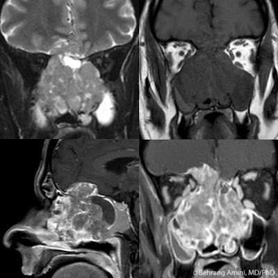
- Esthesioneuroblastoma (shown above): Arises in the superior nasal vault and extends through the cribriform plate. May have calcium. Cyst formation at the interface with the brain is characteristic (seen above).
- Carcinoma: Squamous cell or sinonasal undifferentiated carcinoma
- Nasal dermal sinus: Dermoid or epidermoid between tip of nose and apex of crista galli. With or without a sinus into the frontonasal region through an enlarged foramen cecum. Epidermoid will be low signal on T1, while dermoid will be high signal.
- Cephalocele: Smooth defect in the cribriform plate allows extension of brain into the nasal cavity or the sinuses.
- Bone lesions: Fibrous dysplasia, ossifying fibroma, metastases.
- Melanoma: Arises from melanocytes in sinonasal mucosa.
- Lymphoma: Low T2 signal with homogeneous enhancement.
- Sarcoidosis: Nonspecific appearance.
- Olfactory Schwannoma: Nonspecific appearance. Only interesting fact about these rare lesions is that the olfactory nerve does not contain Schwann cells, and no one knows where these Schwannomas come from (there are theories, but there is no use wasting neurons remembering speculation).
References
Amador AR, Santonja C, Del Pozo JM, Ortiz L. Olfactory schwannoma. Eur Radiol. 2002 Apr;12(4):742-4.Saturday, March 5, 2011
Placental Surface Cyst
A placental surface cyst, also known as a membranous or subchorionic cyst, are cystic lesions located close to the placental cord insertion site. While some regard them to be of little clinical significance, others stress the need for clinical and sonographic follow-up, as cysts larger than 4.5 cm or more than three in number seem to have a higher frequency of intrauterine growth restriction.
On sonography, they are simple cysts without vascular flow on color Doppler. MRI may show a fluid–fluid level with marked shading of the dependent portion on T2-weighted images suggesting the presence of hemorrhagic fluid.
The image above shows a thin-walled cyst within the amniotic. The lesion has some internal debris (red arrow). The cyst is close to the umbilical cordinsertion into the placenta (blue arrows).
References
Motooria K, Kiharab M, Shozub M, Itoa H. MR imaging of placental surface cyst. European Journal of Radiology Extra. 2009; 71(3): e121-e122.Friday, March 4, 2011
Cystic Bronchiectasis
Lower lobes:
- Viral infection: In children
- Primary ciliary dyskinesia: 50% associated with situs inversus (Kartagener syndrome)
- Immunodeficiency: For example, agammaglobulinemia
- Williams-Campbell syndrome: Deficiency in cartilage of the third- to sixth-order bronchi. Usually presents in childhood with coughing and wheezing and recurrent pneumonias. Expiratory CT may reveal collapse of bronchi with distal air trapping and normal-caliber trachea and central bronchi.
- Tracheobronchomegaly: Also known as Mounier-Kuhn syndrome. Rare congenital abnormality of the trachea and main bronchi.
- Cystic fibrosis:
- Allergic bronchopulmonary aspergillosis:
- Tuberculosis:
- Endobronchial tumor:
- Bronchial atresia:
Thursday, March 3, 2011
Fatty Replacement of the Pancreas
Wednesday, March 2, 2011
Metaphyseal Cupping
Metaphyseal cupping refers to the inward bulging of the metaphysis. Focal metaphyseal cupping can occur in response to injury to the central portion of the physis (e.g., from trauma, iatrogenic or otherwise; infection; radiation; etc.).
More generalized metaphyseal cupping can occur in:
- Rickets: Widened and frayed metaphyses. Look for widening of the physes.
- Nutritional: 3-6 months of age. May be seen with exclusively breast-fed babies who don't get vitamin D supplements.
- Vitamin D malabsorption: Liver disease, biliary obstruction, inflammatory bowel disease, celiac disease, etc.
- Renal:
- Hereditary: Vitamin-D dependent types (vitamin D dependent rickets types I and II) and Vitamin-D resistant types (familial hypophosphatemic rickets and hereditary hypophosphatemic rickets with hypercalciuria).
- Medications: Antacids, anticonvulsants, steroids, loop diuretics.
- Sickle Cell Anemia:
- Achondroplasia: Associated with flaring and medial slanting of the femoral metaphyses early on with progression to an inverted V configuration later.
- Hypervitaminosis A: Distal femur typically involved. Hyperostosis can also be seen (more pronounced in ulna and metatarsals). Increased intracranial pressure and subsequent widening of the sutures.
- Metaphyseal dysplasias: For example, Schmid type (milder, autosomal dominant), Jansen type (more severe).
References
- Caffey J. Traumatic cupping of the metaphyses of growing bones. Am J Roentgenol Radium Ther Nucl Med. 1970 Mar;108(3):451-60.
- Nield LS, Mahajan P, Joshi A, Kamat D. Rickets: not a disease of the past. Am Fam Physician. 2006 Aug 15;74(4):619-26.
- Sprogue PL. Epiphyseo-metaphyseal cupping following infantile scurvy. Pediatr Radiol. 1976 Feb 13;4(2):122-3.
Labels:
Boards,
Musculoskeletal,
Pediatric radiology
Tuesday, March 1, 2011
Vascular Malformations of the Brain
- Arteriovenous malformation (shown above): Feeding arteries, draining veins, and intervening nidus of abnormal vessels. Frequently diagnosed after hemorrhage. CT may show high-attenuation and enhancing serpentine structures corresponding to enlarged, tortuous vessels. Focal calcification and lack of mass effect are characteristic. MRI will show flow voids and evidence of hemoglobin breakdown. Perilesion gliosis appears as T2-hyperintensity. Angiography is the gold standard for excluding an arteriovenous malformation and for assessing the size of the nidus. The patient shown above presented with cerebellar hemorrhage. MRI showed heterogeneous T1 and T2 signal due to hemorrhage associated with hemosiderin deposition. Angiography reveals vessels from branches of the left superior cerebellar artery and left posterior inferior cerebellar artery (pink and blue arrows, respectively) feeding a nidus (white arrow), and draining into the transverse sinus (green arrow). The superior cerebellar artery is displaced superiorly by the cerebellar hemorrhage.
- Dural arteriovenous fistula: Arteriovenous fistula in the meninges supplied partly or wholly by dural arteries and drained by dural sinuses and/or leptomeningeal venous channels. They most frequently involve the cavernous or transverse/sigmoid sinuses. CTA may show dilated cortical or subcortical vessels. May be complicated by hemorrhage, in which case there will be parenchymal hematoma with or without subarachnoid blood. The pattern may simulate a hemorrhagic venous infarction (look for a thrombosed venous sinus). Contrast-enhanced MRI and MRA is the best noninvasive modality, but angiography is the gold standard.
- Cavernous angioma: Well-demarcated, high-attenuation lesion on CT due to hemorrhage or calcification. Contrast enhancement is inconsistently seen. Heterogeneous T1 and T2 signal consistent with subacute hemorrhage. Hemosiderin rin ("black halo") typical. Gradient-echo good for detecting small lesions. Differentiated from hemorrhagic tumor by absence of surrounding vasogenic edema. Usually not seen on angiography (used to be called "occult arteriovenous malformations").
- Developmental venous anomaly: Also known as venous angioma and medullary venous malformation. Purely venous anomaly. CT may reveal calcifications if associated with a cavernous angioma (see above). Acute parenchymal hemorrhage is rare. CTA reveals numerous linear or dot-like foci of enhancement that converge on an enlarged tubular draining vein (Medusa head). T1-weighted images can be be normal in small anomalies. Flow voids may be seen on T1- and T2-weighted images. Contrast-enhanced images reveal similar findings as CTA. MRV will help show the drainage pattern of the draining vein.
- Capillary telangiectasia: Small, abnormally dilated capillaries within otherwise normal brain tissue. Presence of normal brain tissue between the capillaries distinguishes it from cavernous angioma pathologically. Most commonly occur in the pons. Usually not visible on CT (even post-contrast). On MRI, there is lacelike and subtle enhancement with little or no signal abnormality on T2-weighted images. They may be hypointense or isointense on T1-weighted images. There is signal loss on gradient echo sequences.
References
Byrne JV. Cerebrovascular malformations. Eur Radiol. 2005 Mar;15(3):448-52.
Labels:
Boards,
Interventional radiology,
Neuroradiology
Subscribe to:
Posts (Atom)