Sunday, July 31, 2011

Camptodactyly

Camptodactyly, from the combination of the Greek words for bent (kamptos) and finger (daktylos), refers to a permanent flexion deformity at the proximal interphalangeal joint of one or more fingers. The deformity is painless and non-neurogenic and most commonly affects the small finger (camptodactyly 5).

The cause of camptodactyly is not known. It may be related to a fascial abnormality at the proximal interphalangeal joint or due to contracture of the flexor digitorum superficialis.

Camptodactyly occurs with an incidence is less than 1% and may be sporadic or familial. It is commonly isolated, but may also be associated with any number of conditions, including oculodentodigital syndrome, orodigitofacial syndrome, Trisomy 13-15, ankyloglossia superior, osteo-onychodysplasia, congenital heart disease, absence of the pectoral muscle, Marfan syndrome, and radioulnar synostosis.

Radiographs show a flexion deformity at the proximal interphalangeal joint witha compensatory hyperextension distally.

Camptodactyly has been illustrated by Botticelli and da Vinci. In his sketch of his hands as an old man, we can see camptodactyly in his small finger.


Camptodactyly should not be confused with Clinodactyly. While both most commonly occur at the small finger, clinodactyly refers to the curvature of a finger in the mediolateral plane (i.e., in a radial or ulnar direction).

References

Poznanski AK, Pratt GB, Manson G, Weiss L. Clinodactyly, camptodactyly, Kirner's deformity, and other crooked fingers. Radiology. 1969 Sep;93(3):573-82.

Saturday, July 30, 2011

Intramuscular Myxoma

Intramuscular myxomas are benign proliferations of fibroblasts with abundant myxoid stroma. MRI findings correlate with the appearance of these tumors at pathology and can suggest the diagnosis. Patients are typically women older than 40, who present with a painless mass.

Pathologically, intramuscular myxomas are gelatinous masses without hemorrhage or necrosis. This is reflected as predominantly high signal intensity on T2-weighted images and low signal intensity on T1-weighted images.

Fibrous bands may be seen within the tumors on pathology. These appear as mildly enhancing internal linear strands (pink arrow) and may lead to a heterogeneous appearance on MRI.

Increased vascularity is not a feature of these tumors, a feature reflected in the mild or absent internal enhancement seen on post-contrast images. Areas of cellularity in the tumor present as patchy internal enhancement.

Intratumoral cysts can be seen in some cases (not seen here).

Myxomas tend to extend into the adjacent muscle, dividing it into small groups of fibers, a process that can lead to focal muscle atrophy. The surrounding muscle may also be edematous and T2-hyperintense (blue arrow).

Peripherally, these tumors have a pseudocapsule that represents condensation of fibrous tissue. The pseduocapsule is about 1 mm – 2 mm in thickness and is discontinuous at the junction of the tumor with the muscle. The thin pseudocapsule enhances on post-contrast images.

Finally, fat cells around the pseudocapsule present as areas of T1 hyperintensity around the tumor (white arrow).

Radiographic findings can be normal or reveal a nonspecific soft tissue mass. Calcifications can rarely be seen. Scintigraphic studies may show mild uptake due to the tumor's moderate vascularity. Ultrasound may reveal a multicystic hypoechoic or anechoic mass with well-defined margins. Similarly, CT reveals a well-defined mass with intermediate homogeneous attenuation.

While MR findings of a cystic intramuscular mass with peripheral and variable internal enhancement suggest the diagnosis of an intramuscular myxoma, a perilesional fat ring and the presence of edema in adjacent muscle are the most distinctive features of these tumors.

In some cases, however, biopsy may be necessary to exclude a malignant tumor with myxoid components, such as, myxoid peripheral nerve sheath tumors, extraskeletal myxoid chondrosarcoma, myxoid liposarcoma, myxofibrosarcoma, myxoid leiomyosarcoma, low-grade fibromyxoid sarcoma, and cellular myxoma.

References

Luna A, Martinez S, Bossen E. Magnetic resonance imaging of intramuscular myxoma with histological comparison and a review of the literature. Skeletal Radiol. 2005 Jan;34(1):19-28.

Friday, July 29, 2011

Fossae in the Inferior Rami of the Pubis

Fossae can occasionally be seen at the inferior pubic rami. These are presumably related to the origin of the adductuor muscles, but I can't find any reference to this entity aside from a passing mention in Keats as "fossae in the inferior rami of the pubis."

References

  • Keats TE and Anderson MW. Atlas of Normal Roentgen Variants That May Simulate Disease. 8th edition, page 468; Mosby (2004).

Thursday, July 28, 2011

Pseudotumor Deltoideus

Pseudotumor deltoideus refers to bony irregularity or lucency at the deltoid muscle insertion on the proximal humerus. In the appropriate clinical setting, this finding may be mistaken for a neoplastic process. Smooth, well-defined borders and the characteristic location on the lateral aspect of the humerus at the level of deltoid insertion can help steer the diagnosis away from neoplasm.

Cortical thickening may be due to irregularity and slight expansion at the level of the deltoid insertion, while lucency has been suggested to represent a form of a herniation pit. Bone scans may show slightly increased uptake.

The image above shows both cortical irregularity (pink arrow) and a well-defined lucency (white arrow) at the lateral aspect of the humerus at the level of deltoid insertion. CT and PET/CT fusion images show the area of cortical thickening and an intracortical lucency. No increased FDG activity is seen.

References

  • Keats TE and Anderson MW. Atlas of Normal Roentgen Variants That May Simulate Disease. 8th edition, pages 521-523; Mosby (2004).
  • Morgan H, Damron T, Cohen H, Allen M. Pseudotumor deltoideus: a previously undescribed anatomic variant at the deltoid insertion site. Skeletal Radiol. 2001 Sep;30(9):512-8.

Wednesday, July 27, 2011

Piezoelectricity in Bone

Piezoelectricity (from the Greek piezo or piezein, to squeeze or press) refers to the ability of certain materials to generate electrical potentials in response to mechanical stress. This phenomenon may be familiar to musicians as the basis of piezoelectric pickups in acoustic-electric instruments.

Crystals such as quartz, as well as biologic tissue such as bone have been shown to generate electrical potentials in response to mechanical stress. For example, recordings from the human tibia during walking have shown generated electric potentials as high as 300 mV. These electrical potentials have been shown to be generated by shear forces of collagen and deformation of fluid-filled Haversian and Volkmann channels.

Because electromagnetic fields have been shown to affect cell division rates, tissue growth, and wound repair, alterations in bone architecture due to mechanical loading are thought to be mediated by piezoelectricity. Tissues, such as bone, that can generate endogenous electrical signals show a higher capacity to regenerate. This phenomenon is thought to be mediated at the level of receptors at the cell membrane.

References

  • Isaacson BM, Bloebaum RD. Bone bioelectricity: what have we learned in the past 160 years? J Biomed Mater Res A. 2010 Dec 15;95(4):1270-9.

Tuesday, July 26, 2011

Fat in the Dural Sinuses

Fat deposition in the dural sinuses can be an incidental finding, with the most common location being the cavernous sinus, torcular herophili, the straight sinus, inferior sagittal sinus, superior sagittal sinus (seen above), and transverse sinus (in decreasing order of frequency). It has been suggested that these deposits represent normal adipose tissue in the dural sinuses and not lipomas.

References

Horsburgh A. Incidental fat in the dural sinuses. Neuroradiology. 2009 Nov;51(11):787-8.

Monday, July 25, 2011

Facial Nerve Hemangioma

Hemangioma of the facial nerve is a rare benign entity, representing less than 1% of temporal bone tumors. The majority of patients present in the 3rd to 6th decade of life.

The most common location for facial nerve hemangiomas is the geniculate fossa followed by the internal auditory canal. This distribution corresponds to the density of capillary plexus surrounding the facial nerve (highest in the region of the geniculate ganglion).

These hemangiomas range in size from 2 mm to 20 mm, but are usually symptomatic early (usually < 10 mm). Hemangiomas in the geniculate fossa usually present with slowly progressive facial nerve paralysis. Invasion of the internal auditory canal and cochlear otic capsule can result in sensorineural hearing loss and pulsatile tinnitus.

CT reveals a lesion with irregular and amorphous margins. The typical honeycomb appearance of hemangiomas is pathognomonic, but is seen in about 50% of cases. Without these spicules, the differential widens to include schwannoma and, less commonly, meningioma.

MRI reveals variable signal intensity on T1-weighted images and increased signal intensity on T2-weighted images with hypointense foci corresponding to the ossific matrix of these lesions (the honeycomb spicules mentioned above). There is usually avid contrast enhancement. The differential consideration with these MRI findings includes normal facial nerve enhancement, Bell palsy, schwannoma, and perineural spread of a parotid malignancy.

Our case shows soft tissue prominence and enlargement of the anterior genu of the right facial nerve in the region of the geniculate ganglion. Spicules are seen associated with this lesion, best appreciated on the sagittal reconstructions.

References

Mijangos SV, Meltzer DE. Case 171: facial nerve hemangioma. Radiology. 2011 Jul;260(1):296-301.

Sunday, July 24, 2011

Osteosarcoma: Prognosis at Initial Diagnosis

Kaste et al and Bieling et al. have shown that measurements of osteosarcoma at diagnosis can be correlated with prognosis. The anteroposterior dimension of the tumor (tumor depth) and absolute tumor volume (see below for calculation) have been shown to be predictive of overall survival and event-free survival in patients with osteosarcoma.

Tumor depth is defined as the horizontal extent of osteosarcoma on a lateral radiograph. The risk of death has been shown to increase approximately 1.5 times for each centimeter increase in tumor depth. A 5-year event-free survival rate of about 80% was seen in patients with tumor depth ≤ 5 cm, while those with tumor depth of > 5 cm had an event-free survival rate of about 55%.

Absolute tumor volume is calculated as an ellipsoid: 0.52 x tumor length x tumor width x tumor height, all measured on radiographs. The 5-year overall survival rate for patients with tumor volume ≤ 150 cm3 has been shown to be about 85%, while that of patients with tumor volume > 150 cm3 has been shown to be about 60%. Looked at in another way, patients with absolute tumor volumes > 150 cm3 have been shown to be 3.6 times more likely to die compared to those with smaller tumor volumes.

References

Saturday, July 23, 2011

Synovial Chondromatosis of the Temporomandibular Joint

Synovial chondromatosis is the benign chondroid metaplasia of the synovium. Cartilaginous nodules form at the synovium and may detach to form loose bodies within the joint. Ossification of these bodies changes the name from chondromatosis to osteochondromatosis.

Synovial chondromatosis usually occurs in large joints such as the knee or shoulder, and has a male predominance. Involvement of the temporomandibular joint is rare and has a female predominance. In the temporomandibular joint, it is usually located on the right with a right-to-left ratio of 4 to 1.

Patients present with restricted movement, as well as preauricular swelling and pain.

Radiographs may reveal widening of the joint space, limitation of motion, irregularity of the joint surfaces, calcified loose bodies, and sclerosis of the glenoid fossa and mandibular condyle. When all these findings are present, a diagnosis of synovial chondromatosis can be made on radiographs; however, some or even all of these findings (except for calcified bodies) can also be seen with osteoarthritis.

On CT and MR, the diagnosis is more straightforward with demonstration of cartilagenous or ossified objects within the joint with joint effusion and erosions of the joint surfaces.

References

  • Herzog S, Mafee M. Synovial chondromatosis of the TMJ: MR and CT findings. AJNR Am J Neuroradiol. 1990 Jul-Aug;11(4):742-5.
  • Koyama J, Ito J, Hayashi T, Kobayashi F. Synovial chondromatosis in the temporomandibular joint complicated by displacement and calcification of the articular disk: report of two cases. AJNR Am J Neuroradiol. 2001 Jun-Jul;22(6):1203-6.
  • Manco LG, DeLuke DM. CT diagnosis of synovial chondromatosis of the temporomandibular joint. AJR Am J Roentgenol. 1987 Mar;148(3):574-6.

Friday, July 22, 2011

Extended Pattern of Uptake

Extended pattern of uptake, also referred to as contiguous bone activity, is a potential diagnostic pitfall in interpreting bone scans where there is increased uptake in normal bone adjacent to a focal lesion.

This uptake in bone definitely beyond the proven limit of a focal bone lesion can be seen in both benign and malignant conditions and is thought to be related to hyperemia. It has been reported in the setting of trauma, osteomyelitis, benign giant cell tumor, avascular necrosis, and thrombophlebitis, as well as malignancies like primary bone tumors (e.g., osteosarcoma), metastatic disease (rare), and soft-tissue neoplasms.

The images above are from a patient with a giant cell tumor of the lateral proximal tibia complicated by a fracture. The bone scan shows intense uptake in the proximal tibia and mild/moderate uptake in the distal femur.

References

  • Chew FS, Hudson TM. Radionuclide bone scanning of osteosarcoma: falsely extended uptake patterns. AJR Am J Roentgenol. 1982 Jul;139(1):49-54.
  • Thrall JH, Geslien GE, Corcoron RJ, Johnson MC. Abnormal radionuclide deposition patterns adjacent to focal skeletal lesions. Radiology. 1975 Jun;115(3):659-63.

Thursday, July 21, 2011

Pseudosubluxation

Pseudosubluxation is an anatomic variant where there is anterior positioning of the body of C2 on C3. It is a normal finding seen in up to 20% of children under 16 years of age and is caused by ligamentous laxity in this age group.

This physiologic laxity can make it difficult to recognize true pathologic displacement of C2 on C3 in children. A line along the posterior arches of C1, C2 and C3 can be used to help make this distinction on lateral radiographs. In the majority of cases, this posterior cervical line passes through the anterior cortex of the posterior arch of C2; however, the posterior cervical line can also be normal if it passes just behind the anterior cortex of the posterior arch of C2, touches the anterior aspect of the anterior cortex of the posterior arch of C2, or if it comes within 1 mm of the anterior cortex of the posterior arch of C2.

Special thanks to Dr. Hansel Otero for the image.

References

  • Cattell HS, Filtzer DL. Pseudosubluxation and other normal variations in the cervical spine in children. A study of one hundred and sixty children. J Bone Joint Surg Am. 1965 Oct;47(7):1295-309.
  • Swischuk LE. Anterior displacement of C2 in children: physiologic or pathologic. Radiology. 1977 Mar;122(3):759-63.

Wednesday, July 20, 2011

Hardcastle Syndrome

Hardcastle syndrome refers to diaphyseal medullary stenosis associated with malignant fibrous histiocytoma of the bone. It is a rare autosomal dominant condition that has recently be localized to chromosome 9 (9p21-22).

The major radiographic feature is diaphyseal medullary narrowing with overlying cortical thickening. Some patients also have lucent linear striations in the metaphysis that run parallel to the long axis of the bone and do not extend to the epiphysis. Cyst-like metaphyseal lesions can be seen in isolation or in proximity to these linear striations.

These findings symmetrically affect the long tubular bones of the upper and lower extremities and are not seen in the skull, spine, or pelvis.

Fractures also occur with high frequency and tend to occur with minimum trauma. Delayed union of fractures is typically seen.

There is a high (35%) tendency for transformation into malignant fibrous histiocytoma (originally classified as medullary fibrosarcoma), usually between the second to fifth decades of life. The malignant transformation occurs in the metaphysis or diaphysis, presumably at the sites of bone infarction.

Non-skeletal abnormalities can include early cataracts and mental retardation.

References

  • Hardcastle P, Nade S, Arnold W. Hereditary bone dysplasia with malignant change. Report of three families. J Bone Joint Surg Am. 1986 Sep;68(7):1079-89.
  • Martignetti JA, Desnick RJ, Aliprandis E, Norton KI, Hardcastle P, Nade S, Gelb BD. Diaphyseal medullary stenosis with malignant fibrous histiocytoma: a hereditary bone dysplasia/cancer syndrome maps to 9p21-22. Am J Hum Genet. 1999 Mar;64(3):801-7.
  • Norton KI, Wagreich JM, Granowetter L, Martignetti JA. Diaphyseal medullary stenosis (sclerosis) with bone malignancy (malignant fibrous histiocytoma): Hardcastle syndrome. Pediatr Radiol. 1996 Sep;26(9):675-7.

Tuesday, July 19, 2011

Buttress Periosteal Reaction

Buttress or buttressed periosteal reaction refers to beaklike solid periosteal new bone formation that can be seen with benign and malignant lesions. It is the interrupted version of the solid periosteal reaction.

It can be seen in the setting of saucerization of bone by a non-mineralized tumor (e.g., periosteal chondroma), whereby the periosteum reacts adjacent to the saucerization, giving a buttressed appearance.

The buttressed appearance can also be seen with an expanding intramedullary process (e.g., aneurysmal bone cyst) that breaks through an area of solid periosteal reaction. This may or may not represent malignant transformation of a previously benign process.

The buttress periosteal reaction should be distinguished from the Codman triangle, which is seen at the periphery of a lesion as an elevation of interrupted periosteum with one or more layers of new bone.

References

  • Miller TT. Bone tumors and tumorlike conditions: analysis with conventional radiography. Radiology. 2008 Mar;246(3):662-74.
  • Ragsdale BD, Madewell JE, Sweet DE. Radiologic and pathologic analysis of solitary bone lesions. Part II: periosteal reactions. Radiol Clin North Am. 1981 Dec;19(4):749-83.

Monday, July 18, 2011

Osteoid Osteoma

Osteoid osteomas are common, benign, bone-forming tumors that are characterized by small size (< 1.5 cm), limited growth potential, and disproportionate pain. Histologically, they are composed of osteoid and woven bone and are often surrounded by a rim of plump osteoblasts, findings that can also be seen in osteoblastomas.

Patients are young, more often white and male, and present with pain that is initially intermittent and mild that eventually becomes more constant and severe. The pain is usually more intense at night and usually (75% of the time) responds to aspirin. The pain may precede radiographic findings, and may be referred to a nearby joint.

When in the spine, patients commonly have scoliosis, which is less likely to occur with an osteoblastoma involving the spine. In fact, osteoblastoma is the most common cause of painful scoliosis in adolescents. Unlike osteoblastomas of the spine, osteoid osteomas are rarely associated with neurological symptoms.

Before continuing, a word is warranted on usage of the term "nidus." Properly, the nidus refers to the lucent lesion of the osteoid osteoma itself, and not the central mineralization that can be seen in some osteoid osteomas.

Three types of osteoid osteomas have been described by Edeiken et al:
  • Cortical/classic: The most common type, seen in 75% of cases. A lucent lesion, the nidus, is located in the center of reactive sclerosis and periosteal reaction. The periosteal reaction is usually solid, but may be lamellated. Tends to affect the tibia and femur.
  • Cancellous/medullary (shown above): About 25% of cases. Tends to affect the femoral neck, small bones of the hands and feet, and the spine. Sclerosis, if present, is mild or moderate. The lucent nidus is not necessarily in the center of the sclerosis, which may be located some distance away. In the spine, the differential diagnosis would include osteoblastoma.
  • Subperiosteal: The rarest type. Presents as soft tissue mass adjacent to bone, which may demonstrate pressure erosion or resorption, but little or no significant reactive sclerosis. Tends to affect the medial aspect of the femoral neck, the hands and feet, and the talar neck. Patients with a painful joint may initially be evaluated with ultrasound, and the finding of cortical irregularity and focal synovitis should raise the possibility of an intraarticular osteoid osteoma.

    Subperiosteal osteoid osteomas may actually represent the early stage of a cortical osteoid osteoma, with the subperiosteal getting incorporated into the cortex by periosteal new bone formation.
Depending on their relationship to the joint, osteoid osteomas can also be classified as intraarticular. Intraarticular osteoid osteomas are generally of the cancellous or subperiosteal type, and most commonly occur at the hip. Imaging findings may be subtle due to the absence of periosteal reaction. Indeed, when the nidus is within 1 cm of the articular surface, little or no periosteal reaction may be present. Imaging studies may reveal nonspecific joint effusion, synovitis, and regional osteoporosis. Radiographs may be normal at first.

Bone scintigraphy reveals increased activity on all three phases. The double density sign describes focal increased uptake (nidus) surrounded by a larger area of increased uptake. The double-density sign can help differentiate an osteoid osteoma from osteomyelitis. In osteomyelitis, there may be an area of decreased uptake corresponding to the lucent sinus or abscess.

On CT, the nidus of a cortical or cancellous lesion is a smooth low-attenuation lesion with or without surrounding sclerosis and periosteal reaction. The nidus may contain central mineralization, which may be punctate, amorphous, ringlike, or dense.

MRI is not as good as CT for detection of osteoid osteomas in about 2/3 of cases. MRI reveals the nidus to be intermediate signal intensity on T1-weighted images, and intermediate-to-high on T2-weighted images. Central mineralization, if present, can cause low signal intensity. The surrounding marrow may show increased T2 signal. Lesions show early enhancement with early washout compared to the adjacent marrow on dynamic MRI.

The images above depict a cancellous osteoid osteoma presenting as a low-attenuation lesion with central mineralization, located within the femoral neck. Note the mild surrounding sclerosis. The mineralized center is low signal intensity on the T1- and T2-weighted images and is located within an intermediate signal intensity nidus, which is surrounded by bone marrow edema.

Treatment consists of radiofrequency ablation, which has a success rate similar to that of surgery. The target is the lucent nidus. After treatment, the surrounding sclerosis may regress.

References

  • Ebrahim FS, Jacobson JA, Lin J, Housner JA, Hayes CW, Resnick D. Intraarticular osteoid osteoma: sonographic findings in three patients with radiographic, CT, and MR imaging correlation. AJR Am J Roentgenol. 2001 Dec;177(6):1391-5.
  • Edeiken J, DePalma AF, Hodes PJ. Osteoid osteoma. (Roentgenographic emphasis). Clin Orthop Relat Res. 1966 Nov-Dec;49:201-6.
  • Helms CA, Hattner RS, Vogler JB 3rd. Osteoid osteoma: radionuclide diagnosis. Radiology. 1984 Jun;151(3):779-84.
  • Kransdord MJ and Murphey MD. Chapter 14: Osseous tumors. in Imaging of Bone Tumors and Tumor-Like Lesions. Davies AM, Sundaram M, and James SLJ (eds). Springer-Verlag Berlin Heidelberg (2009); pp 261-269.
  • Pettine KA, Klassen RA. Osteoid-osteoma and osteoblastoma of the spine. J Bone Joint Surg Am. 1986 Mar;68(3):354-61.

Sunday, July 17, 2011

Osteoblastoma

Osteoblastomas are benign bone-forming neoplasms that produce woven bone spicules bordered by osteoblasts, histologic features that are similar to those of osteoid osteomas. Osteoblastomas have an extremely rich vascular supply and an aneurysmal bone cyst component is seen in up of 15% of cases.

Different forms of osteoblastoma have been described. Epithelioid osteoblastomas, also known as aggressive or malignant osteoblastomas, have plump osteoblasts with prominent nuceloli and can be difficult to distinguish from osteosarcoma. They have been described as borderline lesions between benign osteoblastoma and osteosarcoma, and have a locally destructive growth pattern and a high recurrence rate after curettage.

Toxic osteoblastomas are rare and associated with systemic symptoms (anorexia, weight loss, cachexia, and fever).

The term conventional osteoblastoma is therefore used to differentiate the traditional osteoblastoma from these varieties.

Conventional osteoblastomas typically affect adolescents and young adults, males more frequently than females. Patients present with dull localized pain that is less likely to respond to non-steroidal anti-inflammatory medications than osteoid osteomas. The pain is also less likely to get worse at night.

Conventional osteoblastomas affect the spine in 1/3 of cases, where they more commonly affect the posterior processes and may extend into the vertebral body (isolated vertebral body involvement is rare). Compared to osteoid osteomas, osteoblastomas affecting the spine are more likely to present with neurological symptoms. Scoliosis, on the other hand, is less likely to occur with osteoblastomas compared to osteoid osteomas.

Osteoblastomas can also affect the diaphyses and metaphyses of long bones, where they are more often proximal.

Radiographs and CT may reveal one of three appearances:
  • Lucent lesion with surrounding sclerosis. The appearance is similar to that of the osteoid osteoma, but the lesions are larger (> 1.5 cm). This is the most common appearance in the long bones, where solid continuous periosteal reaction can be seen, sometimes distant from the lesion.
  • Expansile lucency with a sclerotic rim and internal calcifications that may simulate a chondroid matrix. This is the most common form in the spine.
  • Aggressive expansile lesion with bone destruction, soft tissue extension, and matrix calcifications that may simulate a chondroid matrix.
The diagnosis of osteoblastoma in the long bones is difficult, being correctly made in less than 1/3 of cases (compared to 2/3 in spinal lesions). Various degrees of mineralization may be present, and an agrressive lamellated or spiculated periosteal reaction may be present.

As may be expected from their highly vascular nature and periosteal reaction, bone scans demonstrate marked uptake.

Findings on conventional angiography are nonspecific and variable. Some osteoblastomas show no abnormality at all on conventional angiography. Those that do may reveal a contrast blush in the capillary and/or venous phase, regular or irregular vascularity, and/or large draining veins.

As with other bone tumors, the MRI appearance is nonspecific and may be misleadingly aggressive. Lesions have intermediate signal intensity on T1-weighted images and variable signal intensity on T2-weighted images. Extensive peri-tumoral T2 hyperintensity can be seen. An aneurysmal bone cyst component is seen in up to 15% of cases.

In the spine, the main differential consideration is osteoid osteoma. Osteoblastomas tend to have multifocal mineralization compared to the central mineralization that may be seen in osteoid osteomas. In addition, osteoblastomas tend to be expansile and may have a soft tissue component. Finally, serial imaging reveals slow growth, which is distinguished from the stability of osteoid osteomas.

Osteoblastomas of the spine are usually curetted with allograft or autograft packing. Aggressive and larger lesions are typically resected en block. Conventional osteoblastomas may recur in up to 25% of cases, while aggressive osteoblastomas have close to a 50% recurrence rate.

References

  • Dorfman HD, Weiss SW. Borderline osteoblastic tumors: problems in the differential diagnosis of aggressive osteoblastoma and low-grade osteosarcoma. Semin Diagn Pathol. 1984 Aug;1(3):215-34.
  • Eisenberg RL. Bubbly lesions of bone. AJR Am J Roentgenol. 2009 Aug;193(2):W79-94.
  • Kransdord MJ and Murphey MD. Chapter 14: Osseous tumors. in Imaging of Bone Tumors and Tumor-Like Lesions. Davies AM, Sundaram M, and James SLJ (eds). Springer-Verlag Berlin Heidelberg (2009); pp 269-274.
  • Kroon HM, Schurmans J. Osteoblastoma: clinical and radiologic findings in 98 new cases. Radiology. 1990 Jun;175(3):783-90.
  • Kumar R, Guinto FC Jr, Madewell JE, David R, Shirkhoda A. Expansile bone lesions of the vertebra. Radiographics. 1988 Jul;8(4):749-69.
  • Strickland B. The value of arteriography in the diagnosis of bone tumours. Br J Radiol. 1959 Nov;32:705-13.

Saturday, July 16, 2011

FDG-PET and Peripheral T-Cell Lymphomas


T-cell neoplasms are broadly divided by the World Health Organization into precursor T-cell neoplasms and peripheral T-cell neoplasms. Peripheral T-cell neoplasms are further divided into peripheral T-cell leukemias and peripheral T-cell lymphomas. The latter is the subject of this post.

Peripheral T-cell lymphomas are uncommon neoplasma that make up about 10%-25% of all cases of non-Hodgkin lymphoma, although the prevalence has geographic variability. T-cell lymphomas tend to have a poorer prognosis than B-cell lymphomas, but vary in their behavior, from indolent to aggressive.

Peripheral T-cell lymphomas can be subdivided into primary nodal disease and primary systemic (extranodal) disease. The main clinicopathologic presentations of peripheral T-cell lymphomas include:

Extranodal
  • Extranodal NK/T-cell lymphoma, nasal type: Previously known as angiocentric lymphoma and lethal midline granuloma, this is the most common peripheral T-cell lymphoma in Asian patients, and has been associated with the Epstein-Barr virus. The nasal cavity or paranasal sinuses are often involved, with tumor necrosis a common feature, but identical tumors can also be found in the skin, lymph nodes, and viscera, sometimes in isolation.
  • Enteropathy-type T-cell lymphoma: This rare subtype typically occurs in elderly patients with a history of celiac disease or dermatitis herpetiformis. PET scan may reveal FDG-avid lesions in the gastrointestinal tract and mesenteric lymph nodes.
  • Subcutaneous panniculitis-like T-cell lymphoma: Multifocal subcutaneous infiltrates of malignant T lymphocytes are associated with benign histiocytic inflammatory cells. CT may show poorly defined subcutaneous lesions ranging in size from a few centimeters to large confluent infiltration. The omental fat can also be involved. The lesions are FDG-avid. Inflammatory disorders of the subcutaneous tissues, such as erythema nodosum and erythema induratum, and inflammatory or infectious panniculitis can have a similar appearance.
  • Mycosis fungoides (shown above)/Sézary syndrome: This is a cutaneous T-cell lymphoma that exists in three clinical phases: patches, plaques, and tumors. PET can demonstrate plaques and tumors, but particular attention has to be devoted to the skin. On CT, the appearance varies from cutaneous thickening to exophytic soft-tissue masses (see above image). FDG-avid adenopathy can be seen in some patients.

    Sézary syndrome is the leukemic variant of mycosis fungoides, with characteristic Sézary cells found in the peripheral blood. Patients present with diffuse erythroderma; however, PET may not always show diffusely increased cutaneous FDG uptake. When present, diffusely increased cutaneous FDG uptake is similar to the appearance of nonattenuation-correction PET emission images. Patients typically have FDG-avid lymphadenopathy; however, low-grade FDG uptake in lymph nodes may represent benign dermatopathic lymphadenitis (reactive enlargement of lymph nodes draining areas of disrupted skin) and be mistaken for positive nodal disease.
  • Anaplastic large cell lymphoma, primary cutaneous type: Part of the spectrum of disease that includes lymphomatoid papulosis (benign). Patients typically present with persistent cutaneous nodules 1 cm – 2 cm in size, and, in ~10% of cases, extracutaneous disease.
  • Hepatosplenic T-cell lymphoma: This is a rare, recently described subtype with an aggressive course and poor prognosis. Patients have hepatosplenomegaly without lymphadenopathy and significant cytopenia.
Nodal
  • Anaplastic large cell lymphoma, primary systemic type: The majority have FDG activity in the lymph nodes. Patients may also have foci of cutaneous or subcutaneous FDG uptake, as well as visceral disease. Expression of anaplastic lymphoma kinase (ALK) protein suggests a better prognosis than the other peripheral T-cell lymphomas. However, no obvious association has been found between expression of this protein and FDG avidity of tumors.
  • Angioimmunoblastic T-cell lymphoma: This is a distinct subtype that presents with lymphadenopathy, hepatosplenomegaly, skin rash, and constitutional symptoms, with evidence of dysregulation of the immune system (hypergammaglobulinemia, Coombs-positive hemolytic anemia, and immunodeficiency). As suggested by the name, patients have FDG-avid adenopathy. Some patients may have visceral involvement, including the spleen. FDG-avid skin lesions are typically not seen.
  • Peripheral T-cell lymphoma, not otherwise specified: No specific clinicopathologic features to permit further classification. The majority have FDG-avid lesions and FDG-avid lymphadenopathy. Patients may also have cutaneous and visceral disease.

References

Friday, July 15, 2011

Chronic Expanding Hematoma

The term chronic expanding hematoma refers to hematomas that increase in size for over a month after initial hemorrhage. Similar lesions in the bones and soft tissues in patients with hemophilia are called hemophilic pseudotumors.

The persistence and growth of these lesions is thought to be due to the irritative properties of blood and its breakdown products, causing repeated capillary injury and episodic bleeding into the main cavity.

It is unclear why most hematomas spontaneously resolve, while some persist and develop vascular ingrowth, and others calcify peripherally.

Chronic expanding hematomas have been reported in the soft tissues of the extremities, body wall musculature, and visceral organs.

Radiographs reveal a soft tissue mass with or without calcifications and adjacent bone destruction or pressure erosion. CT shows a heterogeneous or homogeneous low-attenuation mass that can have peripheral or internal calcifications and adjacent bone destruction or pressure erosion.

On MRI, chronic expanding hematomas have a pseudocapsule that is low signal intensity of T1- and T2-weighted sequences and is composed of fibrous tissue, hemosiderin deposits and iron-laden macrophages. The pseudocapsule can enhance.

Centrally, they have areas of heterogeneous signal intensity on both T1-weighted and T2-weighted sequences that correspond to a mixture of blood breakdown products; loose connective tissue; granulation tissue with capillary ingrowth; inflammatory tissue; and cavitary collections of necrotic debris, fibrin and blood clots.

Contrast enhancement within benign hematomas is rare, but has been reported. It implies the presence of viable tissue, an intact vascular supply, and abnormal capillary permeability within the central portion of the hematoma.

The most important differential consideration is a hemorrhagic soft tissue sarcoma, which can have a similar appearance on non-contrast MRI. The presence of gadolinium enhancement helps point toward neoplasm.

References

  • Aoki T, Nakata H, Watanabe H, Maeda H, Toyonaga T, Hashimoto H, Nakamura T. The radiological findings in chronic expanding hematoma. Skeletal Radiol. 1999 Jul;28(7):396-401.
  • Batista GR, Rocha Oliveira PC, Alcantara FP, de Teles IG, Senna Najjar YJ, Kalil RK. Chronic diploic hematoma mimicking malignancy on imaging. Skeletal Radiol. 2011 Apr;40(4):475-9.
  • Reid JD, Kommareddi S, Lankerani M, Park MC. Chronic expanding hematomas. A clinicopathologic entity. JAMA. 1980 Nov 28;244(21):2441-2.

Thursday, July 14, 2011

Normal FDG Uptake in the Lingual Tonsils

Accurate interpretation of PET images requires familiarity with the normal patterns of uptake and intensities. In the head and neck, intense FDG activity can be seen in the components of Waldeyer's ring. More than 95% of normal subjects have FDG activity greater than blood pool in the palatine tonsils (mean SUV = 3.48) and lingual tonsils (mean SUV = 3.11). This physiologic uptake has an inverse relationship with age for the palatine tonsils.

The image above shows fused (top row) and maximum intensity projections in a patient with no disease in the head and neck. There is intense FDG activity above blood pool (SUVmax = 7).

References

Wednesday, July 13, 2011

Chondroblastic Osteosarcoma

Chondroblastic osteosarcoma is defined as a high-grade bone tumor with a substantial volume (up to 90%) of tissue with a chondrosarcomatous phenotype next to osteoid-forming areas. It is a rare tumor with a reported overall prevalence between 5% to 25% of all osteosarcomas and with a similar age distribution to osteosarcomas. The familiar periosteal osteosarcoma is a moderately differentiated chondroblastic osteosarcoma.

The problem with chondroblastic osteosarcoma is sampling error during biopsy due to the high chondrosarcomatous component, which may lead to misdiagnosis and mistreatment.

Therefore, chondroblastic osteosarcoma has to be considered when the diagnosis of chondrosarcoma is entertained in a relatively young patient without a previous history of benign cartilaginous tumor such as an enchondroma.

References

Geirnaerdt MJ, Bloem JL, van der Woude HJ, Taminiau AH, Nooy MA, Hogendoorn PC. Chondroblastic osteosarcoma: characterisation by gadolinium-enhanced MR imaging correlated with histopathology. Skeletal Radiol. 1998 Mar;27(3):145-53.

Tuesday, July 12, 2011

Popliteal Artery Aneurysm

A popliteal artery with a diameter larger 7 mm is considered aneurysmal.

Aneurysms can be either true or false. True aneurysms are most commonly atherosclerotic, but can also be associated with connective tissue diseases, such as Marfan or Ehlers-Danlos syndromes, and with pregnancy. True popliteal aneurysms are the most common peripheral arterial aneurysms and are associated with aneurysms in other locations. An abdominal aortic aneurysm is seen in up to 50% of patients with popliteal artery aneurysms, and bilateral popliteal artery aneurysms are seen in up to 70% of cases. In contrast, popliteal artery aneurysms are present in ~15% of patients with abdominal aortic aneurysm. Therefore, patients with a popliteal artery aneurysm should be evaluated for an abdominal aortic aneurysm and for a contralateral popliteal artery aneurysm.

False aneurysms (pseudoaneurysms) can be caused by trauma or infection (mycotic aneurysm).

About 45% of patients with popliteal artery aneurysms are asymptomatic at the time of diagnosis. Symptomatic patients can present with lower-extremity ischemia, rest pain, or severe ischemia associated with thrombosis or embolization. Complications of untreated aneurysms are serious, with a high amputation rate with development of complications. Complications include thrombosis, distal embolization, and rupture (rare) and occur in up to 30% of aneurysms. For these reasons, it is recommended that patients with asymptomatic popliteal artery aneurysms undergo surgical repair, unless the patient is not a good surgical candidate.

Patients who present with acute thrombosis are treated with thrombolytic therapy to recanalize the distal popliteal and trifurcation vessels as targets for bypass surgery.

Diagnosis is most commonly made with ultrasound, which can help determine the patency of the aneurysm and whether the aneurysm contains thrombus. Color Doppler can differentiate an aneurysm from a popliteal mass such as a Baker cyst.

Conventional angiography may not not be helpful, especially if the artery is occluded. CTA and MRA can delineate the aneurysm sac and mural thrombus.

Case courtesy of Dr. Hansel Otero of Tufts Medical Center.

References

Wright LB, Matchett WJ, Cruz CP, James CA, Culp WC, Eidt JF, McCowan TC. Popliteal artery disease: diagnosis and treatment. Radiographics. 2004 Mar-Apr;24(2):467-79.

Monday, July 11, 2011

The Omovertebral Bone

The omovertebral bone is an abnormal bone that extends from the medial border of the scapula to the spinous process, lamina, or transverse process of C5, C6, or C7. It is seen in 25% to 50% of children with a Sprengel deformity (failure of scapular descent).

The articulation of the omovertebral bone with the spine can be osseous or cartilaginous, while the articulation with the scapula can be either osseous, cartilaginous, or ligamentous.

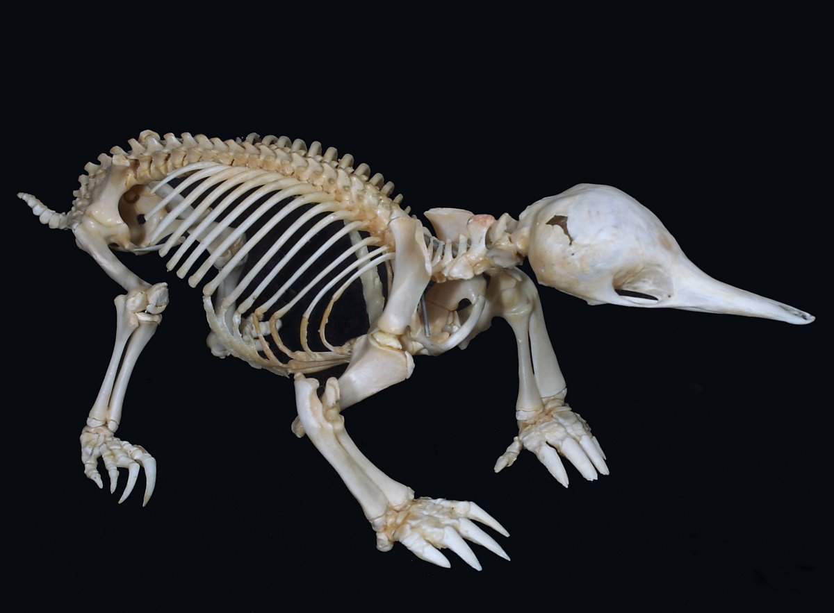
The omovertebral bone is thought to be of scapular origin, as an overgrowth of the epiphysis on the posterior border of the scapula. Amphibians have a very large suprascapular extension that is separated from the scapula by cartilage or a joint. Thornback skates have a distinct bone, the suprascapula, that is fused to the spine and articulates with the scapula. Reptiles and some early mammals (cynodonts and monotremes) have large suprascapular extensions of the scapula that project over the spine and are in close proximity to the spinous processes.


In monotremes, such as platypus (above) and echidna (below), this suprascapular extension may extend across the midline and overlie the other suprascapular extension.


Report of a patient with an omovertebral bone not associated with a Sprengel deformity has cast some doubt on the theory of a scapular origin of the bone. Other theories include a vertebral origin, as an outgrowth of the spinous process; or an origin independent of either the spine or scapula (either as an ossification of the connective tissues between the spine and scapula, or as a rudimentary scapula related to early clefting of the embryo).

References

  • Azouz EM. CT demonstration of omovertebral bone. Pediatr Radiol. 2007 Apr;37(4):404.
  • Williams MS. Developmental anomalies of the scapula-the "omo"st forgotten bone. Am J Med Genet A. 2003 Aug 1;120A(4):583-7.

Sunday, July 10, 2011

Pulmonary Vein Thrombosis

Pulmonary vein thrombosis can be idiopathic or be seen with primary and secondary tumors of the lung or as a rare early complication of lobectomy and lung transplantation. If unrecognized, it can lead to pulmonary gangrene, peripheral embolization, pulmonary hypertension, and pulmonary edema.

Conventional pulmonary angiography may allow a presumptive diagnosis of pulmonary vein thrombosis in the setting of a normal arterial phase is normal by revealing absent venous outflow from the affected lung. Improper image acquisition times can lead to a false positive study in cases of delayed opacification of normal pulmonary veins (for example, as seen with atelectasis, neoplasm, and other causes of shunting of pulmonary blood flow).

CTA and MRA can demonstrate the thrombus. MRI may be able to differentiate bland from tumor thrombus, with the latter demonstrating enhancement and abnormal signal extending through the vessel wall.

The case above is from a young patient with Hodgkin disease, who presented with left-sided pleuritic chest pain a month after sternotomy for mediastinal exploration and biopsy of a mediastinal tumor (pink arrow). CTA performed to rule out pulmonary arterial embolism showed a filling defect in a segmental branch of the right inferior pulmonary vein (white arrows).

References

  • Gyves-Ray KM, Spizarny DL, Gross BH. Unilateral pulmonary edema due to postlobectomy pulmonary vein thrombosis. AJR Am J Roentgenol. 1987 Jun;148(6):1079-80.
  • Selvidge SD, Gavant ML. Idiopathic pulmonary vein thrombosis: detection by CT and MR imaging. AJR Am J Roentgenol. 1999 Jun;172(6):1639-41.

Saturday, July 9, 2011

Bennett Lesion

The Bennett lesion, first described in 1941, refers to extra-articular ossification of the posterior inferior glenoid. The ossification is thought to represent reactive bone formation from traction of the posterior band of the inferior glenohumeral ligament due to the repetitive stresses involved with overhead throwing.

The posterior ossification is associated with a posterior labral injury and posterior articular surface rotator cuff damage.

The Bennett lesion is usually asymptomatic. Throwing motion may be limited by fracture of the ossification, by a fibrous union at its base, or by capsular contracture due to the ossification.

The extra-articular ossification of the Bennett lesion is not well demonstrated on conventional frontal radiographs of the shoulder. A special view, developed by Bennett's x-ray technologist (WR Mitchell) uses a frontal projection with external rotation of the humerus and a 5-degree cephalad tilt of the x-ray tube to rotate the head of the humerus and the glenoid to a position that allows visualization of the ossification (image in Bennett's article below).

CT and MRI demonstrate the crescentic extra-articular ossification extending from the posteroinferior glenoid to the posterior labrum. Reactive sclerosis may also be seen in the posteroinferior glenoid itself. Posterior humeral subluxation can also be present. MRI can reveal labral tears, located in the posterior superior labrum.

References

  • Bennett GE. Shoulder and Elbow Lesions Distinctive of Baseball Players. Ann Surg. 1947 Jul;126(1):107-10.
  • Wright RW, Paletta GA Jr. Prevalence of the Bennett lesion of the shoulder in major league pitchers. Am J Sports Med. 2004 Jan-Feb;32(1):121-4.
  • Ferrari JD, Ferrari DA, Coumas J, Pappas AM. Posterior ossification of the shoulder: the Bennett lesion. Etiology, diagnosis, and treatment. Am J Sports Med. 1994 Mar-Apr;22(2):171-6.

Friday, July 8, 2011

Flexor Carpi Ulnaris Calcium Hydroxyapatite Deposition

The most common site of calcium hydroxyapatite deposition in the wrist is in the flexor carpi ulnaris tendon. Flexor carpi ulnaris calcium hydroxyapatite deposition is also known as peritendinitis calcarea, periarthritis calcarea, calcific tendinitis, calcific bursitis, and calcareous tendinitis and bursitis.

Calcium hydroxyapatite deposits can be found anywhere along the tendon, but most commonly occur near its attachment to the pisiform. Lateral radiographs characteristically reveal amorphous calcification proximal to the pisiform. Localized obliteration of the adjacent soft tissue fascial planes can be seen in the acute phase.

Case courtesy of the New England Baptist Hospital.

References

  • Gandee RW, Harrison RB, Dee PM. Peritendinitis calcarea of flexor carpi ulnaris. AJR Am J Roentgenol. 1979 Dec;133(6):1139-41.
  • Steinbach LS. Calcium pyrophosphate dihydrate and calcium hydroxyapatite crystal deposition diseases: imaging perspectives. Radiol Clin North Am. 2004 Jan;42(1):185-205, vii.

Thursday, July 7, 2011

Dermoid Mesh Sign

The dermoid mesh sign is a specific sign for an ovarian teratoma on ultrasound, appearing as a tangle of entwined linear interfaces that represent hair fibers floating in the cyst.

Special thanks to Dr. Hansel Otero and Tufts Medical Center for the images.

References

Malde HM, Kedar RP, Chadha D, Nayak S. Dermoid mesh: a sonographic sign of ovarian teratoma. AJR Am J Roentgenol. 1992 Dec;159(6):1349-50.

Wednesday, July 6, 2011

Brown Tumor

Brown tumors, also known as osteoclastomas, represent a reparative cellular process with localized accumulations of fibrous tissue and giant cells that can replace bone and even produce osseous expansion. In the setting of hyperparathyroidism, bone resorption by osteoclasts results in holes in the bone that become confluent and filled with hemorrhage, reparative granulation tissue, and active, vascular, proliferating fibrous tissue. The name brown tumor comes from the brownish color of the mass due to its vascularity, hemorrhage, and hemosiderin deposition.

Brown tumors are seen in 3% of patients with hyperparathyroidism and 1.5% of patients with secondary hyperparathyroidism, but due to the higher incidence of secondary hyperparathyroidism, when a brown tumor is encountered it is more likely to be associated with secondary hyperparathyroidism.

Brown tumors of hyperparathyroidism may mimic a true tumor on imaging and histology. Radiographs reveal central, slightly expansile, lightly septated, lucent lesions with distinct geographic margins. Cortical thinning and fracture may be seen; however, cortical destruction should not be present.

CT reveals a lytic lesion with an associated soft-tissue attenuation component Conventional angiography reveals a hypervascular mass, a feature that is seen as intense activity on nuclear medicine bone scan

The appearance of brown tumors on MRI depends on the relative proportion of its tissue components. The lesions may be solid, cystic, or mixed. Solid components are intermediate to low intensity on T1- and T2-weighted images, while the cystic components are hyperintense on T2-weighted images and may have fluid-fluid levels. Contrast administration reveals enhancement of the solid component and septa. Cortical destruction and soft-tissue extension, not readily apparent on radiographs, can be seen in solid lesions with MRI.

Brown tumors involving the spine and cranium tend to be solid, while lesions in the extremities and pelvis tend to be mixed or cystic.

With correction of the underlying metabolic abnormality, rapid healing with sclerosis is typical. Cyst formation may also be seen.

Differential considerations for a solitary brown tumors include: aneurysmal bone cyst, giant cell tumor, solitary bone cyst, and giant cell reparative granuloma.

With mutliple brown tumors, osteolytic metastases, multiple myeloma, Langerhans cell histiocystosis, leukemia, multiple bone cysts, multiple non-ossifying fibromas, and fibrous dysplasia may be considered; however, MRI can narrow the differential, as few lesions are osteolytic on radiographs and T2-hypointense on MRI. Seeking out radiographic findings seen in hyperparathyroidism is also helpful.

The images above show small kidneys (yellow arrows) and a lytic lesion with sclerotic geographic margins in the left femoral head. No cortical destruction is seen. The appearance is nonspecific, but in a patient with hyperparathyroidism, the diagnosis of brown tumor was suggested.

References

  • Chew FS, Huang-Hellinger F. Brown tumor. AJR Am J Roentgenol. 1993 Apr;160(4):752.
  • Hong WS, Sung MS, Chun KA, Kim JY, Park SW, Lee KH, Lim HW, Lim YS, Yoo WJ, Chung MH. Emphasis on the MR imaging findings of brown tumor: a report of five cases. Skeletal Radiol. 2011 Feb;40(2):205-13.

Tuesday, July 5, 2011

Infectious Tenosynovitis

Infectious (septic) tenosynovitis is infection within the closed space of the tendon sheath. It is usually is the result of penetrating trauma (animal or human bites) or extension of infection from adjacent tissues, although disseminated infection may also occur with some organisms (e.g., N gonorrhoeae).

Infectious tenosynovitis most frequently occurs in the tendons and tendon sheaths of the flexor muscles of the digits of the hand. Because tendons in the hand are next to or communicate with structures such as bursae and potential spaces, the spread of infection is a possibility. As an example, consider the radial and ulnar bursae, which are intimately associated with the flexor tendons of the thumb and small finger, respectively, and can become infected when nearby flexor tendons are infected. Because up to 80% of individuals have an anatomic communication between these bursae, infection in one can spread through this bursal communication to the opposite side of the hand (horseshoe abscess).

Failure to promptly treat flexor tendon sheath infection may lead to tendon necrosis and proximal spread. Adhesions and scarring may also result, leading to loss of mobility and contractures.

MRI usually reveals T1-hypointense and T2-hyperintense fluid distending the tendon sheath; however, the signal characteristics of the fluid can vary depending on the presence of debris, gas, or blood. In addition, the tendons lose their normal low signal intensity and become thickened and indistinct; the synovial lining of the tendon sheath thickens and becomes indistinct; and there is surrounding edema. Synovial enhancement is usually intense.

Case courtesy of the New England Baptist Hospital.

References

  • Small LN, Ross JJ. Suppurative tenosynovitis and septic bursitis. Infect Dis Clin North Am. 2005 Dec;19(4):991-1005, xi.
  • Turecki MB, Taljanovic MS, Stubbs AY, Graham AR, Holden DA, Hunter TB, Rogers LF. Imaging of musculoskeletal soft tissue infections. Skeletal Radiol. 2010 Oct;39(10):957-71.

Monday, July 4, 2011

Aneurysmal Bone Cyst

Aneurysmal bone cysts are benign lesions of unknown origin. They can be classified as primary or secondary, with the latter representing aneurysmal cystic change superimposed on a preexisting bone lesion. In ~30% of cases, a preexisting lesion can be identified. More common preexisting lesions include giant cell tumor (30% of cases), osteoblastoma, angioma, and chondroblastoma. Uncommonly, the precursor lesion is fibrous dysplasia, fibrous histiocytoma, Langerhans cell histiocytosis, osteosarcoma, and even metastasis.

The age distribution of secondary aneurysmal bone cysts is determined by that of the underlying tumor. Primary aneurysmal bone cysts usually occur in patients younger than 20 years of age and tend to occur in the metaphyses of the long bones of the lower limbs. Epiphyseal extension can occur after fusion of the growth plate.

Flat bones of the pelvis and the spine can also be affected. In the spine, both the vertebral body and posterior process are involved in the majority of cases. More than one vertebra may be involved in about 40% of cases, and thoracic spine lesions can be accompanied by rib lesions. Surface aneurysmal bone cysts (those arising from subperiosteal and cortical regions) do not tend to affect the flat bones.

Capanna et al classified aneurysmal bone cysts in the long bones based on their location.
  • Type 1: Central with little expansion.
  • Type 2: Central with expansion and cortical thinning.
  • Type 3: Eccentric with involvement of only one cortex.
  • Type 4: Subperiosteal with outward growth and little or no superficial cortical erosion. Usually occur in the diaphysis.
  • Type 5: Subperiosteal with both inward and outward growth and cortical destruction. Usually occur in the diaphysis.

Etiology

Various etiologies have been suggested for aneurysmal bone cysts, including 1) reaction to a vascular phenomenon (e.g., arteriovenous malformation, thrombosis of a large vein leading to intramedullary venous engorgement), 2) neoplasm (USP6 and CDH11 oncogenes in primary aneurysmal bone cysts), and 3) trauma (especially surface aneurysmal bone cysts).

Imaging

On radiographs, we usually see a geographic lesion with well-defined borders and a narrow zone of transition, although the radiographic appearance varies with the phase of the lesion (see below). The margin can be sharp but not sclerotic in 55% of cases, sclerotic in 30% of cases, and indistinct in 15%.
  • Initial phase: Osteolysis and periosteal elevation.
  • Active growth phase: Rapid enlargement and bone destruction. There may be poor demarcation of the lesion with an invisible external border and other aggressive features (Codman triangle, lamellated periosteal reaction). Most patients are symptomatic.
  • Stabilization phase: Thin shell of periosteal bone, septal ossification, and mature periosteal reaction (cortical/periosteal buttress at the interface with normal bone). This is the classic appearance of aneurysmal bone cysts.
  • Healing phase: Gradual ossification of the aneurysmal bone cyst. Matrix mineralization and trabeculation may be seen. May be the same thing as the solid variant of aneurysmal bone cysts (see below).
CT reveals a lesion with a thin surrounding shell of bone. A thin shell of soft tissue attenuation, representing the fibrous periosteum, can also be seen. An extraosseous mass is rare. About 1/3 of lesions will have fluid-fluid levels on CT, with the higher attenuation material layering dependently.

MRI reveals fluid-fluid levels, septae, a low-signal intensity rim of periosteum, surrounding edema, and peirpheral and septal enhancement.

Primary aneurysmal bone cysts have thin septae and minimal or no solid component. Secondary aneurysmal bone cysts tend to have nodular septae and a larger solid component. In a study of 83 lesions with fluid-fluid levels, when cystic spaces (fluid-fluid levels) occupied less than 1/3 of the lesion, more than 2/3 of the lesions were malignant, and half of these malignant lesions were osteosarcomas. If more than 2/3 of the lesion contained cystic spaces (fluid-fluid levels), 90% of the lesions were benign. If the entirety of the lesion contained cystic spaces (fluid-fluid levels), 100% of the lesions were benign. Biopsy should target the solid component

More recently, it has been suggested that T1 signal higher than skeletal muscle in the antidependent layer may point to malignancy, with the thought that the high T1 signal represents recent hemorrhage and liquefied, necrotic tumor, compared to serous fluid and old blood seen in benign lesions.

Solid Variant

Also known as an extragnathic giant cell reparative granuloma, solid aneurysmal bone cysts are uncommon lesions that are histologically identical to the solid components of aneurysmal bone cysts without the blood-filled cavities.

The affected age group is similar to regular aneurysmal bone cysts, but the distribution is slightly different, with metaphyseal and diaphyseal locations being more equal in incidence. Histologically, the lesions are characterized by intraosseous hemorrhage and may represent the healing stage of a conventional aneurysmal bone cyst.

On MRI, the lesions are predominantly solid with high signal on T1- and T2-weighted images and surrounding edema, which may be striking.

References

  • Capanna R, Bettelli G, Biagini R, Ruggieri P, Bertoni F, Campanacci M. Aneurysmal cysts of long bones. Ital J Orthop Traumatol. 1985 Dec;11(4):409-17.
  • O'Donnell PG. Chapter 24: Cystic bone lesions. in Imaging of Bone Tumors and Tumor-Like Lesions. Davies AM, Sundaram M, and James SLJ (eds). Springer-Verlag Berlin Heidelberg (2009); pp 430-438.
  • O'Donnell P, Saifuddin A. The prevalence and diagnostic significance of fluid-fluid levels in focal lesions of bone. Skeletal Radiol. 2004 Jun;33(6):330-6.
  • Parman LM, Murphey MD. Alphabet soup: cystic lesions of bone. Semin Musculoskelet Radiol. 2000;4(1):89-101.
  • Rodallec MH, Feydy A, Larousserie F, Anract P, Campagna R, Babinet A, Zins M, Drapé JL. Diagnostic imaging of solitary tumors of the spine: what to do and say. Radiographics. 2008 Jul-Aug;28(4):1019-41.

Sunday, July 3, 2011

Supracondylar Process of the Humerus

The supracondylar process of the humerus is a bony projection from the anteromedial aspect of the distal humeral diaphysis that can be shaped as a spine or a tubercle. It is typically 5 cm - 7 cm proximal to the medial epicondyle and is often connected to the medial epicondyle by a fibrous band (ligament of Struthers). Seen in about 1% of the population, the supracondylar process is more commonly seen in Caucasians, in males, and on the left.

Synonyms include: supracondylar spur, supracondyloid process, supratrochlear process, epicondylar process, epicondylic process, and avian spur.

In some cases, a fibrous band called the ligament of Struthers arises from the supracondylar process and attaches to the medial epicondyle, forming a foramen through which the median nerve and brachial artery may pass. A case of the neurovascular bundle passing superficial to the ligament of Struthers has also been reported.

The tubercle shape of the supracondylar process is more likely to be associated with the ligament of Struthers than the spine shape. The supracondylar process is also associated with a high origin of the pronator teres muscle, which may have an attachment to the ligament of Struthers or the process itself. Some of the lower fibers of the coracobrachialis muscle may also arise from the supracondylar process or ligament of Struthers.

While usually asymptomatic, patients may present with symptoms related to a fracture of the process or compression of the median nerve and brachial artery by the foramen formed by the ligament of Struthers. Less commonly, there may be compression of a variant ulnar artery and rarely compression of the ulnar nerve. Symptoms tend to increase with pronation of the forearm.

Radiographs reveal a bony projection arising from the anteromedial margin of the distal humerus that is best demonstrated on an oblique, internally rotated projection of the humerus. Unlike aosteochondromas, which are oriented away from the joint, the supracondylar process is directed toward the joint.

The process seems to be a homologue of a bony arch found in the same location on the humeri of cats, certain monkeys, and some other animals. The ligament of Struthers may be a remnant of the tendon of the latissimo-condyloideus, a muscle that extends from the humeral attachment of the latissimus dorsi to the medial epicondyle in certain climbing animals. I couldn't find a reference describing why the process is referred to as an avian spur.

References

  • Ay S, Bektas U, Yilmaz C, Diren B. An unusual supracondylar process syndrome. J Hand Surg Am. 2002 Sep;27(5):913-5.
  • Gunther SF, DiPasquale D, Martin R. Struthers' ligament and associated median nerve variations in a cadaveric specimen. Yale J Biol Med. 1993 May-Jun;66(3):203-8.
  • Jelev L, Georgiev GP. Unusual high-origin of the pronator teres muscle from a Struthers' ligament coexisting with a variation of the musculocutaneous nerve. Rom J Morphol Embryol. 2009;50(3):497-9.
  • Kessel L, Rang M. Supracondylar spur of the humerus. J Bone Joint Surg Br. 1966 Nov;48(4):765-9.
  • Natsis K. Supracondylar process of the humerus: study on 375 Caucasian subjects in Cologne, Germany. Clin Anat. 2008 Mar;21(2):138-41.
  • Sener E, Takka S, Cila E. Supracondylar process syndrome. Arch Orthop Trauma Surg. 1998;117(6-7):418-9.
  • Yazar F, Acar HI. Supracondylar process with a high origin of the radial artery. Clin Anat. 2006 Nov;19(8):730-1.

Saturday, July 2, 2011

Pirifomis Muscle Syndrome

Piriformis muscle syndrome (also piriformis syndrome) is a controversial entity, which, in the absence of reproducible and reliable diagnostic criteria, remains a diagnosis of exclusion.

It is thought to be due to compression of the sciatic nerve through hypertrophy, spasm, contracture, or inflammation and scarring of the piriformis muscle. While the initiating insult is usually unknown, trauma (with resultant muscle injury, hematoma, and scarring) or acute or chronic piriformis muscle stretching associated with gait disturbances can serve as the initial insult leading to the common endpoint of piriformis muscle syndrome.

Imaging can be helpful by delineating the relationship between the sciatic nerve and the piriformis muscle. The diagnosis of piriformis muscle syndrome can be inferred from alterations in the appearance of the sciatic nerve, with sciatic nerve edema being associated with symptoms of piriformis muscle syndrome in ~90% of patients.

Muscle asymmetry, on the other hand, appears to be less useful. Muscle anomalies and significant variations in size of the piriformis muscle in asymptomatic individuals limit the role of imaging in assessing piriformis muscle abnormalities. One study found that muscle asymmetry by itself had a specificity of ~65% and sensitivity of ~45% in identification of patients with piriformis muscle syndrome who responded well to piriformis surgery (that is, they had muscle-based piriformis muscle syndrome).

Use of both piriformis muscle asymmetry (hypertrophy or atrophy) and sciatic nerve edema can lead to ~90% specificity and ~65% sensitivity in predicting response to piriformis surgery.

References

Petchprapa CN, Rosenberg ZS, Sconfienza LM, Cavalcanti CF, Vieira RL, Zember JS. MR imaging of entrapment neuropathies of the lower extremity. Part 1. The pelvis and hip. Radiographics. 2010 Jul-Aug;30(4):983-1000.

Friday, July 1, 2011

Calcaneal Intraosseous Lipoma

Intraosseous or medullary lipomas are rare benign neoplasms of bone that are most commonly seen in the metaphyses of long bones (e.g., proximal femur and the fibula) and in the calcaneus. They are histologically characterized by mature lipocytes in a background of fibroblasts with occasional foci of fat necrosis.

As many as 2/3 of patients with intraosseous lipomas may be symptomatic, presenting with pain and/or soft tissue swelling. These lesions frequently involute spontaneously through infarction, calcification, and cyst and reactive bone formation.

Radiographs reveal a lucent lesion surrounded by a thin, well-defined sclerotic margin. Lobulations and thin septa may also be present. Intraosseous lipomas tend to be expansile when located in smaller bones such as the fibula. While an aggressive appearance is rarely encountered, these lesions are typically benign in appearance without cortical destruction or periosteal reaction.

While this radiographic appearance is usually nonspecific, intraosseous lipomas in the calcaneous have a characteristic appearance.

In the calcaneus, the lesion is almost invariably located in the tangle between major trabecular groups in the same location as simple cysts. A central calcified nidus is typically seen with intraosseous lipomas, but not in simple cysts. Lesions without central calcifications can be further evaluated with CT or MRI to demonstrate internal fat content.

CT can also reveal areas of higher attenuation corresponding to hemorrhage and cyst formation. MRI reveals areas of low T1 and high T2 signal intensity suggestive of necrosis and cyst formation. Areas of T1 hyperintensity and T2 hypointensity may also be seen, corresponding to foci of hemorrhage.

Case courtesy of Dr. Damon J. Spitz and New England Baptist Hospital.

References