- Subarachnoid hemorrhage: May have enlarged ventricles and intraventricular blood.
- Cellular material: Pus, tuberculosis, sarcoid, cancer. May have enlarged ventricles.
- Intracranial hypotension: Dilated dural sinuses, normal-sized ventricles.
- Global anoxia: Diffuse loss of gray-white differentiation, white cerebellum sign, normal-sized ventricles.
- Contrast: From myelogram or intravenous contrast leakage in patients with renal failure.
Monday, February 28, 2011
Diffusely Increased Attenuation of the Subarachnoid Space
Sunday, February 27, 2011
Anomalous High Origin of the Right Coronary Artery
 The ostium of the right coronary artery is normally located on the right aortic sinus. A rare abnormality is a high origin above the sinotubular junction (arrows). This anomaly has been reported in association with ventricular septal defect and patent ductus arteriosus (not present in our patient).
If the abnormal artery travels between aorta and right ventricular outflow tract, there is a risk of acute myocardial infarction or sudden death. It is also important to note this finding to avoid inadvertent ligation during surgery. Anomalous high origin of the right coronary artery may also be associated with unsuccessful induction of cardiac arrest by cardioplegia.
References
- Alpaslan M, Onrat E. Anomalous origin of right coronary artery above the sinus of Valsalva: observation by transthoracic echocardiography. J Am Soc Echocardiogr. 2002 Mar;15(3):264-6.
- Tarhan A, Kehlibar T, Yilmaz M, Arslan Y, Pancaroglu C, Yigit S, Ozler A. Right coronary artery with high takeoff. Ann Thorac Surg. 2007 May;83(5):1867-9.
Saturday, February 26, 2011
Paplpable Solid Mass in a Pregnant or Lactating Woman
- Fibroadenoma: May grow rapidly during pregnancy
- Lactating adenoma: Palplable, mobile, and with rapid growth.
- Tubular adenoma: Rare benign tumor found in young women. May enlarge during pregnancy and lactation
- Focal mastitis:
- Galactocele: Appears during lactation or within a few months after cessation of lactation. Mammographic and ultrasound appearance vary depending on the amount of fat.
- Lobular hyperplasia: Normal breast tissue with lactational change
- Cancer:
Friday, February 25, 2011
Hepatic Lesions in Infants

- Infantile hemangioma: Early peripheral enhancement with nodular puddling of contrast on later phases.
- Hepatoblastoma (shown above): Heterogeneous echogenicity with or without a spoke-wheel appearance (related to fibrous septa). Acoustic shadowing may be seen from calcifications. Typically hypervascular on Doppler. On CT, heterogeneous, predominantly low-attenuation, with or without calcifications. Heterogeneous enhancement, less than normal liver parenchyma. Low T1 signal with areas of high signal related to hemorrhage. High T2 signal with areas of hemorrhage and necrosis. Markedly elevated AFP.
- Mesenchymal hamartoma of the liver: Usually a multicystic, multilocular mass with enhancement of only the septa and solid portions. Solid lesions are less common and are hypovascular at dynamic contrast-enhanced imaging.
- Metastatic neuroblastoma: Associated with elevated levels of urinary catecholamines. Enhance less than the adjacent liver.
References
Chung EM, Cube R, Lewis RB, Conran RM. From the archives of the AFIP: Pediatric liver masses: radiologic-pathologic correlation part 1. Benign tumors. Radiographics. 2010 May;30(3):801-26.Thursday, February 24, 2011
Corkscrews in Radiology
 Some of the more famous corkscrews in radiology include:
- Corkscrew esophagus (shown above): Seen in diffuse esophageal spasm.
- Corkscrew proximal small bowel (shown below): Appearance of the duodenum and jejunum in midgut volvulus.
- Corkscrew hepatic arteries: Due to increased arterial flow and decreased liver size in cirrhosis.
- Corkscrew collaterals: Represent collaterals in the vasa vasorum of occluded arteries in Buerger Disease. Seen in the extremities.
- Corkscrew elongation of the ulnar artery: Seen in hypothenar hammer syndrome. Refers to alternating areas of stenosis and ectasia of the ulnar artery (image from the AJR reference).

References
- Hammond DC, Matloub HS, Yousif NJ, Sanger JR. The corkscrew sign in hypothenar hammer syndrome. J Hand Surg Br. 1993 Dec;18(6):767-9.
- Molvar CA, Funaki BS. AJR teaching file: Carpenter with cold, numb fingers. AJR Am J Roentgenol. 2009 Sep;193(3 Suppl):S46-8.
Wednesday, February 23, 2011
Epidermal Inclusion Cyst
 Epidermal inclusion cysts are common lesions of the skin, but relatively uncommon in the breast. They are filled with lamellated keratin formed through inclusion of keratinizing squamous epithelium within the dermis. The typical appearance is a smooth, round nodule attached to the skin.
On mammography, they appear as circumscribed lesions and may have calcifictions. On ultrasound, they are solid, circumscribed, and complex with extension into the dermis (seen in the grayscale image of patient A). An onion-ring appearance with alternating concentric hyperechoic and hypoechoic rings corresponding to multiple layers of keratin has also been described.
Malignant transformation is rare, but excision is recommended by some.
References
Crystal P, Shaco-Levy R. Concentric rings within a breast mass on sonography: lamellated keratin in an epidermal inclusion cyst. AJR Am J Roentgenol. 2005 Mar;184(3 Suppl):S47-8.Tuesday, February 22, 2011
Celery Stalk Metaphysis
Celery stalk metaphysis, also known as celery stalking, refers to irregular and frayed metaaphyseal margins with bands of radiolucency in the long bones. This appearance can be seen with congenital rubella, cytomegalovirus, toxoplasma, or syphilis infection.
To confuse matters, there is a celery stalk appearance in osteopathia striata, a rare asymptomatic condition or unknown heredity. Dense linear striations extend from the metaphyses into the diaphyses of tubular bones (the celery) or in a fanlike or in a "sunburst" pattern in flat bones such as the ilium. Usually bilateral.
Finally, these should not be confused with the celery stalk sign of the anterior cruciate ligament, discussed earlier.
References
- Pediatric radiology: the requisites By Johan G. Blickman, Bruce R. Parker, Patrick D. Barnes
- Roche CJ, O'Keeffe DP, Lee WK, Duddalwar VA, Torreggiani WC, Curtis JM. Selections from the buffet of food signs in radiology. Radiographics. 2002 Nov-Dec;22(6):1369-84.
Monday, February 21, 2011
Punctate or Stippled Epiphyses
 Punctate epiphyses contain small epiphyseal calcifications, are present at birth, and were classically associated with chondrodysplasia punctata. A variety of conditions can present with punctate epiphyses, however. Some of these include:
- Chondrodysplasia punctata: Multiple variants of chondrodysplasia punctata exist. A patient with Conradi–Hünermann syndrome is shown above.
- Vitamin K disorders: Warfarin embryopathy, Vitamin K reductase deficiency. Exposure to warfarin between the 6th and 9th weeks of gestation. The distal phalanges are very short and often shaped like an inverted triangle.
- Zellweger syndrome: Also known as cerebro-hepatorenal syndrome. Peroxisomal disorder. Severely hypotonic infants with a high forehead, hypertelorism, epicanthic folds, and shallow supraorbital ridges. May have Brushfield spots. Club foot deformity is common. Puncta occur primarily in the patella. Cortical cystic disease in the kidneys. Migrational disorders in the brain.
- Trisomies: 21 and 18
- Drug exposure: Warfarin (see above), alcohol, hydantoin, phenacetin.
References
- Poznanski AK. Punctate epiphyses: a radiological sign not a disease. Pediatr Radiol. 1994;24(6):418-24, 436.
- Savarirayan R. Common phenotype and etiology in warfarin embryopathy and X-linked chondrodysplasia punctata (CDPX). Pediatr Radiol. 1999 May;29(5):322.
Sunday, February 20, 2011
Important MRI Findings for Gynecological Cancer
| Cancer | Finding | Reason |
| Cervical | Parametrial extension | Stage IIB: Nonsurgical. Radiation ± chemotherapy |
| Cervical | Pelvic side wall invasion | Stage IIIB: Poorer prognosis |
| Endometrial | Deep (>50%) myometrial invasion | Stage IB*: Increased (~50%) risk of nodal metastases. Will need radical lymph node resection. |
| Endometrial | Cervical stromal invasion | Stage II*: Requires preoperative radiation therapy or a different surgical plan (i.e., radical hysterectomy instead of total abdominal hysterectomy) |
References
- Beddy P, O'Neill AC, Yamamoto AK, Addley HC, Reinhold C, Sala E. FIGO Staging System for Endometrial Cancer: Added Benefits of MR Imaging. Radiographics. 2012 Jan;32(1):241-54.
- Sala E, Wakely S, Senior E, Lomas D. MRI of malignant neoplasms of the uterine corpus and cervix. AJR Am J Roentgenol. 2007 Jun;188(6):1577-87.
Saturday, February 19, 2011
Clumped Enhancement on Breast MRI
 Clumped enhancement is a type of nonmasslike enhancement in which there is an aggregate of enhancing masses or foci that may be confluent in a cobblestone pattern. Clumped enhancement is suspicious for ductal carcinoma in situ (DCIS) and biopsy is generally recommended.
While the most commonly reported MR manifestation of DCIS is clumped nonmasslike enhancement in a ductal, linear, segmental, or regional distribution, other patterns have also been observed. These include a linear-ductal pattern of enhancement; segmental, focal, or regional area of enhancement; diffuse enhancement; and an enhancing mass.
High-grade DCIS more commonly manifests as an enhancing mass than do intermediate- or low-grade DCIS.
References
- Liberman L, Morris EA, Dershaw DD, Abramson AF, Tan LK. Ductal enhancement on MR imaging of the breast. AJR Am J Roentgenol. 2003 Aug;181(2):519-25.
- Mossa-Basha M, Fundaro GM, Shah BA, Ali S, Pantelic MV. Ductal carcinoma in situ of the breast: MR imaging findings with histopathologic correlation. Radiographics. 2010 Oct;30(6):1673-87.
Friday, February 18, 2011
Adenomatoid Tumor of the Epididymis
Adenomatoid tumors are the most common tumors of the epididymis (although rare overall) and have a predilection for the lower pole of the epididymis. They are usually unilateral (more often on the left) and most commonly affect men older than 20 years of age. 30% of patients present with pain.
Ultrasound findings are nonspecific and variable. While most are isoechoic relative to the adjacent normal epididymis, predominantly cystic adenomatoid tumors may also be seen. In contrast to the usually round testicular adenomatoid tumors, adenomatoid tumors of the epididymis are usually oval.
Differential considerations include subclinical chronic infection and sperm cell granuloma post vasectomy.
References
Akbar SA, Sayyed TA, Jafri SZ, Hasteh F, Neill JS. Multimodality imaging of paratesticular neoplasms and their rare mimics. Radiographics. 2003 Nov-Dec;23(6):1461-76.Thursday, February 17, 2011
Testicular Rupture
 Testicular rupture is defined by interruption of the tunica albuginea, with hemorrhage and extrusion of testicular contents into the scrotal sac. The finding of testicular rupture necessitates emergent surgery, as more than 80% can be salvaged if surgery is performed within 72 hours of testicular injury.
US demonstrate a heterogeneous echotexture of the testis, testicular contour abnormality, and disruption of the tunica albuginea. The normal tunica albuginea appears on ultrasound as two parallel hyperechoic lines outlining the testis. In the case shown above, the tunica albuginea is relatively intact in the upper part of the testis, but is lost in the lower part.
Focal hyperechoic or hypoechoic areas in the testicular parenchyma correspond to areas of hemorrhage or infarction. Scrotal wall thickening and hematoceles may also be seen. Color and duplex Doppler images may show decreased or no flow.
References
- Bhatt S, Dogra VS. Role of US in testicular and scrotal trauma. Radiographics. 2008 Oct;28(6):1617-29.
- Deurdulian C, Mittelstaedt CA, Chong WK, Fielding JR. US of acute scrotal trauma: optimal technique, imaging findings, and management. Radiographics. 2007 Mar-Apr;27(2):357-69.
Wednesday, February 16, 2011
Short 4th Metacarpal
Differential considerations for a short 4th metacarpal include:
- Trauma:
- Normal variant:
- Turner syndrome:
- Pseudohypoparathyroidism:
- Pseudopseudohypoparathyroidism :
- Acrodysostosis: Rare disorder characterized by peripheral dysostosis, nasal hypoplasia, and mental retardation.
- Multiple epiphyseal dysplasia: Hypoplasia of the epiphyseal cartilage of tubular bones with essentially normal vertebrae. Epiphyseal ossification alterations are bilateral, symmetrical, and more marked in the lower extremities. Mild short stature. Early in childhood, there are delayed epiphyseal growth centers, which eventually ossify with an irregular fragmented appearance. Older children will have coxa vara with and increased incidence of slipped capital femoral epiphysis. Young adults and adults have premature osteoarthritic changes, particularly in the hips and knees. A double-layered patella is highly diagnostic for the recessive forms of multiple epiphyseal dysplasia.
References
- Ablow RC, Hsia YE, Brandt IK. Acrodysostosis coinciding with pseudohypoparathyroidism and pseudo-pseudohypoparathyroidism. AJR Am J Roentgenol. 1977 Jan;128(1):95-9.
- Rubenstein JD, Christakis MS. Case 95: fracture of double-layered patella in multiple epiphyseal dysplasia. Radiology. 2006 Jun;239(3):911-3.
Tuesday, February 15, 2011
Midaortic Dysplastic Syndrome
Midaortic dysplastic syndrome refers to hypertension and weakness or absence of the femoral pulses due to diffuse narrowing of the mid thoracoabdominal aorta. There is often involvement of renal and mesenteric arteries.
Midaorticdysplastic syndrome may have an identical appearance to late-phase type II Takayasu arteritis. The two can be differentiated by biopsy by noting inflammatory change in patients with Takayasu arteritis but not in midaortic dysplastic syndrome.
Collateral parietal arteries connect the thoracic aorta and the abdominal aorta, most commonlyfrom the subclavian artery to the internal mammary artery to the superior epigastric artery to the inferior epigastric artery to the external iliac artery.
Patients may be treated with aortic reconstruction using prosthetic or autologous venous grafts.
References
Sebastià C, Quiroga S, Boyé R, Perez-Lafuente M, Castellà E, Alvarez-Castells A. Aortic stenosis: spectrum of diseases depicted at multisection CT. Radiographics. 2003 Oct;23 Spec No:S79-91Monday, February 14, 2011
Renal Masses in Children
| Age < 1 year | Age > 1 year |
| Hydronephrosis | Hydronephrosis |
| Cystic renal disease | Cystic renal disease |
| Mesoblastic nephroma | Wilms tumor |
| Renal vein thrombosis | Hematoma |
| Multilocular cystic nephroma: Benign, but removed since appearance can be mimicked by Wilms tumor | |
| Lymphoma: Hypoechoic and hypoattenuating | |
| Rhabdoid tumor: More prone to have brain and bone metastases |
Sunday, February 13, 2011
Unilateral Dense Pedicle

- Spondylolysis: Stress sclerosis on the contralateral side results in a dense pedicle. The case presented above demonstrates a dense left L5 pedicle (white arrow) and an old right L5 pars fracture (pink arrow).
- Benign tumors: Osteoid osteoma and osteoblastoma may cause a painful rigid scoliosis, with the lesion found on the concave side of the curve. Bone island uncommonly affects the pedicles.
- Malignant tumors: Blastic metastases (most common malignant cause of a unilateral dense pedicle). Other malignant tumors rarely present with a unilateral dense pedicle (e.g., lymphoma, multiple myeloma, Ewing sarcoma).
- Iatrogenic: Laminectomy (most commonly contralateral to the dense pedicle)
- Infection: Rarely affects the pedicle in isolation.
- Agenetic or hypoplastic pedicle or facet: The dense pedicle in on the opposite side to the agenetic or hypoplastic pedicle or facet.
- Miscellaneous: Tuberous sclerosis has an affinity for the posterior elements when it occurs in the spine, with multiple sclerotic pedicles being a frequent finding. Rarely Paget disease, fibrous dysplasia, and sarcoidosis, may affect the pedicle in isolation.
References
Yochum TR, Sellers LT, Oppenheimer DA, Peterson CK, Kirton CW, Dal Mas EC, Anderson AL. The sclerotic pedicle--how many causes are there? Skeletal Radiol. 1990;19(6):411-7.Saturday, February 12, 2011
Absent Urinary Bladder on Prenatal Ultrasound
An absent urinary bladder on prenatal ultrasound may be due to a transiently empty bladder or bilateral renal issues, including:
- Multicystic dysplastic kidney: Cysts of variable size and shape, nonmedial location of large cysts, absence of an identifiable renal sinus, lack of communication between cysts, and minimal surrounding parenchyma.
- Autosomal recessive polycystic kidney disease: Enlarged, echogenic kidneys.
- Renal agenesis: Self explanatory.
Friday, February 11, 2011
Maisonneuve Fracture of the Fibula
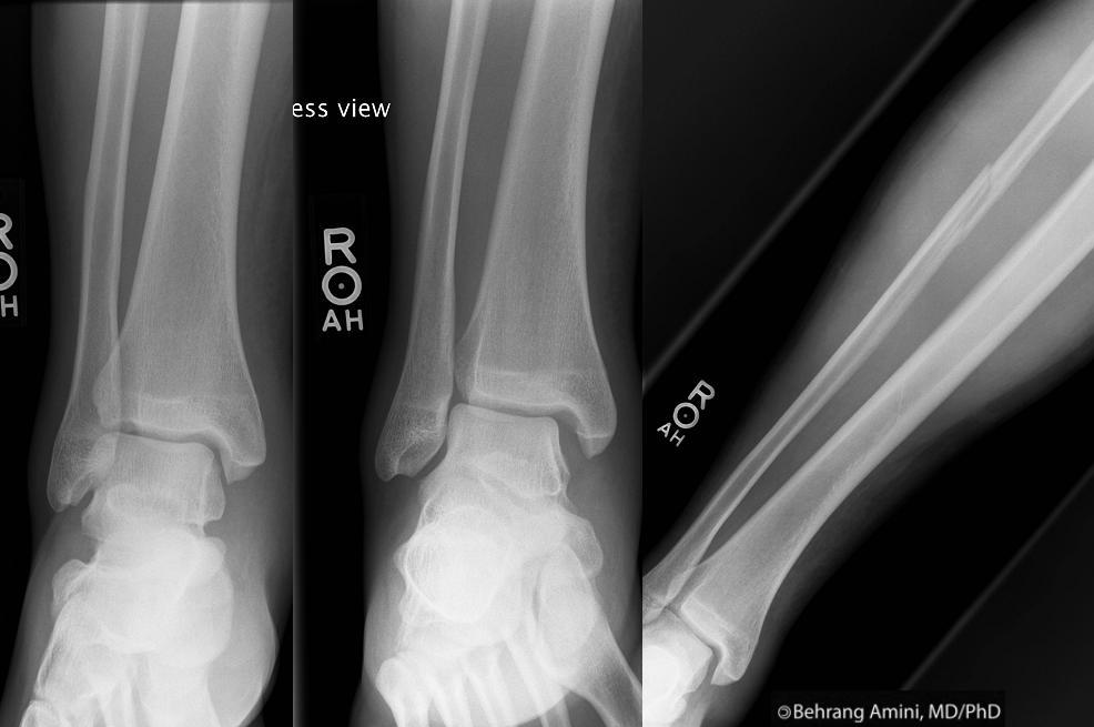 A Maisonneuve fracture is a spiral proximal fibular fracture associated with an ankle joint injury and represents a pronation-external rotation injury that results a medial injury (medial malleolar avulsion or deltoid ligament tear), a posterior injury (posterior malleolar fracture or posterior tibiofibular ligament tear), and a tear of the anterior tibiofibular ligament and interosseous membrane to the level of the fibular fracture.
A Maisonneuve fracture should be suspected whenever there is lateral talar displacement or tibiofibular widening without a distal fibular fracture or in the case of an "isolated" posterior malleolar fracture.
The patient in the above case has some soft tissue swelling medially in the first panel. Stress views (medial panel) show widening of the medial clear space (i.e., lateral talar dsiplacement). A frontal radiograph of the tibia and fibula reveals a fracture of the proximal-to-mid fibula.
References
Hanson JA, Fotoohi M, Wilson AJ. Maisonneuve fracture of the fibula: implications for imaging ankle injury. AJR Am J Roentgenol. 1999 Sep;173(3):702.Thursday, February 10, 2011
Mild Ventriculomegaly on Prenatal Ultrasound
Mild ventriculomegaly (atrial width between 10 mm to 20 mm) can be seen in association with the following conditions and should prompt close evaluation of the posterior fossa.
- Chiari II malformation: Important to recognize due to the high association with open neural tube defects. Inspection of the posterior fossa will reveal a small posterior fossa with effacement of the cisterna magna and a banana-shaped cerebellum. The lemon sign (flattened or inwardly scalloped frontal bones) is a less specific finding.
- Dandy-Walker: Hypogenic cerebellar vermis and communication of the cisterna magna with the 4th ventricle.
- Agenesis of the corpus callosum: Look for tear-drop shaped lateral ventricles.
- Infection:
- Chromosomal abnormalities:
- Isolated ventriculomegaly:
Wednesday, February 9, 2011
Peroneocalcaneus Internus Muscle
 The peroneocalcaneus internus muscle (arrow) originates along the inner aspect of the inferior fibula, below the origin of the flexor hallucis longus origin. The peroneocalcaneus internus muscle descends posterior and lateral to the flexor hallucis longus muscle and displaces it anteriorly and medially. Its tendon inserts onto a small tubercle on the medial aspect of the calcaneus below the sustentaculum tali.
Most patients with a peroneocalcaneus internus muscle are asymptomatic; however, there may be encroachment on the neurovascular bundle in the tarsal tunnel in some.
References
- Mellado JM, Rosenberg ZS, Beltran J, Colon E. The peroneocalcaneus internus muscle: MR imaging features. AJR Am J Roentgenol. 1997 Aug;169(2):585-8.
- Sookur PA, Naraghi AM, Bleakney RR, Jalan R, Chan O, White LM. Accessory muscles: anatomy, symptoms, and radiologic evaluation. Radiographics. 2008 Mar-Apr;28(2):481-99.
Tuesday, February 8, 2011
Thoracic Mass on Fetal Ultrasound

- Congenital pulmonary airways malformation (CPAM): Most common of the congenital thoracic masses. Imaging appearance depends on the nature of the cysts. Fetal ultrasound of large-cyst CPAMs have variable-sized anechoic lesions intertwined with echogenic soft tissue. At fetal MR, there are T2-hyperintense unilocular or multilocular lesions with discrete walls. Small-cyst CPAMs are echogenic with multiple small cysts and have a variable appearance on T2-weighted images depending on the cystic and solid components. Microcystic CPAMs are homogeneously echogenic and are homogeneously hyperintense solid masses on T2-weighted images.
- Bronchopulmonary sequestration: Extralobar form presents as a homogeneous hyperechoic mass in a paraspinal location, usually in the left lower thorax, mimicking a microcystic CPAM. A feeding artery originating from the descending aorta at color Doppler helps distinguish the two. On MR, extralobar sequestration presents as a solid, well-defined, uniformly hyperintense mass on T2-weighted images. Intralobar sequestration is usually diagnosed in childhood or adulthood.
- Hybrid lesion: Combination of CPAM with bronchopulmonary sequestration. The image above demonstrates a T2-hyperintense lesion in the right hemithorax. Pathology showed combined features of intralobar sequestration and congenital pulmonary adenomatoid malformation.
- Congenital lobar emphysema: Collapsed airway acts as a one-way valve, resulting in air trapping and expanded alveoli with intact walls. The intact alveolar walls make the term emphysema technically inaccurate, and some people refer to this condition as congenital lobar overinflation. Left upper lobe is involved in about 50% of cases. Fetal ultrasound may show a homogeneously hyperechoic mass that is homogeneously high signal on T2-weighted images.
- Congenital diaphragmatic hernia: Usually left sided. Look for stomach and bowel next to the heart.
- Congenital high airway obstruction syndrome: Rare. Caused by laryngeal or tracheal atresia, tracheal stenosis or web, or extrinsic compression (e.g., double aortic arch). Results in outflow obstruction of the fetal lung fluid and pulmonary hyperplasia. Prenatal US shows symmetrically enlarged echogenic lungs, dilated and fluid-filled trachea and bronchi, and inverted hemidiaphragms. The large lungs compress and anteriorly displace the heart. Fetal MRI shows enlarged, T2-hyperintense lungs with flattened or inverted hemidiaphragms.
- Bronchial atresia: Rare. Focal obliteration of a segmental, subsegmental, or lobar bronchus with dilated and mucus-filled bronchi distal to the stenosis. There will be an echogenic lesion that is homogeneously high signal on T2-weighted images.
- Bronchogenic cyst: Foregut duplication cysts. Usually in the mediastinum near the carina, but may occur within the lung parenchyma, pleura, or diaphragm.
- Cystic teratoma:
References
Biyyam DR, Chapman T, Ferguson MR, Deutsch G, Dighe MK. Congenital lung abnormalities: embryologic features, prenatal diagnosis, and postnatal radiologic-pathologic correlation. Radiographics. 2010 Oct;30(6):1721-38.Monday, February 7, 2011
Pseudodefect of the Capitulum
 An abrupt contour change can be observed at the posterolateral margin of the capitulum at the junction of anterior capitulum and posterior lateral epicondyle. This contour change can be mistaken for an osteochondral lesion and has been termed the pseudodefect of the capitulum/capitellum.
The pseudodefect is most obvious on coronal images through the posterior aspect of the capitulum and on sagittal images on more lateral images.
The pseudodefect can be differentiated from an osteochondral lesion by noting that the latter occurs along the normally smooth convex surface of the anterior aspect of the capitulum and is associated with adjacent bone marrow edema. Increased bone marrow signal on T2-weighted images, however, can be simulated by filling of the pseudodefect with joint fluid or intraarticular contrast, as seen in this case.
References
- Chung CB. Chapter 12. In Chung CB and Steinbach LS. MRI of the Upper Extremity: Shoulder, Elbow, Wrist, and Hand. Lippincott Williams & Wilkins. 2010. p 462.
- Mulligan M. Medical terminology. AJR Am J Roentgenol. 2006 May;186(5):E10; discussion E10.
- Rosenberg ZS, Beltran J, Cheung YY. Pseudodefect of the capitellum: potential MR imaging pitfall. Radiology. 1994 Jun;191(3):821-3.
Sunday, February 6, 2011
Cavities, Coccidioidomycosis, and Food
Cavity formation is a common manifestation of acute and chronic pulmonary coccidioidomycosis. It is thought to result from necrosis of a coccidioidal granuloma or in areas of previous consolidation.
The cavities in coccidioidomycosis may be thin-walled "grape skin" cysts (more common) or thick-walled "dough nut" cavities. The thick-walled cavities may revert to thin-walled cysts with rapid inflation and deflation, a sequence very suggestive of coccidioidal infection.
While most cavities associated with coccidioidomycosis are silent and of little clinical significance, they may harbor aspergillosis or coccidioidal mycetoma, which may dissect from the cavity and cause empyema, pneumothorax, or bronchopleural or bronchocutaneous fistulas. They may also be secondarily infected by bacteria, leading to pyogenic lung abscesses.
A much rarer form of cavity in coccidioidomycosis is chronic cavitary coccidioidal pneumonia, which resembles apical fibrotic tuberculosis.
References
- Capone D, Marchiori E, Wanke B, Dantas KE, Cavalcanti MA, Deus Filho A, Escuissato DL, Warszawiak D. Acute pulmonary coccidioidomycosis: CT findings from 15 patients. Br J Radiol. 2008 Sep;81(969):721-4.
- McGahan JP, Graves DS, Palmer PE, Stadalnik RC, Dublin AB. Classic and contemporary imaging of coccidioidomycosis. AJR Am J Roentgenol. 1981 Feb;136(2):393-404.
Saturday, February 5, 2011
Choanal Atresia
 The posterior choanae (funnel in Greek), also known as the posterior nasal apertures in English, are the holes that connect the nasal cavity with the nasopharynx. They are separated by the vomer (plowshare in Latin), the thin flat bone that forms the posteroinferior portion of the nasal septum.
Choanal atresia refers to the obstruction of one or both of these posterior choanae. This obstruction can be either bony or membranous. Because babies are obligate nose breathers, bilateral obstruction leads to severe respiratory distress. Unilateral choanal atresia, on the other hand, is ususally first diagnosed when the child is older.
The diagnosis can be made in the clinic by failure to pass a nasogastric tube. CT is needed, however, to differentiate membranous from bony atresia and to define the length of the obstruction, distinctions that determine the surgical management.
In bony choanal atresia, there is bony narrowing of the posterior choanae, < 3.4 mm (blue arrows); enlargement of the vomer (pink arrow); and inward bowing and thickening of the lateral walls of the nasal cavity (white arrows), which are apposed or fused to the vomer. These findings are not seen in membranous atresia.
A potential pitfall in estimating the thickness of the membranous atretic segment is the accumulation of secretions in the nose, which may give a falsely thick appearance of the atretic segment. CT, therefore, should be done after properly suctioning the nose and applying a topical vasoconstrictor.
References
Slovis TL, Renfro B, Watts FB, Kuhns LR, Belenky W, Spoylar J. Choanal atresia: precise CT evaluation. Radiology. 1985 May;155(2):345-8.Friday, February 4, 2011
Cystic Lung Lesions
 Differential considerations for multiple cystic lung lesions (wall thickness < 2 mm):
- Langerhans cell histiocytosis: Upper lobe predominance with sparing of the costophrenic angles. Multiple nodules with cysts later. Nodules progressively cavitate to become thick-walled cavitary nodules and then thin-walled cysts.
- Lymphangioleiomyomatosis: Primarily affects women of childbearing age. Indistinguishable from pulmonary involvement in tuberous sclerosis. Increased lung volumes, well-defined, smooth, and regular cysts interspersed with normal intervening lung. Diffuse distribution is in contrast to Langerhans cell histiocytosis.
Nodules typically absent. Lymphangioleiomyomatosis may be associated lymphadenopathy and renal angiomyolipomas.
- Lymphoid Interstitial Pneumonia (LIP): Idiopathic form is rare. More commonly associated with AIDS, autoimmune disorders, and collagen–vascular diseases (especially Sjögren syndrome). Chest radiographs reveal a nonspecific fine linear or reticulonodular pattern. The dominant high-resolution CT feature is ground-glass attenuation, which when combined with thin-walled perivascular cysts in the mid lung zones is highly suggestive of lymphoid interstitial pneumonia. The cysts are usually less numerous than in lymphangioleiomyomatosis or Langerhans cell histiocytosis. Diffuse or multifocal ground-glass opacities can be seen early on, but cysts may be the only finding in more chronic cases and after therapy. Lymphoid interstitial pneumonia may be associated lymphadenopathy.
- Metastases: While typically thick-walled, thin-walled cysts can occur with sarcoma, squamous cell carcinoma, transitional cell carcinoma of the bladder, melanoma, and less commonly with lymphoma. Cysts are usually of different sizes and have a basal predominance.
- Pneumatoceles: Caused by infection (pneumococcus, Escherichia coli, Klebsiella, and Staphylococcus, Pneumocystis jiroveci, Coccidioidomycosis), trauma, or hydrocarbon fluid inhalation. Can be thick-walled in the acute phase. Spontaneous resolution is the rule with infectious pneumatoceles.
- Tracheobronchial papillomatosis: Lung parenchymal involvement by tracheobronchial papillomatosis is rare, but shows up on board exams. Cysts typically have a posterior predominance. Look for papillomas of the airway on CT.
- Neurofibromatosis Type 1: Multifocal lung cysts with an upper lobe predominance are a rare manifestation. Look for subcutaneous and intercostal neurofibromas, ribbonlike ribs, meningoceles, mediastinal masses, and pulmonary fibrosis.
- Congenital pulmonary airway malformation: Rare.
- Birt-Hogg-Dubé syndrome: Rare autosomal-dominant multiorgan disorder that affects the skin, kidneys, and lungs. Patients can present with characteristic skin lesions, solid renal tumors, and multiple lung cysts of variable size in a lower lobe distribution.
References
- Cantin L, Bankier AA, Eisenberg RL. Multiple cystlike lung lesions in the adult. AJR Am J Roentgenol. 2010 Jan;194(1):W1-W11.
- Koyama M, Johkoh T, Honda O, Tsubamoto M, Kozuka T, Tomiyama N, Hamada S, Nakamura H, Akira M, Ichikado K, Fujimoto K, Rikimaru T, Tateishi U, Müller NL. hronic cystic lung disease: diagnostic accuracy of high-resolution CT in 92 patients. AJR Am J Roentgenol. 2003 Mar;180(3):827-35.
Thursday, February 3, 2011
Kidner Procedure
 The Kidner procedure is a surgical technique for treatment of a symptomatic accessory navicular bone. The original procedure involved dissection of the tendon fibers attaching to the cuboid and metatarsals, releasing the insertion of the posterior tibial tendon from the accessory navicular with a thin sliver of bone, removing the accessory navicular (if type II) or the accessory navicular and a portion of the scaphoid (if type III), and attaching the tendon with bone sliver to the undersurface of the navicular body.
Since the original description in 1929, modifications have been introduced, including the use of suture anchors (shown above), biotenodesis screws, or bone tunnels to reattach the posterior tibial tendon.
References
- Kidner, FC. The prehallux (accessory scaphoid) in its relation to flat foot. J. Bone Joint Surg. 1929 11(4):831–837.
- Kopp FJ, Marcus RE. Clinical outcome of surgical treatment of the symptomatic accessory navicular. Foot Ankle Int. 2004 Jan;25(1):27-30.
- Scott AT, Sabesan VJ, Saluta JR, Wilson MA, Easley ME. Fusion versus excision of the symptomatic Type II accessory navicular: a prospective study. Foot Ankle Int. 2009 Jan;30(1):10-5.
Wednesday, February 2, 2011
Hyperenhancing Lymph Nodes
Differential considerations for hyperenhancing lymph nodes include:
- Castleman disease
- Kaposi sarcoma:
- Hypervascular metastases: For example, thyroid cancer.
- Kimura disease: Multiple subcutaneous nodules, cervical adenopathy, salivary gland adenitis, and eosinophilia.
- Angioimmunoblastic T-cell lymphoma:
Tuesday, February 1, 2011
Prominent Vascular Remnants in the Calcaneus
 A subtalar focus of signal abnormality within the calcaneus near the insertion of the cervical and interosseous ligaments represents a benign finding consistent with prominent vascular remnants and should not be confused with pathology. Histologically, this represents thinned cortical bone surrounded by fatty marrow and numerous dilated vascular channels.
On MRI, there is a focus of increased T2 and decreased T1 signal within the calcaneus in a characteristic subtalar location.
The main differential consideration is a subtalar subchondral cyst, which would have a similar appearance but would have to be subarticular.
References
Fleming JL 2nd, Dodd L, Helms CA. Prominent vascular remnants in the calcaneus simulating a lesion on MRI of the ankle: findings in 67 patients with cadaveric correlation. AJR Am J Roentgenol. 2005 Dec;185(6):1449-52.
Subscribe to:
Posts (Atom)