Canal wall down mastoidectomy involves removing the posterior wall of the external auditory canal to allow communication of the mastoid air cells and the external auditory canal. The drawback with canal wall down mastoidectomy is that the need for water restriction and frequent ear wax removal to avoid infection.
Canal wall up mastoidectomy leaves the external auditory canal intact and removes the trabeculated bone of the mastoid. Water restriction is not necessary, but there is a higher incidence of recurrent or residual disease.
Friday, July 31, 2009
Thursday, July 30, 2009
Apperance of Hemorrhagic Contusion on MRI

| Acute | Chronic | |
| T1 | Heterogeneous isointense to hyperintense |
Atrophy |
| T2 | Hyperintense | |
| FLAIR | Edema: hyperintense subarachnoid hemorrhage: hyperintense |
Hypointense hemosiderin in scar and in cystic encephalomalacia Hyperintense in areas of demyelination and gliosis |
| GRE | Hypointense hemorrhage | Hypointense hemosiderin in scar |
| DWI | Hyperintense with decreased ADC in areas of cell death Isointense with increased ADC vasogenic edema |
Wednesday, July 29, 2009
Hyperattenuating Pericardial Collection
 The differential diagnosis of a pericardial collection with attenuation greater than that of water includes the following:
- malignancy
- blood
- pus
- effusion associated with hypothyroidism
References
Wang ZJ. CT and MR Imaging of Pericardial Disease. RadioGraphics. 2003(23); S167-S180.Tuesday, July 28, 2009
Pelvic Digit
A pelvic digit (also known as pelvic rib) refers to the development of bone in the soft tissues of the pelvis or abdominal wall. It can have one or more pseudoarticulations, making it look like a finger.
The main differential considerations are heterotopic ossification and avulsion fracture.
The main differential considerations are heterotopic ossification and avulsion fracture.
Reference
Van Breuseghem I. The pelvic digit: a harmless "eleventh" finger. Br J Radiol. 2006 Sep;79(945):e106-7.Monday, July 27, 2009
Biconcave Vertebral Bodies
- Hyperparathyroidism, primary or secondary
- Metastatic disease
- Osteomalacia, rickets
- Osteoporosis
- Paget disease
- Schmorl nodes
- Sickle cell disease (seen in ~10% of patients with sickle-cell anemia)
- Gaucher disease
References
- Gamuts
- Ejindu VC, et al. Musculoskeletal Manifestations of Sickle Cell Disease. RadioGraphics. 2007; 27:1005-1021.
Sunday, July 26, 2009
Left-Sided Superior Vena Cava
A left-sided superior vena cava is the result of a persistent left common cardinal vein. The left SVC originates from junction of left internal jugular and subclavian veins and travels along the left side of the mediastinum to (usually) drain into the coronary sinus. On rare occasions, the drainage may be to the left atrium, in which case multiple severe cardiac anomalies are also seen.
The majority of cases are associated with an absent left brachiocephalic vein. A smaller-than-normal, or, less commonly, an absent right SVC may also be seen.
The majority of cases are associated with an absent left brachiocephalic vein. A smaller-than-normal, or, less commonly, an absent right SVC may also be seen.
Saturday, July 25, 2009
Symptoms Related to the Superior Colliculus
Either I've started over-calling pineal lesions and my attendings are letting it slide, or we've had a rash of pineal masses that touch the superior colliculus.
The superior colliculus receives retinal fibers and sends out fibers to motor centers responsible for directing eye movement, head turn, and arm-reach. Dysfunction of the superior colliculus is seen in Parinaud syndrome and progressive supranuclear palsy.
Tumors of the pineal gland can compress the superior colliculus and cause Parinaud syndrome: paralysis of upward gaze, convergence retraction nystagmus, light-near dissociation of the pupils (Argyll-Robertson pupil), and lid retraction (Collier sign).
Progressive supranuclear palsy is a degenerative disorder that affects the basal ganglia and brainstem. It is one of the Parkinson plus diseases, the plus in this case involves eye movements, the most distinctive being vertical gaze palsy.
References
Eric R Eggenberger and Zeba F Vanek. Progressive supranuclear palsy. eMedicine; Feb 20, 2007.Friday, July 24, 2009
The Colon Cutoff Sign
The colon cutoff sign refers to the abrupt termination of gas within the proximal colon at the level of splenic flexure, seen on abdominal radiographs, CT, and barium enema in patients with acute pancreatitis. Usually, decompression of the distal colon is also seen.
It is thought that inflammatory mediators from acute pancreatitis extend into the phrenicocolic ligament, resulting in spasm and/or mechanical narrowing of the colon as it returns to the retroperitoneum. The effect is accentuated by focal ileus from the same inflammatory mediators.
The phrenicocolic ligament (sustentaculum linealis) is a peritoneal fold that extends from the splenic flexure to the diaphragm at the level of the eleventh rib.
Reference
Pickhardt PJ. The Colon Cutoff Sign. Radiology. 2000 May;215(2):387-9.Thursday, July 23, 2009
Postablation Tubal Sterilization Syndrome
 Postablation tubal sterilization syndrome is cyclic pelvic pain with or without vaginal
bleeding seen women with tubal sterilization following endometrial ablation.
Postablation tubal sterilization syndrome is cyclic pelvic pain with or without vaginal
bleeding seen women with tubal sterilization following endometrial ablation.
It is thought that after endometrial ablation, retained or regenerating endometrium at the cornua continues to cyclically produce menstrual blood, which gets trapped by endometrial synechia caused by ablation. The retained blood can reflux into the blocked fallopian tubes and cause hematosalpinx.
These ultrasound images show complex material within the endometrial cavity, consistent with retained blood. There is a dilated tube-like structure (better appreciated on real-time imaging) in the left adnexal region, consistent with hematosalpinx.
Reference
Huang SY, et al. Postablation tubal sterilization syndrome. Taiwan J Obstet Gynecol. 2008 Mar;47(1):120-2. (PDF)Wednesday, July 22, 2009
Ansa Pancreatica
 Ansa pancreatica is a rare variant of the pancreatic duct, where a the dorsal duct (of Santorini) forms an inferior loop and is connected with a side branch of the ventral duct, located in the uncinate process. Some believe that ansa pancreatica is associated with a higher incidence of chronic pancreatitis.
Tuesday, July 21, 2009
Chronic Recurrent Multifocal Osteomyelitis
Chronic recurrent multifocal osteomyelitis (CRMO) is a bone disorder of children and young adults that is characterized by chronic, multifocal, aseptic inflammation with relapses and exacerbations. While typically self-limited, if untreated, CRMO can cause growth disturbance, early physeal fusion, and pathologic fractures.
The typical imaging findings of CRMO include lytic and sclerotic lesions in the metaphyses of long bones and the medial clavicles, vertebral bodies, pelvis, ribs, and mandible. Hematogenous osteomyelitis also tends to involve the metaphyses and metaphyseal equivalents; however, involvement of the medial clavicles is rare in hematogenous osteomyelitis.
Radiographs initially show osteolytic lesions adjacent to the physis in the metaphysis. Sclerosis then develops around the lytic lesions. Chronic lesions may be predominantly sclerotic and demonstrate hyperostosis. During exacerbations, radiographs may show new lytic lesions with periosteal reaction.
Differential diagnosis:
- subacute and chronic infectious osteomyelitis
- histiocytosis
- hypophosphatasia
- leukemia, lymphoma, and Ewing sarcoma
References
Khanna G, et al. Imaging of Chronic Recurrent Multifocal Osteomyelitis. Radiographics. 2009;29(4):1159-77.Monday, July 20, 2009
Secondary Cleft Sign
The "primary" pubic symphyseal cleft is a midline physiologic space that develops in the fibrocartilage of the pubic symphysis in maturity. The secondary cleft is the pathologic extension of this space inferolaterally and has been suggested as a marker of injury in athletes with groin pain (athletic pubalgia). It is thought to represent a defect in the rectus abdominis tendon insertion that extends into the adductor tendon origins.
The sign may be seen during fluoroscopic arthrography or on fluid-sensitive sequences at MR. In one study, it was seen in approximately 50% of patients who were referred to a subspecialty surgeon for groin pain. Its sensitivity and specificity as an indicator of rectus abdominis muscle injury were 57% and 60%, respectively.
The sign may be seen during fluoroscopic arthrography or on fluid-sensitive sequences at MR. In one study, it was seen in approximately 50% of patients who were referred to a subspecialty surgeon for groin pain. Its sensitivity and specificity as an indicator of rectus abdominis muscle injury were 57% and 60%, respectively.
References
- Brennan D, et al. Secondary cleft sign as a marker of injury in athletes with groin pain: MR image appearance and interpretation. Radiology. 2005 Apr;235(1):162-7.
- Zoga AC, et al. Athletic pubalgia and the "sports hernia": MR imaging findings. Radiology. 2008 Jun;247(3):797-807.
Sunday, July 19, 2009
Supracondylar Process
The supracondylar process, also known as an avian spur, is a bony spur that arises from the anterior cortex of the humerus and extends inferiorly. The supracondylar process may be joined to the medial epicondyle by the ligament of Struthers.
On rare occasions, it can present as a neuropathy of the median nerve at the level of the distal humerus. Patients may present with median nerve symptoms on extension of the elbow. Differential considerations include other conditions that cause compression of the median nerve:
On rare occasions, it can present as a neuropathy of the median nerve at the level of the distal humerus. Patients may present with median nerve symptoms on extension of the elbow. Differential considerations include other conditions that cause compression of the median nerve:
- high bifurcation of the brachial artery,
- high origin of the pronator teres muscle, and
- anomalous insertion of the coracobrachialis muscle
References
Andreisek G, et al.. Peripheral Neuropathies of the Median, Radial, and Ulnar Nerves: MR Imaging Features. RadioGraphics. 2006; 26: 1267-1287.Saturday, July 18, 2009
Lipomatous Hypertrophy of the Interatrial Septum
 Lipomatous hypertrophy of the interatrial septum (LHIS) is the overgrowth of the fat normally present in the interatrial septum. The right and left atria are separated by this fatty deposit. The fossa ovalis is spared in greater than 90% of patients, giving the appearance of a dumbbell.
Reference
- Cardiac Imaging: The Requisites, 3rd edition. p 149
- Heyer CM, et al. Lipomatous Hypertrophy of the Interatrial Septum: A Prospective Study of Incidence, Imaging Findings, and Clinical Symptoms. Chest 2003; 124(6): 2068-2073.
Friday, July 17, 2009
Currarino Syndrome
Currarino syndrome is a form of caudal regression syndrome that is also known as ASP triad (Anorectal malformation, Sacrococcygeal osseous defect, and Presacral mass). The presacral mass may be a teratoma, anterior sacral meningocele, dermoid cyst, hamartoma, or enteric duplication cyst.
Reference
Kocaoglu M and Frush DP. Pediatric Presacral Masses. RadioGraphics 2006; 26: 833-857.Thursday, July 16, 2009
Anterior and Lateral Meningoceles
Anterior meningoceles are rare and usually seen in the thoracic or sacral spine. They usually occur in association neurofibromatosis type 1 or Currarino or Marfan syndromes, but can also be seen in isolation.
Lateral meningoceles can be seen in the thoracic, and less commonly, cervical spines, and are also seen in association with neurofibromatosis type 1.
Lateral meningoceles can be seen in the thoracic, and less commonly, cervical spines, and are also seen in association with neurofibromatosis type 1.
References
- Onera AY, et al. Isolated True Anterior Thoracic Meningocele. AJNR 2004 (25):1828-1830.
Wednesday, July 15, 2009
NASCET Criteria for Carotid Stenosis
 The North American Symptomatic Carotid Endarterectomy Trial (NASCET) is a method of quantifying internal carotid artery stenosis. The diameter of the stenotic segment is divided by the diameter of a normal, distal segment of internal carotid artery (where walls are parallel) and subtracted from 1.
References
Ota H, et al. Quantitative Vascular Measurements in Arterial Occlusive Disease. RadioGraphics 2005;25:1141-1158.Tuesday, July 14, 2009
Hypertrophic Pyloric Stenosis
Hypertrophic pyloric stenosis (HPS) is thickening of the pyloric muscle that can lead to near-complete gastric outlet obstruction.
 Radiographs may show a distended stomach and decreased bowel gas distally. This 5-week-old boy presented with progressively worsening vomiting, which is now projectile. The supine radiograph of the abdomen shows mild distention of the stomach. There is gas in the rest of the gastrointestinal tract, and the bowel pattern is nonobstructive.
 Ultrasound shows a hypertrophied muscle and decreased gastric emptying on real-time imaging. HPS is generally considered when the pyloric single wall thickness is greater than 3 mm and the pyloric channel length is greater than 15 mm.
Overlying gas may obscure the pylorus, and the baby is best evaluated in the right lateral decubitus position. Administration of fluid in this position can also help when measurements are not 100% convincing ("normal" values vary among sources and affect sensitivity and specificity). If fluid is seen to pass to the duodenal bulb, HPS becomes less likely.
An abdominal ultrasound in the same patient showed abnormal thickening of pylorus up to 4.2 mm and abnormal elongation of the antropyloric region of at least 1.9 cm. A small amount of pedialyte was given orally, but there was no significant passage of fluid through the pylorus.
References
Hayden CK, Jr. et al.. Ultrasound: The definitive imaging modality in pyloric stenosis. RadioGraphics 1984; 4: 517-530.Monday, July 13, 2009
Combination Marrow Scan and Indium-Labeled White Blood Cell Scan
The combination of a In-111 white blood cell scan and Tc-sulfur colloid scintigraphy is very sensitive and specific for osteomyelitis in prosthetic devices.
Leukocytes and sulfur colloid both accumulate within bone marrow, whereas only leukocytes accumulate at sites of infection. Visualization of Tc-sulfur colloid and labeled leukocytes in the same location indicates presence of marrow. An area of labeled leukocyte activity without corresponding accumulation of sulfur colloid indicates infection.
Leukocytes and sulfur colloid both accumulate within bone marrow, whereas only leukocytes accumulate at sites of infection. Visualization of Tc-sulfur colloid and labeled leukocytes in the same location indicates presence of marrow. An area of labeled leukocyte activity without corresponding accumulation of sulfur colloid indicates infection.
References
Love C, Tomas MB, Marwin SE, Pugliese PV, Palestro CJ. Role of nuclear medicine in diagnosis of the infected joint replacement. Radiographics. 2001 Sep-Oct;21(5):1229-38.Sunday, July 12, 2009
Osteitis Pubis
Osteitis pubis (OP) is the non-infectious inflammation of the pubic symphysis. It can be caused by childbirth, suprapubic surgery, athletic activities, trauma, and rheumatologic disorders. Osteitis pubis is a self-limiting process, which resolves without treatment. NSAIDS are the treatment of choice, but symphyseal injection of steroids and anesthetics may also be performed.
Radiographs will show sclerosis and lysis around the pubic symphysis (usually 1 month following onset of symptoms) and widening of the pubic symphysis (greater than 1 cm). Stability of the joint can be assessed by taking radiographs with the patient alternating standing on each foot (flamingo views). The joint is considered unstable if there is more than 2 mm of cephalad translation of the superior pubic ramus.
MRI will show bone marrow edema spanning the pubic symphysis, symphyseal fluid, and peripubic soft-tissue edema.
The main differential consideration is osteomyelitis. Bone destruction may be seen in active osteomyelitis. Bony bridging and symphyseal ankylosis may be seen in both OP and osteomyelitis. The two entities are differentiated based on clinical grounds, but biopsy and culture may be required for definitive diagnosis.
References
- GibbonWW and Hession PR. Diseases of the pubis and pubic symphysis: MR imaging appearances. AJR Am J Roentgenol 1997;169(3):849–853.
- Allen KL and Fried GW. Osteitis Pubis. eMedicine. Jul 28, 2009.
Saturday, July 11, 2009
Arrhythmogenic Right Ventricular Dysplasia
 Arrhythmogenic right ventricular dysplasia (ARVD) refers to the fibrofatty infiltration of the right ventricular myocardium, leading to focal or global right ventricular motion abnormalities or focal aneurysms. The distribution of the fibrofatty infiltration is patchy, making biopsy unreliable for diagnosis.
MRI may show transmural fatty infiltration of the right ventricular myocardium, which is neither specific or sensitive. Delayed post-contrast images may show enhancement of the fibrosis. Cine images may show right ventricular wall motion abnormalities (most sensitive), thinning of the myocardium, and right ventricular dilatation.
Diagnosis relies on satisfying major and minor criteria
- 2 major criteria,
- 1 major and 2 minor criteria, or
- 4 minor criteria
- Autopsy or surgically proven disease in a family member
- Fibrofatty replacement of the right ventricular myocardium in the patient
- Right ventricular aneurysm (arrow in image, most specific)
- Severe segmental or global right ventricular dilatation
- Epsilon waves on ECG
- Suspected family history of ARVD or family history of sudden cardiac death
- ECG abnormalities in the right precordial leads (V1-V3)
- Mild segmental or global right ventricular wall motion abnormality
- Antiarrhythmic agents: First choice.
- Catheter ablation: Alternative in patients refractory to antiarrhythmic agents and with localized disease. Also improves the effectiveness of antiarrhythmic agents.
- Implantable cardioverter defibrillator: For patients intolerant of antiarrhythmic agents and at serious risk for sudden death.
- Surgery: Last resort. Initially ventriculotomy, followed by total disconnection of the right ventricular free wall. Cardiac transplantation in special cases.
Reference
- Abbara S and Miller SW. Chapter 9: Pericardial and Myocardial Disease. in Cardiac Imaging: The Requisites (third ed). pp 290-291.
- Kayser HW, van der Wall EE, Sivananthan MU, Plein S, Bloomer TN, de Roos A. Diagnosis of arrhythmogenic right ventricular dysplasia: a review. Radiographics. 2002 May-Jun;22(3):639-48.
Friday, July 10, 2009
Acute Stroke
| Hyperacute <6 hrs |
Acute 6 hrs-2 days |
Subacute 2 days-2 wks |
Chronic >2 wks |
|
| DWI | hyper | HYPER | hyper | iso |
| ADC | hypo | HYPO | iso | hyper |
| T2 | iso | hyper | hyper | hyper |
Thursday, July 9, 2009
Heart Seen on Whole-Body Imaging
The heart demonstrates radiotracer uptake on the following scans:
Thallium: Will see GI uptake and some kidney uptake
Sestamibi: Will see GI uptake and more intense kidney uptake
MIBG: No GI uptake
PET: Can also see kidneys
Thallium: Will see GI uptake and some kidney uptake
Sestamibi: Will see GI uptake and more intense kidney uptake
MIBG: No GI uptake
PET: Can also see kidneys
Wednesday, July 8, 2009
The Rim Sign on HIDA
 The rim sign refers to an area of increased hepatic activity surrounding the gallbladder on HIDA. It is also known as a rim of increased hepatic activity (RIHA), and is thought to be due to the spread of inflammation from the gallbladder to the liver.
The rim sign is very specific (up to 100%) and has a high positive predictive value (up to 100%) for complicated acute cholecystitis (ulceration, necrosis, fibrous exudation, perforation, empyema, or gangrene). Unfortunately, its sensitivity and negative predictive value are only 45% and 39%, respectively.
Because of its high specificity and positive predictive value for complicated acute cholecystitis, it has been suggested that 2- to 4-hour delayed images for excluding chronic cholecystitis can be skipped when the rim sign is present with nonvisualization of the gallbladder.
Special thanks to Jon Sweany for corrections.
References
Gregory K. Meekin, Harvey A. Ziessman and R. Scott Klappenbach. Prognostic Value and Pathophysiologic Significance of the Rim Sign in Cholescintigraphy. The Journal of Nuclear Medicine Vol. 28 No. 11 1679-1682.Tuesday, July 7, 2009
Metanephric Adenoma
Metanephric adenoma is a rare benign renal tumor, accounting for 0.2% of adult renal epithelial tumors. It is a hypovascular solid tumor.
It is a well-demarcated mass that usually shows mild enhancement on CT. It may appear hypointense on T2-weighted images. Differential considerations include papillary renal cell carcinoma and angiomyolipoma with minimal fat.
It is a well-demarcated mass that usually shows mild enhancement on CT. It may appear hypointense on T2-weighted images. Differential considerations include papillary renal cell carcinoma and angiomyolipoma with minimal fat.
Reference
- Silverman SG, Mortele KJ, Tuncali K, Jinzaki M, Cibas ES. Hyperattenuating renal masses: etiologies, pathogenesis, and imaging evaluation. Radiographics. 2007 Jul-Aug;27(4):1131-43.
- Hwang SS and Choi YJ. Metanephric adenoma of the kidney: case report. Abdom Imaging. 2004 May-Jun;29(3):309-11.
Monday, July 6, 2009
Reverse 3 Sign
On upper gastrointestinal series, the reverse 3 sign refers to thickening of the duodenal folds with puckering at the site of the ampulla of Vater. This is due to edema, both from duodenal inflammatory changes, or indirect changes involving the pancreatic head. It's classically associated with pancreatic head adenocarcinoma, but can also be seen with acute pancreatitis and duodenal carcinoma.
Clin Imaging. 1991 Oct-Dec;15(4):283-5.
Reference
Gastrointestinal Radiology: The Requisites (2nd ed). p 85Clin Imaging. 1991 Oct-Dec;15(4):283-5.
Sunday, July 5, 2009
Puestow Procedure
The Puestow procedure is a lateral side-to-side pancreaticojejunostomy that is used for the treatment of chronic pancreatitis. The pancreas is essentially filleted along its long axis from the uncinate process to the tail and connected to a Roux en-Y loop of jejunum.
 The pancreaticojejunal anastomosis can usually be seen on CT. The anastomosed loop of
bowel may be collapsed and empty, creating a soft tissue bulge arising from the anterior aspect of the pancreatic body and/or tail. This may be mistaken for a tumor when collapsed, or an abscess or pseudocyst when full of fluid and/or gas.
The pancreaticojejunal anastomosis can usually be seen on CT. The anastomosed loop of
bowel may be collapsed and empty, creating a soft tissue bulge arising from the anterior aspect of the pancreatic body and/or tail. This may be mistaken for a tumor when collapsed, or an abscess or pseudocyst when full of fluid and/or gas.
The anastomosed loop of bowel may also contain small amounts of fluid or gas, mimicking an abscess, or be filled with refluxed enteric contrast. In some cases, however, the anastomosis may not be clearly seen.
You may also seen peripancreatic stranding, pancreatic calcifications, or gas or enteric contrast within the pancreatic duct. Some patients who also have a choledochoduodenostomy, choledochojejunostomy, hepaticoduodenostomy, or hepaticojejunostomy before or during the Puestow procedure may also demonstrate pneumobilia.
Reference
Freed et al. Abdomen after a Puestow Procedure: Postoperative CT Appearance, Complications, and Potential Pitfalls. Radiology. 1997; 203(3):790Saturday, July 4, 2009
Traumatic Carotid-Cavernous Fistula
The intraorbital contents should be carefully evaluated in the setting of trauma. A carotid-cavernous fistula can be suggested on non-contrast studies by the enlargement of the superior ophthalmic vein and extraocular muscle(s). The cavernous sinus will also be enlarged, but may not be seen on non-contrast CTs. A CTA can be performed to evaluate the cavernous sinus. CTA of the brain may show dilatation and early enhancemnet. Angiography is considered the gold standard for diagnosis and can be used for treatment.
A potential pitfall is isolated dilatation of the superior ophthalmic vein, which can be seen as a normal variant, in cavernous sinus thrombosis, venous varix, and Graves disease.
A potential pitfall is isolated dilatation of the superior ophthalmic vein, which can be seen as a normal variant, in cavernous sinus thrombosis, venous varix, and Graves disease.
Friday, July 3, 2009
Vein of Labbe
The vein of Labbé (also known as the inferior anastomotic vein) is a large superficial vein running from the superficial middle cerebral vein to the transverse sinus. It's found over the mid (60%), posterior (30%), or anterior (10%) aspects of the temporal lobe
The characteristic appearance of thrombosis of the vein of Labbé is hemorrhagic infarction in the lateral aspect of the temporal lobe. Since the drainage territory of the vein of Labbé overlaps the middle cerebral artery territory, occlusion of middle cerebral artery branches may cause a similar appearance.
The two may be differentiating by noting 1) the invariable involvement of the adjacent insular cortex in middle cerebral artery occlusion 2) the propensity of venous occlusions to cause hemorrhagic infarction, and 3) the age of the patient (venous infarctions are more common in young patients).
Reference
- Radiopaedia
- Jones BV. Lobar Hemorrhage from Thrombosis of the Vein of Labbé. Radiology 2003;228:693-696.
Thursday, July 2, 2009
Rugger Jersey Spine
 The "rugger jersey" spine refers to sclerotic bands along the superior and inferior vertebral body endplates of the thoracolumbar spine, giving it the appearance of a rugby jersey.
The sclerotic areas reflect accumulation of excess osteoid. Although sclerotic-appearing, these areas are actually poorly mineralized.
The finding is virtually diagnostic of osteosclerosis seen with secondary hyperparathyroidism of chronic renal failure.
The rugger jersey spine can be confused with the appearance of other diseases:
- Paget disease: The "picture frame" appearance of Paget disease demonstrates thickening of the cortex of the vertebral body on all sides, not just at the at the superior and inferior endplates.
- osteoporosis: Trabecular thinning causes lucency of the middle vertebral body and relative opacity of the superior and inferior endplates, but you may also see wedging and compression of the vertebral bodies
Reference
Wittenberg A. The rugger jersey spine sign. Radiology. 2004 Feb;230(2):491-2.Wednesday, July 1, 2009
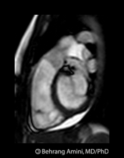
Atrial Myxoma
 Atrial myxomas are the most common primary tumor of the heart. The majority (>80%) are located in the the left atrium attached to atrial septum, usually at the fossa ovalis.
Clinical Issues
The main clinical concern in embolization, which happens more often with villous tumors (about 1/3 of myxomas are vollous). The myxoma may also become infected, resulting in septic emboli. If the myxoma obstructs the mitral or tricuspid valve, symptoms of valvular stenosis may be seen.Imaging
Echocardiography is the modality of choice for evaluation. Plain films are almost always normal. 50% of myxomas in the right atrium calcify (rare in left atrium), which may be seen on radiographs.Differential Diagnosis
- thrombosis
- metastasis
- cardiac lipoma
- lymphoma
Subscribe to:
Posts (Atom)